Introduction
Wegener's granulomatosis was first described by Heinz Klinger in 1931 and subsequently by Frederich Wegener in 1936.Reference Srouji, Andrews, Edwards and Lund1 It is a systemic disease characterised by granulomatous lesions which can affect any organ of the body. The disease may be difficult to diagnose because of its numerous manifestations and lack of specific histological findings.
The classic form of the disease is characterised by a triad comprising: necrotising, granulomatous vasculitis of the upper and lower respiratory tract; focal necrotising glomerulonephritis; and a variable degree of small vessel vasculitis.Reference Lloyd, Lund, Beale and Howard2
Early diagnosis is imperative in order to commence treatment, and is established on the basis of the clinical pattern, laboratory investigations (including urinanalysis, erythrocyte sedimentation rate (ESR) and serum antineutrophil cytoplasmic antibody (ANCA) testing) and histological evaluation. However, not all patients demonstrate the classic triad of clinical disease, and not all biopsy tissue shows the characteristic histopathological triad of vasculitis, necrosis and inflammation.
We present a case of localised Wegener's granulomatosis in a diabetic patient whose clinical picture mimicked skull base osteomyelitis (malignant otitis externa), and we discuss the diagnostic process and treatment.
Case report
A 56-year-old woman presented complaining of right-sided facial palsy for one year and decreased hearing for one month. She had been diagnosed with diabetes mellitus three years previously, and was taking oral hypoglycaemic agents.
Otoscopy revealed an intact but retracted right tympanic membrane, and a left tympanic membrane with a central perforation. On further examination, the patient had a right-sided, House–Brackmann grade II facial palsy.
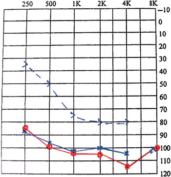
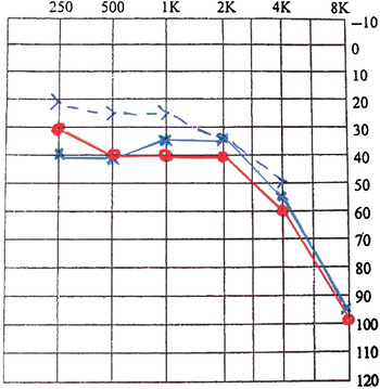
An audiogram revealed bilateral severe to profound, mixed hearing loss (Figure 1).
Pre-treatment audiogram showing bilateral, severe to profound, mixed hearing loss. > = Left ear Bone Conduction threshold; ○ = Right ear Air Conduction threshold; × = Left ear Air Conduction threshold

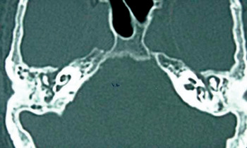
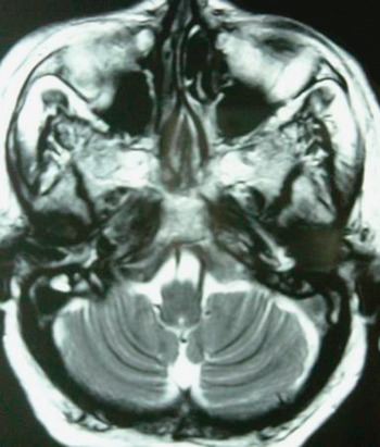
High resolution computed tomography (CT) of the temporal bone showed bilateral sclerosing mastoiditis (Figure 2). A magnetic resonance imaging (MRI) brain scan, undertaken at another institution for facial weakness, revealed ill-defined, patchy, predominantly hypointense lesions in the central and posterior skull base adjoining the clivus and jugular foramen bilaterally (Figure 3).
Axial high resolution computed tomography scan of the temporal bones, showing bilateral sclerosing mastoiditis.

Axial magnetic resonance imaging brain scan showing bilateral patchy, predominantly hypointense lesions in the central and posterior skull base adjoining the clivus and jugular foramen.

Microbial culture of an ear swab resulted in a growth which tested positive for pseudomonas. Laboratory analysis revealed an elevated ESR (65 mm at 1 hour) and an elevated C-reactive protein level (55.23 mg/l). Urinanalysis and chest X-ray were normal.
At this stage, keeping in mind the patient's history of diabetes, pseudomonal growth from the ear swab, hearing loss with facial palsy, poor general condition, high ESR and MRI scan suspicious of skull base infection, a provisional diagnosis of skull base osteomyelitis was made.
The patient was commenced on intravenous antibiotics (ceftazidime 1 g BD), based on microbial culture and sensitivity analysis. A technetium-99m bone scan was scheduled.
Treatment continued for two weeks. However, the patient's signs and symptoms showed no improvement, and her general condition remained poor. A repeated ESR test showed persistent elevation. The technetium-99m bone scan was performed but results were negative, with no evidence of abnormal increased uptake in the region of the skull base.
Given the above clinical picture, the lack of clinical improvement and the persistently raised ESR, the patient was investigated further. Further testing revealed positivity for serum antineutrophil cytoplasmic antibodies (ANCA).
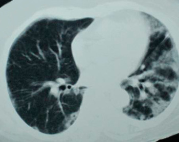
In view of the positive ANCA test, a diagnosis of Wegener's granulomatosis was made. Repeated urinalysis was negative. A chest high resolution CT scan (Figure 4) showed evidence of interstitial lung disease with upper zone predominance.
Axial high resolution computed tomography chest scan showing patchy ‘ground glass’ appearance of the lung fields, indicative of interstitial lung disease.

The patient was commenced on systemic steroid therapy, in the form of prednisolone 60 mg per day.
Over the next two weeks, the patient's general condition improved dramatically, her ESR fell, hearing in both ears improved (from bilateral, severe to profound, mixed hearing loss to bilateral, mild to moderate, conductive tending to sensorineural hearing loss) (Figure 5), and her facial palsy completely recovered.
Post-treatment audiogram showing bilateral, mild to moderate, conductive tending to sensorineural hearing loss. > = Left ear Bone Conduction threshold; ○ = Right ear Air Conduction threshold; × = Left ear Air Conduction threshold

Discussion
Wegener's granulomatosis is a systemic disease with various manifestations involving numerous organ systems. Cases presenting with otological manifestations and facial nerve palsy are uncommon.Reference Nikolaou, Vlachtsis, Daniilidis, Petridis and Daniilidis3 In such patients, a high index of clinical suspicion is thus required to reach the correct diagnosis.
Clinicians should be alert to the possibility of Wegener's granulomatosis in cases with: a constitutional upset out of proportion to the apparent intensity of the local lesion; middle-ear disease which fails to respond to conventional treatment; and a consistently raised ESR.
The course of the disease is very similar in several respects to that of skull base osteomyelitis assumed to be due to pseudomonas infection. The latter condition generally manifests in diabetic patients in the form of otological changes together with cranial nerve palsy, constitutional symptoms and a high ESR.Reference Illum and Thorling4 It is imperative to distinguish between these two diseases, as their treatment differs widely. In our patient, the presence of bilateral otological features (compared with the unilateral features usually seen in skull base osteomyelitis) and a negative bone scan helped us to exclude skull base osteomyelitis.
Once Wegener's granulomatosis is suspected, analysis for antineutrophil cytoplasmic antibodies (ANCA) should be performed. Two ANCA patterns may be seen following indirect immunofluorescence of ethanol-fixed neutrophils: a cytoplasmic pattern and a perinuclear pattern. Positivity for ANCA has been demonstrated to be highly specific for Wegener's granulomatosis.Reference Macias, Wackym and McCabe5 Serum ANCA levels have been found to correlate well with disease activity: ANCA titres fall during treatment and tend to be negative in patients with inactive disease, while a rising ANCA titre is a sensitive indicator of relapse following treatment.Reference Banerjee, Armas and Dempster6 Enzyme-linked immunoadsorbent assay techniques of ANCA analysis have a 60 per cent sensitivity in detecting cases of active locoregional (i.e. limited) disease, and 93 per cent sensitivity in cases of active, generalised disease. These figures rise to 67 and 96 per cent, respectively, if indirect immunofluorescent antibody detection is used.Reference Carrie, Hughes and Watson7 The cytoplasmic ANCA pattern is found less frequently in localised Wegener's granulomatosis (39 per cent) than in generalised Wegener's granulomatosis (86 per cent). One study assessed the utility of combined cytoplasmic and perinuclear pattern ANCA testing in detecting Wegener's granulomatosis, and found a sensitivity of 85 per cent, a specificity of 93 per cent and a positive predictive value of 52 per cent.Reference Schonermarck, Lamprecht, Csernok and Gross8 Patients positive for the perinuclear ANCA pattern have less organ involvement than those with the cytoplasmic ANCA pattern.Reference Schonermarck, Lamprecht, Csernok and Gross8 Our patient had limited Wegener's granulomatosis and a perinuclear ANCA pattern, in keeping with the literature.
Wegener's granulomatosis is a necrotising, granulomatous vasculitis with classical histopathological features. Biopsy specimens taken from the ear are often small, and it is frequently difficult to make a definitive histological diagnosis based on this evidence alone. More recently, it has been stated that a typical or complete histopathological picture is not always essential for the diagnosis of Wegener's granulomatosis, and that, if the clinical picture is suggestive of the diagnosis, a less typical histological picture may be acceptable.Reference Bradley9
Two forms of Wegener's granulomatosis have been described: (1) the classical generalised systemic or diffuse form, which always involves the kidney and causes necrotising glomerulonephritis; and (2) the localised or limited form, without upper respiratory tract or renal involvement.Reference Banerjee, Armas and Dempster6 Our patient belonged to the second category.
• Wegener's granulomatosis in a diabetic patient is reported, with features mimicking skull base osteomyelitis
• Wegener's granulomatosis is a systemic vasculitis with many ENT (and other organ) manifestations
• A high index of clinical suspicion is required to enable early diagnosis, and to distinguish from other diseases (e.g. skull base osteomyelitis in diabetic patients)
Cases of Wegener's granulomatosis which present with otological manifestations and facial paralysis are uncommon. Facial nerve paralysis may occur secondary to compression of the nerve in its middle-ear course, especially in the presence of a dehiscent fallopian canal, or may be due to vasculitis of the nerve microvasculature. The mechanism of sensorineural hearing loss in Wegener's granulomatosis is still unclear. This may be due to vasculitis of cochlear vessels, immune complex deposition in the cochlea, or granulomas directly affecting the auditory nerve.Reference Bibas, Fahy, Sneddon and Bowdler10
The early diagnosis of Wegener's granulomatosis of the ear depends on a high index of clinical suspicion. As the presented case shows, the clinical course of this condition may be very similar to that of skull base osteomyelitis, and it is important to distinguish between the two conditions. Intensive treatment with steroids and immunosuppressants should be commenced once reasonable suspicion of Wegener's granulomatosis has been established. Early diagnosis and treatment reduces the morbidity and mortality of the disease.
Conclusion
Wegener's granulomatosis is a systemic disease with various manifestations. A high index of clinical suspicion is required to reach the correct diagnosis. The clinician should be alerted to the possibility of Wegener's granulomatosis in the presence of: systemic upset out of proportion to the apparent intensity of the local lesion; middle-ear disease failing to respond to conventional treatment; and a consistently raised erythrocyte sedimentation rate.
Once Wegener's granulomatosis is suspected, analysis of antineutrophil cytoplasmic antibodies should be performed. Early diagnosis and treatment reduces the morbidity and mortality of the disease.