Introduction
Endoscopic sinus surgery is widely used in the treatment of patients with chronic rhinosinusitis. The indications for endoscopic surgery have been expanded to include other nasal diseases, such as post-operative sinus cysts, cerebrospinal fluid fistulas, blow-out fractures of the orbital floor, odontogenic maxillary cysts, decompression of Grave's ophthalmopathy, ligation of the sphenopalatine artery and resection of benign neoplasms of the nasal cavity.Reference Seno, Ogawa, Shibayama, Ogawa, Fukui and Owaki1, Reference Seno, Arikata, Sakurai, Owaki, Fukui and Suzuki2 Improvements in endoscopic technology, such as powered instrumentation and computer-assisted surgery, now enable the surgeon to perform safer and more accurate operations. Preliminary reports highlight the potential efficacy of endoscopic surgery for the treatment of carefully selected cases of malignant sinonasal tumours.Reference Stammberger, Anderhuber, Walch and Papaefthymiou3–Reference Villaret, Yakirevitch, Bizzoni, Bosio, Bignami and Pistochini13 However, the indications for endoscopic surgery as treatment for sinonasal malignancy are limited and remain undefined.
In this retrospective study, we present our experiences of endoscopic surgery used in the treatment of malignant sinonasal tumour. The endoscopic approach avoids external scars, preserves the bony structure of the facial skeleton and produces excellent functional results. The current report evaluates the outcome and morbidity of endoscopic resection with or without radiotherapy and/or chemotherapy.
Materials and methods
Surgical resections of malignant sinonasal tumours were performed in 35 patients in our university hospital, between 2000 and 2009. Classical external approaches were used in 24 patients. The transnasal endoscopic approach was performed in 11 patients, however, recurrent cases (3 patients) and palliative cases (2 patients) were excluded. The current report focuses on the remaining 6 patients with previously untreated tumours (4 men and 2 women, aged 38–76 years).
Pre-operative diagnosis testing involved endoscopic evaluation, computed tomography (CT) and magnetic resonance imaging (MRI). Endoscopic biopsies were taken from all patients. The histopathological diagnoses varied and included squamous cell carcinoma, inverted papilloma with squamous cell carcinoma, olfactory neuroblastoma, chordoma, extramedullary plasmacytoma and haemangiopericytoma. All six patients had previously been diagnosed with malignant tumour on the basis of pre-operative biopsies.
All endoscopic surgery was performed under general anaesthesia with controlled hypotension. Before the endoscopic resection, the area around the lesion was infiltrated with 0.5 per cent xylocaine with adrenaline. After making the incision around the tumour (incorporating an appropriate safety margin) using a KTP laser, the mucoperiosteal flap was elevated. The tumour and the adjacent mucosa and bone were then removed. The surgical technique varied slightly between individual patients depending on the site of tumour origin and the extent of the lesion. Frozen sections were used to determine the tumour margin. Precise visual evaluation was maintained at all stages of the operation. A CT-guided navigation system was used in four of the six cases.
Pre-operative angiographic embolisation was performed 48 hours prior to the operation in the patient with a haemangiopericytoma.
Following the endoscopic resection, all patients underwent a strict follow-up protocol consisting of endoscopic evaluation every three months. Magnetic resonance imaging and CT scans were conducted in cases where there was a clinical suspicion of tumour recurrence.
Results
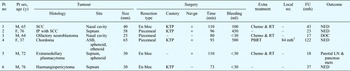
Table I summarises data on the patients, the surgery and the surgical outcomes, for the use of endoscopic surgery to treat malignant sinonasal tumours. Two patients with conditions arising from the nasal septum were treated by endoscopic resection only. Pre-operative chemotherapy and pre-operative or post-operative radiotherapy were performed in three cases. The patient with chordoma received post-operative proton beam radiotherapy. En bloc resection was completed in three cases, and piecemeal resection was performed in three cases. The post-operative course was uneventful in all cases.
Summary data: patients, surgery and outcomes*

* For the treatment of malignant sinonasal tumours using endoscopic resection.
† Treated with proton beam radiotherapy (PBRT). Pt no = patient number; y = years; rec = recurrence; FU = follow up; mth = months; navgn = navigation system; min = minutes; M = male; F = female; SCC = squamous cell carcinoma; += present; – = absent; KTP = KTP laser; Chemo = chemotherapy; RT = radiotherapy; NED = no evidence of disease; IP = inverted papilloma; ASB = anterior skull base; DOC = dead of other causes without disease; LN = lymph nodes; mets = metastases
The follow-up period ranged from 17 to 122 months (mean of 43 months). The patient with chordoma suffered a local recurrence, 84 months after the initial treatment, but this was successfully treated by additional proton beam radiotherapy. The patient with olfactory neuroblastoma died of other causes (without disease). In the patient with extramedullary plasmacytoma, metastases were found in the parotid lymph nodes and pancreas six months after the initial treatment. The other four patients were free of disease.
Case reports
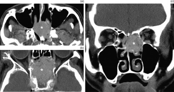
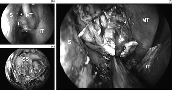
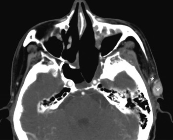
Case one (patient number 5 in Table I) was a 72-year-old man referred to our university hospital complaining of a two-month history of headache. Endoscopic examination revealed a round tumour in the posterior nasal septum. Axial and coronal, enhanced CT images showed an expansive, enhanced tumour in the posterior nasal septum and sphenoid sinus (Figure 1). Endoscopic biopsy revealed a histopathological diagnosis of extramedullary plasmacytoma. Following chemotherapy (cyclophosphamide 1200 mg, vincristine 2 mg, Adriamycin® 80 mg and prednisolone 90 mg) and radiotherapy (50 Gy), the residual tumour was completely removed by endoscopic surgery. After incising around the tumour and surrounding safety margin using a KTP laser, the mucoperiosteal flap was elevated, and blunt separation completed the en bloc removal of the tumour (Figure 2). We were able to preserve most of the mucosa of the sphenoid sinus. The post-operative course was uneventful, with no local recurrence (Figure 3). However, parotid lymph nodes metastases (Figure 3) and distant metastasis in the pancreas were found six months after the surgery.
Axial (a) & (b) and coronal (c), enhanced computed tomography images of case one showing an expansive, enhanced tumour (asterisk) in the posterior nasal septum and sphenoid sinus.

Endoscopic photograph of case one, showing: (a) tumour in the posterior nasal septum and sphenoid sinus; (b) tumour following incision and elevation of the mucoperiosteal flap; and (c) tumour removed by blunt separation, with preservation of sphenoid sinus mucosa. S = septum; MT = middle turbinate; T = tumour; IT = inferior turbinate; SS = sphenoid sinus

Axial, enhanced computed tomography image of case one six months after surgery, showing parotid lymph nodes metastases (asterisks), with no local recurrence in sphenoid sinus.

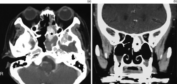
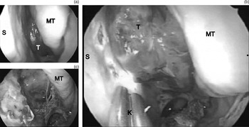
Case two (patient number 6 in Table I) was a 76-year-old man referred to our university hospital with a two-month history of epistaxis. Endoscopic examination revealed a round, red tumour in the posterior nasal septum. Axial and coronal, enhanced CT images showed the enhanced tumour arising from the posterior nasal septum, with part of the tumour invading the sphenoid sinus (Figure 4). Endoscopic biopsy enabled a histopathological diagnosis of haemangiopericytoma. Pre-operative angiographic embolisation was performed 48 hours prior to endoscopic surgery. At the operation, an incision was made around the tumour and surrounding safety margin using a KTP laser (Figure 5). En bloc removal of the tumour was then completed under endoscopic guidance. The post-operative course was uneventful, and no recurrence was noted at 37 months' follow up.
Axial (a) and coronal (b), enhanced computed tomography images of case two, showing the enhanced tumour (asterisk) in the nasal septum. Part of the tumour invades the sphenoid sinus. R = right

Endoscopic photograph of case two, showing: (a) tumour in the posterior nasal septum; (b) incision made around the tumour (using a KTP laser); and (c) complete removal of the tumour. MT = middle turbinate; S = septum; T = tumour; K = KTP laser

Discussion
Endoscopic sinus surgery has been widely used in the last two decades, and endoscopic procedures are now utilised in the treatment of many nasal diseases. Endoscopic procedures have been used in combination with conventional sublabial, transfacial and transcranial approaches for the treatment of malignant sinonasal tumours. The use of a transnasal endoscopic approach has recently been reported in the treatment of specific entities and tumours with limited extensions. Stammberger et al. reported the use of transnasal endoscopic resection for 22 malignant tumours of various histologies.Reference Stammberger, Anderhuber, Walch and Papaefthymiou3 Roh et al. treated 15 malignant tumours with curative intent using the transnasal approach.Reference Roh, Batra, Citardi, Lee, Bolger and Lanza4 More recent reports have evaluated the outcome of endoscopic resection in patients with sinonasal malignancies.Reference Nicolai, Battaglia, Bignami, Bolzoni, Delu and Khrais5–Reference Batra, Citardi, Worley, Lee and Lanza11
Transnasal endoscopic resection of sinonasal malignancy is suitable for carefully selected patients. The indications for this approach are limited and have not been defined. Patient selection is based on the site of tumour origin, the extent of the lesion, the histopathological and morphological characteristics of the tumour, and the experience of the operating surgeon. Pre-operative evaluations with endoscopy, CT, MRI and endoscopic biopsy are critical to determine the most appropriate surgical approach.
We selected patients whose tumours were limited to the nasal cavity, the ethmoid and/or sphenoid sinuses, or the medial wall of the maxillary sinus, without bone erosion at the skull base or lamina papyracea. Relative contraindications included lateral extension into the maxillary sinus and anterior–superior extension into the frontal sinus. We also excluded patients with aggressive malignant tumours such as melanoma, neuroendocrine carcinoma and undifferentiated carcinoma, because of the complexities associated with oncogenic radicality.
The main advantages of the endoscopic approach include the avoidance of facial incisions, the relatively short operation time and the reduction in blood loss. In addition, endoscopic resection allows excellent visualisation of the operating field and precise localisation of the tumour origin. There are no reported serious complications associated with craniofacial resection. The hospital stay is usually shorter than that for patients treated with the conventional external method, and post-operative radiotherapy is not delayed by wound healing.
A disadvantage of this approach is the limited accessibility to endoscopic instruments. However, the improvement in surgical instrumentation and the advent of new equipment, such as microdebriders, high-speed endonasal drills and CT-guided navigation systems, have helped to refine the surgical techniques involved. A CT-guided navigation system is useful to determine the accessibility of the tumour and to identify appropriate surgical margins. We used a CT-guided navigation system in four of our six patients.
Intra-operative bleeding is a major concern in endoscopic tumour resection. The bleeding is usually controlled by electrical cauterisation and temporary packing. We performed intra-operative endoscopic ligation of the sphenopalatine artery in our patient with squamous cell carcinoma (patient number 1 in Table I), and pre-operative angiographic embolisation was used in our patient with a haemangiopericytoma (patient number 6 in Table I). These ligation and embolisation techniques are useful to minimise the intra-operative bleeding.
• Endoscopic resection is useful and effective in some sinonasal malignancy cases
• It can be combined with chemotherapy and/or radiotherapy
• Computed tomography guided navigation systems can determine tumour accessibility and identify surgical margins
• Sphenopalatine artery ligation or angiographic embolisation can minimise bleeding
Another criticism of endoscopic resection is the potential difficulty of performing an en bloc resection. Most of the literature reports endoscopic resections of sinonasal malignancies performed by piecemeal resection.Reference Stammberger, Anderhuber, Walch and Papaefthymiou3, Reference Nicolai, Battaglia, Bignami, Bolzoni, Delu and Khrais5, Reference Nicolai, Castelnuovo, Lombardi, Battaglia, Bignami and Pianta6, Reference Lund, Howard and Wei7, Reference Kühn, Mann and Amedee9–Reference Villaret, Yakirevitch, Bizzoni, Bosio, Bignami and Pistochini13 As sinonasal malignancies are surrounded by bony structures that are a structural barrier to the invasion of tumour cells, piecemeal resection is performed with a constant, progressive assessment of tumour boundaries. If the tumour origin can be determined without debulking, en bloc resection can be performed. We completed en bloc resection in three of our six cases. However, confirmation of the site of the tumour may be more important than striving for en bloc resection. We determined surgical margins by histopathological examination of intra-operative frozen sections, in all cases. A similar strategy has been successfully used for other cases of sinonasal malignancy. McCutcheon et al. reported that the craniofacial approach with piecemeal resection did not appear to compromise survival.Reference McCutcheon, Blacklock, Weber, DeMonte, Moser and Byers14 In addition, Nishino et al. reported the efficacy of conservative piecemeal resection, in combination with radiotherapy and intra-arterial chemotherapy, for the treatment of maxillary sinus carcinoma.Reference Nishino, Miyata, Morita, Ishikawa, Kanazawa and Ichimura15
Conclusion
This preliminary report indicates that endoscopic resection of sinonasal malignancy is useful and effective, with or without adjunctive chemotherapy and/or radiotherapy, when it is properly planned and performed by experienced surgeons. Although our data include only a small number of patients with histological heterogeneity, and a limited follow-up period, we suggest that this procedure is technically feasible in carefully selected patients. It is not the intention of this report to suggest that endoscopic resection is superior to conventional external approaches. However, transnasal endoscopic resection may provide a valid alternative to these approaches in selected cases. In the future, long-term studies using a larger number of patients will be required to further validate the endoscopic approach for the management of sinonasal malignancies.