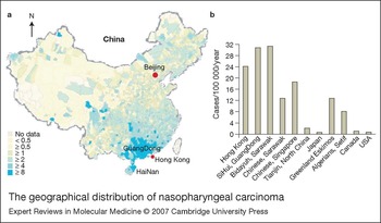


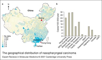
Nasopharyngeal carcinoma (NPC) is a malignancy with an unusual geographical and ethnic distribution across the world (Ref. Reference Vokes, Liebowitz and Weichselbaum1). It was documented more than 100 years ago in the endemic areas in southern China (including GuangDong province and Hong Kong), with a peak incidence of 30 cases per 100 000 per year (∼20% of all cancers), and it remains a serious healthcare problem in these regions. In Hong Kong, NPC is the fourth leading cancer in men, but the leading one in men aged 20–44 years (Hong Kong Cancer Registry 2002, The Hong Kong Hospital Authority; http://www3.ha.org.hk/cancereg/). Earlier work showed that Cantonese ‘boat people’ in southern China had the highest incidence of NPC (54.7/100 000/year), although it is impossible to identify such a pure ethnic community of Cantonese boat people nowadays. Other provinces in mainland China next to GuangDong, such as GuangXi, HaiNan, HuNan and FuJian, also have a relatively high incidence of NPC. Within China, the incidence decreases from south to north with only ∼2 cases per 100 000 per year among men in northern provinces (Ref. Reference Yu and Yuan2) (Fig. 1a).
By contrast, the incidence of NPC in neighbouring oriental countries such as Korea and Japan is very low (Fig. 1b), although in other areas of southeast Asia, including the Philippines, Malaysia and Singapore, its incidence is also relatively high (∼18/100 000/year in the Chinese population in Singapore). A previous study comparing the incidence among different ethnic groups living in Singapore (Chinese, Malays) showed that the Cantonese-speaking group had a much higher NPC incidence than that of the Teochew- or Hokkien-dialect group (Ref. Reference Ho and Ablashi3). A recent interesting study showed that a distinct ethnic group – the native Bidayuh in Sarawak, East Malaysia – has a high incidence of NPC (23.1/100 000/year), comparable with the endemic southern China areas (Ref. Reference Devi4). However, NPC is a rare disease in most countries of the world, except for an intermediate incidence in Alaskan Eskimos and northern Africans in the Mediterranean basin (∼10/100 000/year) (Ref. Reference Yu and Yuan2) (Fig. 1b). Its incidence in the USA and Canada is <1/100 000/year, representing only 0.2% of all tumours.
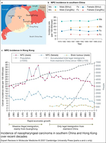
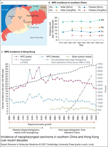
The International Agency for Research on Cancer (IARC) reported no demonstrable change in NPC incidence among Chinese in China or southeast Asia for 50 years before the early 1980s (Ref. Reference Yu and Yuan2). Recently, a widely quoted report showed that the age-standardised incidence rates of NPC have decreased during the past 25 years in Hong Kong (Ref. Reference Lee5). Similarly, in Singapore, little change in the NPC incidence rate from 1973 to 1992 was followed by a slight decrease of NPC incidence rates in both sexes in Singapore Chinese from 1993 to 1997 (Ref. Reference Yu and Yuan2). These drops in NPC incidence in recent years might be partially real, attributed to a gradual change in lifestyle, declined use of salted fish to feed young children and decreased exposure to traditional preserved food after World War II as these areas underwent rapid economic development. However, these results also need to be viewed cautiously in the context of population shifting as a result of constant immigration, as discussed for Hong Kong in detail in Figure 2c. In poorly developed, more closed Cantonese areas (e.g. SiHui in GuangDong and CangWu in GuangXi) that have much more stable populations and also a high NPC incidence, the NPC incidence rates have not changed for 35 years (Ref. Reference Jia6) (Fig. 2b). Thus, in this context, further careful epidemiological studies of the NPC incidence in Hong Kong and Singapore, which consider the possibility of population shifting, are needed.
The geographical distribution of nasopharyngeal carcinoma. (a) Nasopharyngeal carcinoma (NPC) mortality rate (male, world standardised rate) in China in the 1970s (China Cancer Database, Ministry of Science and Technology of China; http://cancernet.cicams.ac.cn). Within China, the mortality rate of NPC shows great variations from north to south. The dark-blue colour indicates regions with significantly higher mortality rates than the national average, which are mainly located in southern China. (b) The annual incidence of NPC worldwide in males. In addition to the association of high incidence of NPC with Cantonese populations, high incidence was also reported recently in the native Bidayuh in Sarawak, East Malaysia. Alaskan Eskimos and northern Africans in the Mediterranean basin show an intermediate prevalence of the disease.
Incidence of nasopharyngeal carcinoma in southern China and Hong Kong over recent decades. (a) More-detailed map of the highest nasopharyngeal carcinoma (NPC) incidence regions (within the shaded circle) in China: these are the Cantonese areas centered around SiHui and GuangZhou, including Hong Kong. (b) The annual incidence of NPC in SiHui and CangWu (1978–2002), which are the poorly developed, more closed Cantonese areas in southern China, with stable populations. The incidence of NPC (both male and female) has not changed for ∼30 years (up to 2002) in SiHui, which has the highest NPC incidence (30/100 000), and in CangWu (on the border between GuangXi and GuangDong), which has a high incidence of 20/100 000. Graph adapted from Ref. Reference Jia6, with permission from Drs W-H. Jia and Y-X. Zeng (State Key Laboratory of Oncology in Southern China, Cancer Center, Sun Yat-sen University, GuangZhou, China). (c) NPC incidence in Hong Kong (1965–2003). Interpretation of NPC incidence in Hong Kong (data from the Hong Kong Cancer Registry, The Hong Kong Hospital Authority; http://www3.ha.org.hk/cancereg/) is complicated by the waves of immigration from mainland China, which have resulted in the population in Hong Kong increasing by 49% from 3.6 million in 1965 to 6.97 million in 2006. From the mid-1960s, as a result of the Cultural Revolution, poor living conditions in China and the rapid growth of the Hong Kong economy, a huge number of illegal immigrants from mainland China flooded to Hong Kong (up to 450 per day). This immigration was mainly from the GuangDong areas near Hong Kong and thus predominantly of Cantonese origin, with a high risk of NPC. The population increased by 1 million from 1972 to 1980, which was the fastest growth period in Hong Kong history after 1945–1946, and this might explain the abnormal peak of NPC incidence in males and females during 1973–1980. By contrast, a stricter immigration policy from 1980, with only legal immigrants allowed to stay and limited issuing of visas (75/day 1983–1993, 105/day in 1993–1995, and 150/day from 1995) led to immigration from all areas of mainland China. From 1990 to 2001, a total of 533 552 such immigrants came to Hong Kong, of which ∼60% were females. It is most likely that this second peak of continuing immigration after 1990s, which was not Cantonese-dominant, diluted the high NPC incidence rate in Hong Kong and contributed to the reported decline of incidence rates in recent years (Ref. Reference Lee5). The incidence of another tumour (brain), which is not specifically associated with Cantonese, as a comparison, has been quite stable in Hong Kong for years.
Histology of NPC
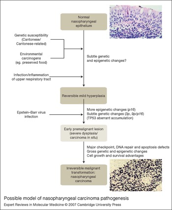
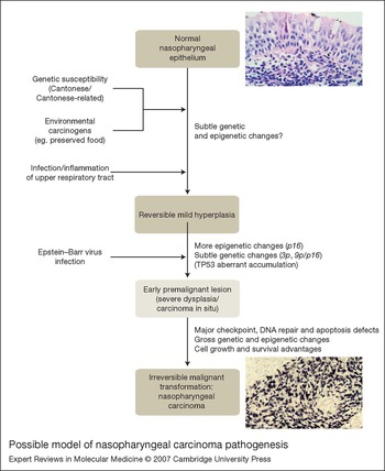
NPC is a squamous cell carcinoma that develops from the epithelium of the nasopharynx (Fig. 3); it normally originates from the fossa of Rosenmuller in the nasopharynx, often in the recess. Histologically, it has a high background of reactive lymphocytes, and used to be called lymphoepithelioma. NPC has been shown to be multifocal in origin in its early pathogenesis in some cases (Ref. Reference Sham7). As inflammation and infection are very common in the upper respiratory tract, mild hyperplasia is common in normal nasopharyngeal epithelium, and is a reversible mild lesion. Early lesions of NPC including severe dysplasia or carcinoma in situ (CIS) have been described, but are difficult to differentiate from each other and extremely rare (reported in 11 out of 5326 biopsies in one report) (Refs Reference Sham7, Reference Cheung8, Reference Pak9, Reference Pathmanathan10). At the molecular level, these lesions are infected with clonal Epstein–Barr virus (EBV) (see below) and express the same EBV viral proteins as NPC, and are therefore already clonal tumours (Ref. Reference Pathmanathan10).
Possible model of nasopharyngeal carcinoma pathogenesis. Although the whole pathogenesis of nasopharyngeal carcinoma (NPC) might take as long as ∼40 years, the transition from early premalignant lesion (severe dysplasia/carcinoma in situ) to an NPC appears to be quick (shown as lighter shading to indicate this short window), probably due to the tumour-promoting potentials of EBV infection. The image of normal nasopharyngeal mucosa is a haematoxylin- and eosin-stained section (method in Ref. Reference Tao33; magnification 400x) kindly provided by Professor Zifen Gao (Department of Pathology, Peking University Health Science Center, China). The image of NPC tumour tissue is a digoxigenin-labelled in situ hybridisation of Epstein–Barr virus EBER-RNA (method in Ref. Reference Tao34; magnification 200x), showing an island of EBER-positive NPC tumour cells, kindly provided by Professor Gopesh Srivastava (Department of Pathology, University of Hong Kong).
The World Health Organization (WHO) classification system (1978) acknowledges three types of NPC based on the differentiation status of tumour cells: type I, differentiated, keratinising squamous cell carcinoma (25% of NPCs in North America; ∼2% of NPCs in southern China) (Refs Reference Marks, Phillips and Menck11, Reference Wei and Sham12); type II, nonkeratinising carcinoma (12% of NPCs in North America; ∼3% of NPCs in southern China); and type III, undifferentiated carcinoma (63% of NPCs in North America; 95% of NPCs in southern China). WHO types II and III can be considered together as undifferentiated carcinomas of the nasopharyngeal type (UCNT), which have similar treatment response and prognosis.
Aetiology of NPC
Although the molecular basis of NPC pathogenesis is still poorly understood, it has been suggested that the pathogenesis and development of NPC is a multistep process (Refs Reference Lo, To and Huang13, Reference Young and Rickinson14) (Fig. 3), similar to that of other carcinomas as elegantly proposed by Vogelstein and Kinzler (Ref. Reference Kinzler and Vogelstein15). Previous epidemiological studies suggest three major aetiological factors for NPC: genetic susceptibility, early-age exposure to chemical carcinogens (particularly Cantonese salted fish), and latent EBV infection (Refs Reference Yu and Yuan2, Reference Raab-Traub16, Reference Tao17).
Genetic susceptibility
The predisposition to NPC among southern Chinese (Cantonese) strongly suggests the involvement of both genetic susceptibility and environmental factors. The high NPC incidence is retained by second-generation Cantonese who migrate to other, nonendemic countries. The NPC clustering in families in endemic areas further suggests a strong ethnic and genetic influence (Refs Reference Jia18, Reference Zeng and Jia19). In fact, ∼10% of NPC cases have a family history of the disease.
Earlier linkage study on Chinese sib pairs with NPC identified an NPC susceptibility locus at the HLA (human leukocyte antigen) region (Ref. Reference Lu20). Study of HLA susceptibility among Chinese has shown that people with HLA A*0207 or B*4601, but not A*0201, have an increased NPC risk (Ref. Reference Hildesheim21). Recent genome-wide linkage analyses of high-risk Chinese familial NPC pedigrees identified two candidate NPC susceptibility loci – 4p15.1-q12 (Ref. Reference Feng22) and 3p21.3 (Ref. Reference Xiong23) – with another suspected locus reported at 5p13-15 [L.F. Hu (MTC, Karolinska Institute, Sweden), pers. commun.]. However, since these chromosomal regions are very large with many candidate genes, the relevant NPC gene(s) are still far from identified.
Environmental factors
For many years, NPC has been reported to be associated with environmental factors other than EBV. The most relevant environmental exposure among Cantonese is salted fish and other preserved foods containing volatile nitrosamines, which are mutagenic chemicals (Ref. Reference Mirvish24). Consumption of salted fish during childhood is related to increased NPC risk in southern Chinese (Refs Reference Yu and Yuan2, Reference Ho, Biggs, de The and Payne25). This has been experimentally supported by animal-model studies showing that rats fed on a diet of salted fish developed carcinomas in the nasal cavity in a dosage-dependent manner (Refs Reference Huang26, Reference Yu27). A recent review of extensive epidemiological literature outlined the association between adulthood intake of preserved and nonpreserved vegetables and NPC risk in multiple countries (Ref. Reference Gallicchio28). Preserved vegetable intake is associated with a twofold increase in NPC risk, while high nonpreserved vegetable intake is associated with a 36% decrease, consistent between vegetable types and countries.
Occupational exposure to toxic pollutants (formaldehyde) in the air or wood dust is also linked to increased NPC incidence (Refs Reference Armstrong29, Reference Hildesheim30). A very recent report on female textile workers in Shanghai, China also revealed increased NPC risk with accumulative exposure to cotton dust, acids, caustics or dyeing processes (Ref. Reference Li31). Tobacco smoking is also mildly associated with increased NPC risk (Ref. Reference Yu and Yuan2). These studies indicate that a prolonged exposure to pollutants contributes to NPC pathogenesis. Other environmental factors associated with NPC risk have also been reported, such as Chinese herb usage, presence of heavy metal (nickel) in the endemic area, alcohol and even fungus infection in the nasal cavity (Ref. Reference Yu and Yuan2).
Epstein–Barr virus (EBV) infection
Unlike all other head and neck squamous cell carcinomas, NPC is strongly associated with EBV, a human B-lymphotropic herpesvirus that is associated with a variety of tumours (Refs Reference Raab-Traub16, Reference Tao17). The link between NPC and EBV was discovered in 1966 from serological studies, and later supported by the demonstration of EBV DNA and EBV nuclear antigen (EBNA) proteins in NPC tumour cells (Ref. Reference zur Hausen32). Clonal viral genome was detected in NPC tumour DNA by Southern blot hybridisation, suggesting that EBV infection occurred before clonal expansion of tumour cells (Ref. Reference Raab-Traub16). It is proposed that EBV plays an important role (probably indispensable in endemic areas) in NPC pathogenesis, since, as mentioned above, the earliest NPC lesion detected (severe dysplasia/CIS) is already EBV-positive, with latent and clonal viral genomes and expressing viral oncoproteins such as latent membrane protein 1 (LMP1) (Ref. Reference Pathmanathan10), while EBV infection is not present in normal nasopharyngeal epithelium nor in low-grade dysplasia (Refs Reference Tao33, Reference Tao34).
The WHO type III NPC is always associated with EBV, regardless of geographical distribution, ethnic origin and local prevalence of the disease (Refs Reference Raab-Traub16, Reference Tao17). The other two types of NPC are less frequently associated in nonendemic areas, with EBV positivity in less than 50% of cases (Refs Reference Raab-Traub16, Reference Nicholls35); however, in endemic NPC areas (Malaysia and Hong Kong), all the type I NPC tumours were EBV-associated (Refs Reference Raab-Traub16, Reference Nicholls35, Reference Pathmanathan36, Reference Zhang37). In NPC tissues, EBV exists in every tumour cell in a latent form, but rarely in the surrounding normal cells (Refs Reference Tao33, Reference Nicholls35, Reference Sam38).
The pattern of EBV latent gene expression in normal and tumour cells can be classified into different forms of latency. The pattern in EBV-transformed lymphoblastoid cell lines (LCLs) is often referred to as latency III, with the expression of six different EBNAs (EBNA1, EBNA2, 3A, 3B, 3C and -LP) from the EBV Cp (and/or Wp) promoter, three LMPs (LMP1, 2A and 2B), the BamHI A rightward transcripts (BARTs), and two abundant small nonpolyadenylated (noncoding) RNAs (EBER 1 and 2) (Refs Reference Young and Rickinson14, Reference Tao17). However, the EBV gene expression pattern in most tumours is either latency I or latency II, except for post-transplant lymphomas and some AIDS-associated lymphomas, which show a latency III pattern. Latency I is characterised by restricted viral gene expression involving only EBNA1, EBERs, LMP2A and BARTs, as observed in Burkitt lymphoma, gastric carcinoma and possibly normal peripheral blood mononuclear cells (PBMCs). Latency II is present in most EBV-positive tumours, including NPC, Hodgkin lymphoma, nasal natural killer (NK)/T-cell lymphoma and some T-cell lymphomas, with the expression of EBNA1 (from the Qp promoter), EBERs, BARTs, BARF1 (except for Hodgkin lymphoma), LMP1 and LMP2 (Refs Reference Young and Rickinson14, Reference Tao17). Recently, multiple EBV-encoded microRNAs (up to 17) have also been detected in EBV-infected B cells and carcinomas including NPC and gastric carcinoma, although the pathobiological functions of these small regulatory RNA molecules are still not very clear (Refs Reference Cai165, Reference Pfeffer166).
In NPC (latency II), EBNA1 and EBERs are expressed in all EBV-positive NPC cases, while expression of the oncoprotein LMP1 is highly variable (Ref. Reference Tao17); this is different from Hodgkin lymphoma and nasal NK/T-cell lymphoma, which almost always express LMP1 (Refs Reference Murray39, Reference Tao40, Reference Chiang41). Expression of EBV viral oncoproteins would confer tumour cell growth and survival advantages, maintaining the malignant phenotype; however, the rate of LMP1 detection in NPC tumour tissues varies substantially (∼20–60% of cases) (Refs Reference Tao17, Reference Pathmanathan36). The only EBV-positive NPC cell line, C666-1, barely expresses LMP1 and the promoter is methylated (Refs Reference Tao17, Reference Cheung42). Expression of LMP2A mRNA has been reported by reverse-transcriptase PCR and more recently by sensitive immunostaining (Ref. Reference Heussinger43). The expression of BARTs and BARF1 [a transforming EBV oncogene (Ref. Reference Sheng44)] was detected in virtually all NPC tumours, suggesting a pathogenic role for these RNAs/proteins in NPC (Refs Reference Chen45, Reference Decaussin46). The expression of the viral early lytic protein BZLF1 has been reported in some cases (Ref. Reference Cochet47), although there is virtually no virus replication in NPC tumour cells, indicating that viral lytic activation, if it exists, is abortive in tumour cells.
Somatic genetic and epigenetic alterations
Cytogenetic changes
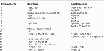
Multiple genetic changes are present in NPC. Earlier cytogenetic analyses of NPC cell lines and xenografts showed hypo- or hyperdiploidy, with further chromosomal abnormalities such as deletion of 3p also observed (Refs Reference Huang48, Reference Huang49). Later, spectral karyotyping was used to characterise the genome-wide genetic alterations in NPC cell lines (Ref. Reference Wong50). Microsatellite analyses also revealed various regions with frequent allelic imbalance in primary tumours (Refs Reference Lo51, Reference Huang52). Comparative genomic hybridisation (CGH) can detect genetic lesions more accurately than conventional cytogenetics, and was also used in NPC (Ref. Reference Wong50). Through these studies, numerous genetic abnormalities have been detected on multiple chromosomal regions in NPC tumours and cell lines. Several minimal deleted regions were mapped to 3p14.1-22, 11q13.3-24, 13q14.3-22, 14q24.3-32.1 and 16q22-23.
As the best resolution of conventional CGH is only ∼10 Mb (Ref. Reference Huang53), higher-resolution array-based CGH has been recently used to detect genome-wide genetic abnormalities in NPC. A commercial CGH microarray containing 58 known oncogenes commonly amplified in cancers was applied to 15 NPC samples (Ref. Reference Hui54). Amplification of a series of oncogenes, including MYCL1, TERC, PIK3CA, NRAS and MYB, was detected. A high-density cDNA microarray containing 21 632 cDNAs was also used to examine gene copy number changes and expression levels in five Chinese NPC cell lines, with the detection of amplification and overexpression of the oncogene EVI1, as well as deletion and downregulation of the transcription regulator RYBP (Ref. Reference Guo55). A recent study using both conventional CGH and array-based CGH detected chromosomal alterations in a series of Mediterranean NPC xenografts and biopsies, including frequent gains associated with overexpression at 1q25-qter and 12p13, and losses at 11q14-q23, 13q12-q31, 14q24-q31 and 3p13-p23. Compared with Asian NPC, Mediterranean NPC has higher frequencies of 1q gain and 13q loss (Ref. Reference Rodriguez56). Another study, using 3 Mb array-based CGH (University of California at San Francisco arrays) containing 1803 BAC (bacterial artificial chromosome) clones was performed on three NPC cell lines, two xenografts and 21 primary tumours (Ref. Reference Hui57). Frequent gains on 1q, 3q, 8q, 11q, 12p and 12q, and losses on 3p, 9p, 11q, 14q and 16q, were found. Moreover, several minimal regions of gains including 3q27.3-28, 8q21-24 and 11q13.1-13.3 were identified, with cyclin D1 (CCND1) verified as an amplified and overexpressed oncogene at 11q13.3. Recently, we also examined the genome-wide chromosomal alterations of NPC cell lines using high-resolution (1 Mb) array-based CGH with Sanger Institute whole-genome arrays containing 3040 BAC clones (Ref. Reference Hurst58) (http://www.sanger.ac.uk/Projects/Microarrays/) (Refs Reference Seng59, Reference Ying60, Reference Ying61). Multiple alterations were detected, notably deletions of 3p12-14, 8p22, 10p and 18q and amplification of 3q26. These loci provide us with good candidate regions for the further identification of candidate tumour suppressor genes and oncogenes associated with NPC.
Table 1 summarises the major genetic alterations commonly detected by microsatellite marker analysis, CGH and array-CGH in NPC cell lines and primary tumours.
Major genetic alterations detected by microsatellite marker analysis, CGH and array-CGH in NPC cell lines and primary tumoursa

aData from Refs Reference Lo, To and Huang13, Reference Tao17, Reference Xiong23, Reference Huang48, Reference Huang49, Reference Wong50, Reference Lo51, Reference Huang52, Reference Huang53, Reference Hui54, Reference Guo55, Reference Rodriguez56, Reference Hui57, Reference Seng59, Reference Ying60, Reference Ying61.
Abbreviations: CGH, comparative genomic hybridisation; NPC, nasopharyngeal carcinoma.
Oncogenes
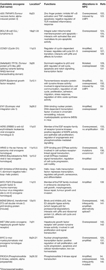
Specific amplifications in NPC identified by cytogenetic or array-based CGH studies have implicated several putative oncogenes in NPC. Oncogenes identified include BCL2, CCND1, EGFR, EVI1, HER2/ERBB2, HRAS, NRAS, MDM2, MYC and PIK3CA, which may show amplification, overexpression or gain-of-function mutations (summarised in Table 2). For some of these genes, such as CCND1, encoding cyclin D1 (Ref. Reference Hui57), functional studies have demonstrated their oncogenic potential in NPC cells.
Candidate oncogenes involved in nasopharyngeal carcinoma pathogenesis

aChromosomal location.
Abbreviations: EGF, epidermal growth factor; FGF, fibroblast growth factor; LMP1, latent membrane protein 1; MAPK, mitogen-activated protein kinase; NF-κB, nuclear factor κB; pRB, retinoblastoma protein; TLR, Toll-like receptor; TNF, tumour necrosis factor.
Tumour suppressor genes (TSGs)
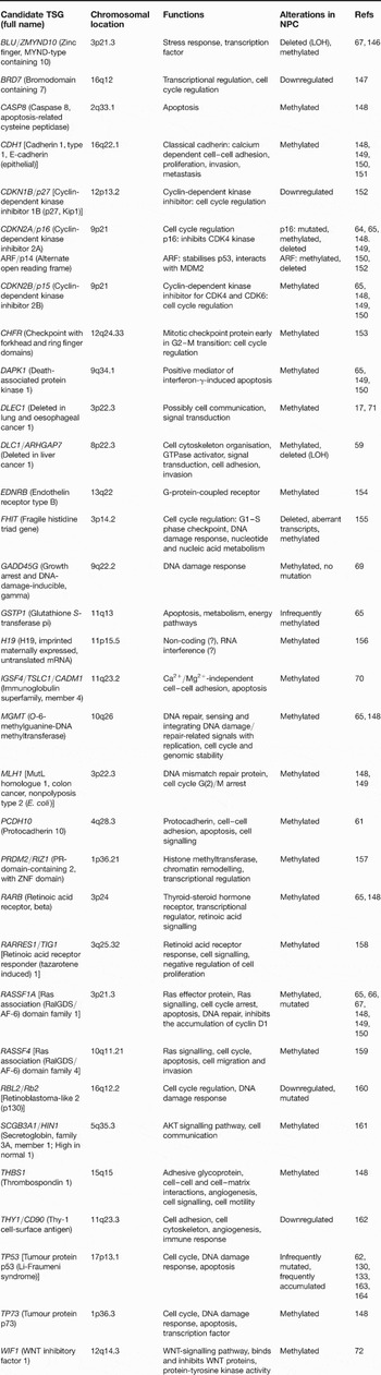
Although alterations of the well-established TSGs (such as TP53, encoding p53, and RB1, encoding the retinoblastoma protein) are relatively rare in NPC tumours (but relatively more common in NPC cell lines) (Refs Reference Effert62, Reference Sun, Hegamyer and Colburn63), multiple genetic and epigenetic abnormalities of various other TSGs have been detected. The most commonly affected TSG regions are 9p21 and 3p, which have as high as 85–95% deletion rates in invasive tumours. TSGs affected include those at 9p21 (p16, p15 and p14ARF) (Refs Reference Huang52, Reference Lo64, Reference Kwong65) and 3p21.3 (RASSF1A, BLU/ZMYND10 and CACNA2D2) (Refs Reference Lo66, Reference Qiu67), which were found to be defective as a result of either promoter methylation, or deletions and mutations or both (Table 3). Ectopic expression of p16, RASSF1A or BLU/ZMYND10 in NPC cell lines lacking expression resulted in significant inhibition of cell growth, colony formation or tumour growth in immunodeficient animals (Refs Reference Tao17, Reference Chow68), providing strong evidence that these genes function as tumour suppressors for NPC.
Candidate tumour suppressor genes involved in nasopharyngeal carcinoma pathogenesis

Abbreviations: LOH, loss of heterozygosity; WNT, wingless-type MMTV integration site family member.
Through more extensive epigenetic studies, many more putative TSGs were found to be silenced by epigenetic alterations in NPC than previously thought, indicating that the molecular pathogenesis of NPC is rather complex. The epigenetic abnormalities of GADD45G (9q22.2) (Ref. Reference Ying69), TSLC1 (11q23.3) (Ref. Reference Lung70), DLC1 (8p22) (Ref. Reference Seng59), DLEC1 (3p22.3) (Refs Reference Tao17, Reference Kwong71), WIF1 (12q14.3) (Ref. Reference Chan72), CHFR, PRDM2/RIZ1, RARRES1/TIG1, SCGB3A1/HIN1, THY1/CD90, and even new genes such as PCDH10 (4q28.3) (Ref. Reference Ying61), have been increasingly reported (Table 3). These abnormalities would disrupt multiple normal cellular regulatory and signalling processes and contribute to the pathogenesis of NPC. Moreover, the methylation of these TSGs could serve as epigenetic biomarkers, as indicated below.
Clinical implications – molecular diagnosis and therapy
Molecular diagnosis
As early detection of NPC will greatly help the treatment and improve the survival of patients, various detection systems have been developed for the early molecular diagnosis of this tumour by taking advantage of the specific presence of EBV. Higher EBV antibody titres, particularly of the IgA class, occur in approximately 90% of NPC patients, but in less than 10% of normal people. These antibody levels rise with the tumour burden regardless of different geographical locations and ethnic groups, which can be seen several years prior to the development of NPC, and correlate with tumour remission and recurrence (Ref. Reference Henle and Henle73). Furthermore, the high copy number of circulating, cell-free EBV DNA in the peripheral blood sera of NPC patients has been successfully used as a powerful diagnostic tumour marker (Refs Reference Chan74, Reference Lo75). Quantitative analysis of cell-free EBV DNA in plasma samples of NPC patients is found to be highly sensitive and specific (96% and 93%, respectively).
In addition, quantitative analysis of EBV DNA or carcinoma-specific viral BARF1 mRNA, or even promoter methylation of multiple TSGs, from brushing samples collected directly from the nasopharynx has been shown to be a noninvasive and sensitive diagnostic test for NPC, allowing direct investigation of disease progression in the nasopharynx (Refs Reference Tune76, Reference Stevens77, Reference Tong78).
In the near future, these molecular diagnostic systems will help to detect early tumour development, monitor early recurrence, and predict prognosis and treatment response of NPC patients.
Radiotherapy and chemotherapy
Following the definitive diagnosis of NPC by endoscopic biopsy, magnetic resonance imaging is the standard imaging for local disease and facilitates high-precision radiotherapy planning, the mainstay treatment for NPC. With the use of intensity-modulated radiation therapy (IMRT), the local control rate exceeds 90% (Refs Reference Lee79, Reference Kam80). However, for locoregionally advanced stages, at which most patients present, there remains significant rates of distant metastases, and attempts to improve outcome by the addition of systemic chemotherapy have met with mixed success. Randomised Phase III studies of pure neoadjuvant or adjuvant chemoradiotherapy have not resulted in improvement in overall survival compared with radiotherapy alone. However, randomised studies of concurrent chemoradiotherapy with or without adjuvant chemotherapy have consistently resulted in improvement in progression-free and overall survival (Refs Reference Rossi81, Reference Chan82, 83, Reference Chua84, Reference Al-Sarraf85, Reference Al-Sarraf86, Reference Ma87, Reference Chi88, Reference Chan89, Reference Chan90, Reference Lin91, Reference Kwong92, Reference Wee93, Reference Lee94). A meta-analysis using original data of 1753 patients demonstrated a highly significant benefit from concurrent chemoradiotherapy (Ref. Reference Baujat95). Hence, the current standard for locoregionally advanced patients is concurrent cisplatin–IMRT with or without adjuvant cisplatin–5-fluorouracil (5-FU). The use of neoadjuvant chemotherapy remains experimental.
Despite improvements in the primary treatment, 30% of patients with locoregionally advanced disease will subsequently fail with distant metastases (Ref. Reference Hui96). The median survival figure after distant metastases is around 9–12 months and is influenced by disease, treatment and patient characteristics. Cisplatin-based chemotherapy is the standard first-line treatment, with response rates of up to 80% (Ref. Reference Ma and Chan97). Novel therapeutics using targeted agents are being actively investigated and early results using the monoclonal antibody against epidermal growth factor receptor (EGFR) (Cetuximab) are promising (Refs Reference Ma and Chan97, Reference Chan98).
Novel therapies
Since EBV is present exclusively in every NPC tumour cell and rarely in normal cells (Refs Reference Tao17, Reference Tao34), it is not only a very specific diagnostic biomarker but also a specific therapeutic target. Various approaches in immunotherapy, gene therapy and epigenetic therapy have been developed to target EBV in NPC cells. Epigenetic changes in TSGs might also provide therapeutic opportunities.
EBV-based immunotherapy
HLA class I-restricted cytotoxic T lymphocytes (CTLs) play a major role in controlling EBV infections, and also in the pathogenesis of EBV-associated tumours including NPC. Adoptive transfer of EBV-specific CTLs has been used for the treatment of EBV-positive tumours, such as post-transplant lymphoproliferative disorders (PTLDs) (Refs Reference Rooney99, Reference Papadopoulos100, Reference Khanna101). As only LMP2A and LMP1, but none of the immunodominant EBV antigens (EBNA2, EBNA3 family), are expressed in NPC (latency II), which is different from the situation in PTLDs (latency III), immunotherapy targeting EBV in NPC might not work as well as in PTLDs. The first trial in NPC patients with advanced disease showed that infusion with autologous EBV CTLs was safe, and led to increased CTL levels and a reduced plasma EBV level, indicating that immune intervention of NPC patients is feasible (Ref. Reference Chua102). Adoptive transfer of allogeneic EBV-specific CTLs boosted the LMP2-specific immune response in an NPC patient with advanced-stage disease (Ref. Reference Comoli103). Later, when ten patients with stage IV NPC received autologous EBV-specific CTLs (which were reactivated and expanded ex vivo from PBMCs stimulated with EBV-transformed autologous LCLs), control of disease progression and increased frequency of LMP2-specific immunity were observed (Ref. Reference Comoli104). A recent trial of using autologous CTLs to treat NPC patients showed remarkable antitumour results. At 19 to 27 months after infusion, four out of ten patients treated in remission from locally advanced disease remained disease-free, while other patients with refractory disease had either complete responses and remained in remission or partial remission or stable disease; only two patients had no response (Ref. Reference Straathof105). Therapeutic vaccination with LMP2-peptide-pulsed dendritic cells also boosted epitope-specific CD8+ T-cell responses and resulted in partial tumour reduction in some NPC patients (Ref. Reference Lin106). The recently developed dual antigen stimulation strategy using a chimaeric construct (part of EBNA1 fused with LMP2) to boost both CD4 + and CD8 + EBV-specific T-cell responses will be of better therapeutic value to NPC (Ref. Reference Taylor107). Thus, the use of EBV-specific immunotherapy along with other standard therapies in the future treatment of NPC looks promising.
Epigenetic therapy
CpG methylation plays important roles in NPC pathogenesis, including the silencing of TSGs and EBV immunodominant antigens (EBNA2, 3A, 3B, 3C) (Refs Reference Ambinder, Robertson and Tao108, Reference Tao and Robertson109). It also suppresses LMP1, immediate-early lytic antigens Zta and Rta, and lytic cycle viral kinases, and thus is an important regulatory process in the EBV life cycle and latency in tumour cells (Refs Reference Tao17, Reference Tao and Robertson109). As CpG methylation is reversible with pharmacological demethylation using epigenetic agents, epigenetic treatment can be explored either as a novel therapeutic strategy or as a combinational intervention with other therapies. Reactivating methylated and silenced cellular genes (mostly TSGs) and immunodominant tumour/viral antigens would be expected to restore normal cell growth control, induce apoptosis in tumour cells, or induce cell immunity. Demethylation would also reactivate the expression of EBV early and lytic genes in latently infected NPC cells, which will lead to further tumour cell death. Furthermore, the lytic cycle viral kinases involved in the phosphorylation of ganciclovir and other antiviral nucleoside analogues that are of therapeutic value to NPC (see below) will also be activated (Refs Reference Moore110, Reference Westphal111).
Epigenetic agents include DNA methyltransferase inhibitors – nucleoside analogues such as 5-aza-2'-deoxycytidine/Decitabine/DAC, 5-azacitidine and the newly developed Zebularine (Ref. Reference Cheng112) – and various histone deacetylase (HDAC) inhibitors (TSA, SAHA and PXD101). Earlier clinical trials using such agents have been carried out on cancer patients with colon, head, neck, renal and lung tumours, with only a partial response observed in some patients (Refs Reference Abele113, Reference van Groeningen114). In our recent clinical trial of azacitidine in patients with NPC and EBV-positive AIDS-associated Burkitt lymphoma (Ref. Reference Chan115), all the latent and early lytic EBV promoters [Cp, Wp, LMP1p (ED-L1), Zp, Rp] examined in post-treatment tumour samples showed significant demethylation, with the reactivation of viral antigen expression (Zta) detected by immunostaining. This study demonstrated the potential of epigenetic therapy for NPC.
EBV-based gene therapy
As targeting EBV is tumour-specific, gene therapy strategies with EBV-specific targeting to deliver cytotoxic proteins have been developed for NPC and other EBV-associated tumours. The EBV C promoter has been used to drive the expression of suicide genes (thymidine kinase), as EBV-specific targeting therapy (Ref. Reference Franken116). Moreover, the utility of EBV oriP to drive p53 (Ref. Reference Li117) or a mutant noncleavable form of FasL (Ref. Reference Chia118) in a replication-deficient adenovirus vector system has also been explored. EBV itself can also be used as a gene therapy vector, with the incorporation of therapeutic genes into a transformation-defective EBV deleted for transforming genes (EBNA2, LMP1) (Ref. Reference Delecluse and Hammerschmidt119). The major problem of gene therapy is how to target every individual tumour cell, although activation of EBV kinases (or delivery of thymidine kinase by gene therapy) together with ganciclovir and AZT will result in bystander killing to surrounding tumour cells (Refs Reference Moore110, Reference Westphal111, Reference Ambinder120).
Conclusions
There has been impressive achievement in understanding the molecular pathogenesis of NPC. Like other solid tumours such as colon, cervical and lung cancers, for which stepwise pathogenic models have been proposed (Refs Reference Kinzler and Vogelstein15, Reference Minna, Roth and Gazdar121), the pathogenesis of NPC is also thought to be a multistep process: indeed, a similar model of NPC pathogenesis has been suggested (Ref. Reference Lo, To and Huang13), although most of the details of individual steps in the model are still missing. The increasing number of genetic and epigenetic changes uncovered by recent studies (Tables 1, 2 and 3) suggests that NPC pathogenic events are more complex than initially thought. Furthermore, NPC has pathobiological features distinct from other solid tumours, suggesting different pathogenic mechanisms, which may not be exactly stepwise as in the linear progression model applied to other common cancers such as colon and cervical cancers. For example, NPC shows a unique peak incidence age plateau of ∼40–50 years, which is distinct from the constantly increasing incidence of colon, lung and gastric cancers with age. As mentioned earlier, NPC does not have clearly defined early pathogenic steps as seen in other common carcinomas like colon, cervical or lung carcinoma. The premalignant/CIS lesion is very rare (<1/1000 cases), and most diagnosed NPC tumours are either preinvasive or invasive lesions. As NPC is strongly viral driven, it is conceivable that early EBV infection in premalignant NPC cells would promote and speed up the malignant transformation to a full-scale carcinoma, leaving only a very short window of premalignant/CIS lesion that is not easily detectable.
Nevertheless, it is now believed that a combination of environmental, genetic, epigenetic and viral factors drives the normal nasopharyngeal epithelial cells to preinvasive, then invasive tumour stages, as marked by various genetic and epigenetic abnormalities (Fig. 3). However, many questions remain to be addressed. When does EBV infect normal nasopharyngeal epithelium? Mild hyperplasia is common and also reversible in normal nasopharyngeal epithelium: is there any subtle genetic and epigenetic change in these lesions? Is the EBV gene expression pattern in the early infection of nasopharyngeal epithelial cells the same as that in NPC? Why is it so difficult to establish EBV-positive NPC cell lines and EBV is lost in virtually all the NPC cell lines except for C666-1, which is a special subline from a xenograft in athymic nude mice?
As for other solid tumours, early diagnosis is still the key to treatment success for NPC. Progress in the molecular studies of this tumour should lead to novel diagnostic and therapeutic strategies, as already indicated by approaches arising from EBV and epigenetic studies of this tumour.
Acknowledgements and funding
We thank: Prof. Gopesh Srivastava and Prof. Zifen Gao for the histology images in Figure 3 (see legend for details); Wei-Hua Jia and Yi-Xin Zeng (State Key Laboratory of Oncology in Southern China, Cancer Center, Sun Yat-Sen University, China), and Lai Shui Ling Karen and Lee Wai Yee (Department of Clinical Oncology, Chinese University of Hong Kong) for providing some NPC epidemiology information of China and Hong Kong; and Yan Cui, Jianming Ying, Ka Man Ng and Kwan Yeung Lee for helping with the references. We are also grateful to the peer reviewers for their helpful comments. The authors are supported by the Michael and Betty Kadoorie Cancer Genetics Research Program (MBKCGRP) Fund, HK RGC Earmarked Grant (CUHK4443/05M) and the Chinese University of Hong Kong.