Introduction
Somatic cell nuclear transfer (SCNT) is a technique in which the nucleus of a somatic cell is transferred to the cytoplasm of an enucleated oocyte, fused and activated. Then the reconstructed embryo is transferred to the oviduct or uterus of a female recipient, and the SCNT embryo can develop into a newborn. With this technique, a terminally differentiated cell can be reprogrammed into a pluripotent embryonic state. SCNT has shown impressive results when applied for example in the breeding of top-quality livestock, the production of transgenic animals, the rescue of endangered animals, and stem cell therapy (Qu et al., Reference Qu, Wang, Zhang and Liu2020a). However, the overall success rate of the somatic cell cloning technology is very low, and the frequency of abnormalities in cloned offspring is high, which limits the application of this technology (Matoba and Zhang, Reference Matoba and Zhang2018). Compared with the fertilized embryo, cloned embryos manifest abnormal epigenetic modification, incomplete reprogramming, a high apoptosis rate and other inferior developmental properties such as low pregnancy rate, fetal placental abnormalities, large-offspring syndrome, immunodeficiency, and early death in the pups (Matoba and Zhang Reference Matoba and Zhang2018; Gouveia et al., Reference Gouveia, Huyser, Egli and Pepper2020). In SCNT embryos, sperm plays no role at all, and we hypothesize that the low efficiency of SCNT might be related to the absence of sperm roles.
Traditionally, sperm is assumed to mainly provide paternal haploid chromatin for the fertilized embryo and to activate the oocyte (Tosti and Ménézo, Reference Tosti and Ménézo2016; Parrington et al., Reference Parrington, Arnoult and Fissore2019). Regulatory factors that guide the development of the embryo after fertilization are mainly provided by the oocyte (Gonzalez-Munoz and Cibelli, Reference Gonzalez-Munoz and Cibelli2018), therefore most previous studies have focused on oocytes to find and identify key regulatory factors related to epigenetic reprogramming and embryonic development. SCNT embryos show an abnormal epigenetic state and incomplete reprogramming. The level of histone acetylation in SCNT embryos is low, and histone deacetylase inhibitors are used to increase the level of histone acetylation, significantly improving SCNT efficiency in some mammals. Tri-methylation of lysine 9 on histone H3 (H3K9me3) plays an important role in modulating cell fate control (Nicetto and Zaret, Reference Nicetto and Zaret2019). A high level of H3K9me3 modifications in SCNT embryos can be adjusted by the overexpression of demethylase (KDM4A or KDM4D) or the downregulation of methyltransferase (Suv39h1 and Suv39h2), which significantly improves the efficiency of SCNT. In recent years, with a deepening of our understanding of sperm, it has been found that proteins, mRNAs, miRNAs and other non-coding RNAs derived from sperm are introduced into the oocyte, where they play an important role in pronucleus formation, cleavage and the activation of the zygote’s genes, and even influence the phenotype of offspring (Zhang et al., Reference Zhang, Shi, Rassoulzadegan, Tuorto and Chen2019; Qu et al., Reference Qu, Wang, Zhang and Liu2020a).
Rabbits are very important laboratory animals in the study of cardiovascular diseases, nervous and mental disorders, cancer and reproductive diseases. After mice and rats, the laboratory rabbit is the third most often used experimental mammal (Honda and Ogura, Reference Honda and Ogura2017; van der Merwe et al., Reference van der Merwe, van der Veeken, Ferraris, Gsell, Himmelreich, Toelen, Ourselin, Melbourne, Vercauteren and Deprest2019; Castellini et al., Reference Castellini, Mattioli, Bosco, Cotozzolo, Cartoni Mancinelli, Rende, Stabile and Pistilli2020). The study of SCNT has a long history in rabbits, and the micromanipulation procedure for mammalian nuclear transfer has been primarily based on research in rabbits (Bromhall, Reference Bromhall1975). The first cloned rabbit was reported in 2002: six rabbits were successfully cloned using cumulus cells as nuclear donors, but the cloning efficiency only was 1.6% (Chesné et al., Reference Chesné, Adenot, Viglietta, Baratte, Boulanger and Renard2002). In this study, to explore the effect of small RNAs in sperm on the efficiency of SCNT, rabbit sperm small RNA was extracted and then injected into cloned embryos. Then, in vitro experiments were carried out to analyze the level of H3K9me3, the blastocyst apoptosis rate and the developmental proficiency of pre-implantation cloned embryos in rabbit.
Materials and methods
Materials
All chemicals and reagents were obtained from Sigma Aldrich (St. Louis, MO, USA) unless specifically stated otherwise.
Sperm collection
Semen was obtained using the false vagina method. A gradient centrifugation medium was prepared as follows: 2 ml of 80% Percoll was added to the bottom of a 15-ml centrifuge tube, and then 2 ml of 40% Percoll was added over the 80% layer. After semen liquefaction, 2 ml of seminal plasma was added to the gradient centrifugation medium, which was centrifuged at 300 g for 20 min. The semen and 40% Percoll layers were sucked out and, after addition of 4 ml of phosphate-buffered saline (PBS), were centrifuged at 300 g for 10 min. The supernatant was aspirated, and then the sediment, which contained sperm, was collected.
Isolation of small RNAs
Isolation of small RNAs was carried using the miRNeasy Micro Kit (QIAGEN, Düsseldorf, Germany) as reported previously. Briefly, TRIzol was preheated to 60°C, and an aliquot of 500 μl TRIzol was added to the samples. Samples were thoroughly mixed by blowing air down a pipette tip, vortexed for 2 min, and incubated at room temperature (RT) for 10 min. Then β-ME was added and the suspension was incubated at 65°C for 40 min, while vortexing to mix. The cells were homogenized by vortexing for 1 min, then the tube was kept at RT for 5 min, after which 140 µl of chloroform was added. The tube was shaken vigorously for 15 s and held at RT for 3 min, then centrifuged for 15 min at 12,000 g at 4°C. After centrifugation, the upper aqueous phase was transferred to a new collection tube; 1.5 volumes of 100% ethanol were added and mixed thoroughly. Up to 700 µl of the sample was pipetted into an RNeasy MinElute spin column in a 2 ml collection tube and centrifuged at 8000 g for 15 s at RT. Then 500 µl Buffer RPE was pipetted onto the spin column and the column was centrifuged for 15 s at 8000 g to wash the column. Subsequently, 500 µl of 80% ethanol was pipetted onto the spin column and centrifuged for 2 min at 8000 g to wash the spin column membrane. The spin column was placed into a new 2 ml collection tube and centrifuged at full speed for 5 min to dry the membrane. The spin column was placed in a new 1.5 ml collection tube, 14 µl RNase-free water were added directly to the centre of the spin column membrane which was then centrifuged for 1 min at full speed to elute the small RNA. The quality of the small RNA was assessed using a NanoDrop spectrophotometer, as described previously. The harvested small RNA was stored at −80°C for RT-PCR or microinjection.
Collection of oocytes
Female rabbits were injected subcutaneously with 80 IU pregnant mare serum gonadotropin (PMSG; SHUSHENG, Ningbo, China) and, after 96 h, 100 IU of chorionic gonadotropin (CG; SHUSHENG) were injected into the ear vein. At 12 h later, the female rabbits were anaesthetised. The fallopian tube was washed with 5 ml of Dulbecco’s phosphate-buffered saline (DPBS) containing 5% fetal bovine serum (FBS). The flushing solution was placed in a 60 mm dish, and oocytes were collected under a stereomicroscope. The oocytes were treated with hyaluronidase for 5 min, and cumulus cells surrounding the oocytes were removed by gently blowing with a pipette. Then the oocytes were transferred to DPBS containing 5% FBS and placed on a hot stage of 37°C for SCNT.
Somatic cell nuclear transfer
Donor cells were prepared as previously reported (Jia et al., Reference Jia, Ding, Shen, Luo, Zhang, Zhou, Ding, Qu and Liu2019). Fibroblasts were cultured to confluency in 96-well plates. The cells were digested with DPBS containing 0.25% (w/v) trypsin, and the reaction terminated by adding Dulbecco’s modified Eagle’s medium containing 10% FBS. Then they were centrifuged at 350 g for 10 min to obtain a cell pellet for SCNT.
Metaphase II (MII) oocytes with first polar bodies were stained with 10 μg/ml Hoechst 33342 for 10 min. Oocytes were placed into M2 medium supplemented with 7.5 μg/ml cytochalasin B (CB) and 10% FBS. Each oocyte was held with a micropipette, and the polar body and the surrounding cytoplasm was removed using another micropipette. The extracted cytoplasm was examined under ultraviolet illumination in another microdrop to confirm successful enucleation. A donor cell was injected into the perivitelline space of the enucleated oocyte, and the complexes of enucleated oocyte and the donor cell were transferred into Cytofusion Medium C (BTX; Holliston, MA, USA). Next, the complexes were fused by applying three direct current (DC) pulses of 3.2 kV/cm for 20 μs each. After fusion, the reconstructed embryos were washed three times for 3 min and allowed to recover for 1 h at 38.5°C and 5% CO2 in air. Then the reconstructed embryos were activated by a combination of electrical stimulation and chemical treatment. The electrical stimulation parameters were the same as those used for fusion. Once the electrical stimulation was completed, the reconstructed embryos were treated with chemical activation medium, which contained 2 mmol/l 6-dimethylaminopurine (6-DMAP) and 5 μg/ml cycloheximide (CHX) for 1 h. SCNT embryos were then cultured in an incubator at 38.5°C and 5% CO2 in air.
Microinjection
Microinjection was performed as reported previously: the injection concentration of sperm small RNAs was adjusted to 100 pg/μl; 2 μl of small RNA solution was loaded into the injection needle and 2 pl of small RNAs were injected into the reconstructed embryo once after activation (Qu et al., Reference Qu, Zuo, Liu, Niu, Zhang, Du, Ma, Qiao, Wang, Zhang, Qing and Wang2019). The injected embryos were then transferred to preheated culture medium and cultured in an incubator at 38.5°C and 5% CO2 in air.
RT-PCR and qPCR
The RT-PCR and qPCR procedure were as described previously (Ma et al., Reference Ma, Zhang, Qiao, Wang, Zuo, Zhang and Wang2018). Reverse-transcription was performed using the Mir-X miRNAs First-Strand Synthesis Kit (Takara, Tokyo, Japan). Two sperm-borne small RNAs, miR-34c and miR-125b, were quantified using the Mir-X™ miRNA qRT-PCR TB Green™ Kit (TaKaRa) and the StepOne Plus Real-Time PCR System (Thermo Fisher Scientific, Waltham, MA, USA). U6 RNA was used for data normalization of the small RNA.
Immunofluorescence staining of H3K9me3
Embryos were incubated in 10 mg/ml Pronase E to remove the zona pellucida and the mucin membrane. Embryos were then fixed with 4% paraformaldehyde for 30 min, washed with 0.2% polyvinyl alcohol and PBS (PVA-PBS), treated with 0.1% Triton X-100 for 20 min, and washed with 0.2% PVA-PBS. The embryos were acidified with hydrochloric acid for 30 min, and then blocked with 5% BSA for 30 min. After that, embryos were incubated at 4°C for 12 h in a 1:200 dilution of H3K9me3 antibody (ABclonal Technology, Wuhan, China). The embryos were washed three times with 0.2% PVA-PBS and transferred to the diluent containing an immunofluorescence second antibody (Beyotime Biotechnology, Beijing, China), at a dilution ratio of 1:200, and incubated at RT for 1 h. The embryos were then transferred to DAPI and incubated for 15 min. Subsequently, embryos were washed and placed onto glass slides, pressed with a cover glass and sealed with nail polish. Slides were examined and photographed by fluorescence microscopy. The relative level of H3K9me3 was analyzed using Image Pro-Plus software (Media Cybernetics, Silver Spring, USA).
Apoptosis detection
The terminal deoxynucleotidyl transferase dUTP nick end labelling (TUNEL) kit (Promega, Madison, WI, USA) was used to analyze the apoptotic cells from the blastocysts. Embryos were fixed with 4% paraformaldehyde for 1 h. The embryos were then washed with 0.2% PVA-PBS. Embryos were equilibrated in E-buffer for 5 min. Following this step, all subsequent steps needed to be executed in the dark. The embryos were transferred to apoptotic dye solution at 37°C for 1 h. Here, 51 μl dye solution contained 45 μl of E-buffer, 5 μl of nucleotide mix and 1 μl of rTDT. SSC (2×) was used to terminate the reaction after 15 min. DAPI staining was then carried on for 3 min. The embryos were washed, transferred to a glass slide, pressed with a cover glass and sealed with nail polish. Then they were examined and photographed using fluorescence microscopy, and analyzed using Image Pro-Plus software.
Statistical analysis
Each experiment was repeated at least three times. The difference between cleavage ratio and blastocyst ratio was analyzed using the chi-squared test. Relative expression of the miR-34c and miR-125b, relative level of the H3K9me3, apoptosis index, and total cell number of the blastocysts were analyzed by one-way analysis of variance. SPSS software was used for statistical analysis.
Results
High expression levels of miR-34c and miR-125b in rabbit sperm
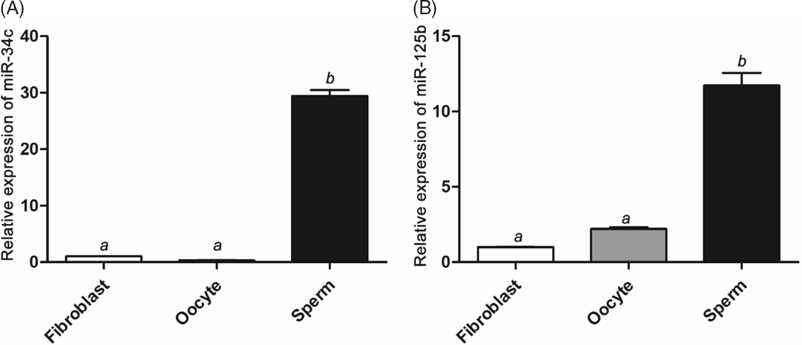
Small RNAs extracted from sperm, fibroblasts and stage MII oocytes were reverse transcribed and quantified. Results showed that the relative expression levels of miR-34c (Fig. 1 A) and miR-125b (Fig. 1 B) in sperm were significantly higher than those in oocytes and fibroblasts (P < 0.05).
Relative expression levels of miR-34c and miR-449b in fibroblast, sperm and oocyte. (A) Quantitative polymerase chain reaction (qPCR) analysis of relative expression levels of miR-34c in fibroblast, sperm, and oocyte. (B) qPCR analysis of relative expression levels of miR-449b in fibroblast, sperm, and oocyte. a,bDifferent letters above the bars indicate significant differences (P < 0.05).

Sperm small RNA downregulates H3K9me3 levels in rabbit SCNT embryo at the 8-cell stage
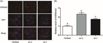
The H3K9me3 levels of embryos at the 8-cell stage were analyzed in each group, and results showed that the level of H3K9me3 in the NT-C group was significantly higher than that in the fertilized group, and that sperm small RNA significantly decreased the H3K9me3 level of SCNT embryos (P < 0.05; Fig. 2).
The level of H3K9me3 in fertilized and SCNT embryos at the 8-cell stage. (A) The staining pattern of tri-methylation of lysine 9 on histone H3 (H3K9me3) in fertilized and somatic cell nuclear transfer (SCNT: NT-C, control; NT-T, treated) embryos at the 8-cell stage. H3K9me3: red; DNA: blue. (B) Relative fluorescence intensities of H3K9me3 at the 8-cell stage. a–cDifferent letters above the bars indicate significant differences (P < 0.05).

Sperm small RNA reduces the apoptosis index of rabbit SCNT blastocysts
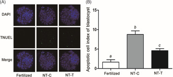
The apoptosis index of rabbit SCNT blastocysts was analyzed in each group, and the result showed that apoptosis index of blastocysts in the NT-C group was significantly higher than that in the fertilized group, and that sperm small RNA significantly reduced the apoptosis index of rabbit SCNT blastocysts (P < 0.05; Fig. 3).
Apoptotic index in Day 4 blastocysts in the fertilized, NT-C and NT-T groups. (A) Apoptotic blastomeres were detected by terminal deoxynucleotidyl transferase dUTP nick end labelling (TUNEL) (green). DNA was stained with 4′,6-diamidino-2-fenylindole (DAPI) (blue) to visualize all blastomeres. (B) Apoptotic index in blastocysts of the fertilized, NT-C (nuclear transfer control) and NT-T (nuclear transfer treated) groups. a–cDifferent letters above the bars indicate significant differences (P < 0.05).

Sperm small RNA increases the total cell number of blastocysts
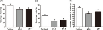
The cleavage ratio (Fig. 4 A) and the blastocyst ratio (Fig. 4 B) of the three groups were analyzed. Results showed that the cleavage ratios of the NT groups were significantly lower than that of the fertilized group (P < 0.05), and that there was no significant difference between the NT-C group and the NT-T group (P > 0.05). The blastocyst ratios of the NT groups were significantly lower than that of the fertilized embryo (P < 0.05). There was no significant difference between the NT-C group and the NT-T group (P > 0.05). The total cell number of SCNT embryos was significantly lower than that of fertilized embryos, and sperm small RNA injection could significantly increase the total cell number of SCNT blastocysts (P < 0.05; Fig. 4 C).
Embryo development ratios and total cell number of blastocysts in fertilized, NT-C and NT-T groups. (A) Cleavage ratios at 24 h in the fertilized, NT-C (nuclear transfer control) and NT-T (nuclear transfer treated) groups. (B) Blastocyst ratios on Day 4 in the fertilized, NT-C and NT-T groups. (C) Total cell number of blastocysts in the fertilized, NT-C and NT-T groups. a–cDifferent letters above the bars indicate significant differences (P < 0.05).

Discussion
Since the birth of the first cloned mammal, ‘Dolly’ the sheep in 1997, various strategies have been adopted to improve cloning efficiency (Wilmut et al., Reference Wilmut, Schnieke, McWhir, Kind and Campbell1997; Matoba and Zhang, Reference Matoba and Zhang2018). The potential of the SCNT embryo could be improved by serum starvation or contact inhibition of the donor cell (Miyamoto et al., Reference Miyamoto, Hoshino, Minami, Yamada and Imai2007; Song et al., Reference Song, Liu, He, Wang, Li, Li, Li, Jiang and Huan2017). Treatment of the donor cells or SCNT embryo with chemical drugs, such as valproic acid, trichostatin A, 5-azacytidine, and others, can rectify abnormal epigenetic modification of the SCNT embryo, and improves the efficiency of animal cloning (Selokar et al., Reference Selokar, St John, Revay, King, Singla and Madan2013; Jennane et al., Reference Jennane, El Haddad, Mahtat, Doghmi and Mikdame2017). Alteration of the expression of some epigenetic-related genes can also be beneficial for the reprogramming of SCNT embryos (Liu et al., Reference Liu, Wang, Gao, Su, Zhang, Xing, Zhou, Yao, An and Zhang2018). In addition, improving the culture medium of SCNT embryos or optimizing the procedure of nuclear transplantation can also improve the SCNT embryo’s development potential (Qu et al., Reference Qu, Wang, Zhang and Liu2020a). Despite a large number of attempts, the efficiency of nuclear transplantation is still very low, indicating that some important regulatory factors may be neglected. Compared with the fertilized embryo, sperm-derived regulators are completely absent in SCNT embryos, which may play an important role in SCNT embryo development. Here, to test this presumption, we studied the effect of sperm-borne small RNA on the development of SCNT pre-implantation embryos.
It has been reported that each mature sperm contains about 10–50 fg of RNA, and that there are approximately 24,000 small RNAs within each normal human spermatozoon (Chen et al., Reference Chen, Yan, Cao, Li, Zhang, Shi, Feng, Peng, Zhang, Zhang, Qian, Duan, Zhai and Zhou2016a; Parthipan et al., Reference Parthipan, Selvaraju, Somashekar, Kolte, Arangasamy and Ravindra2015). Recent studies have reported that sperm RNAs have a direct causal role in transferring acquired traits across generations in mammals by regulating gene expression and the epigenetic state (Schuster et al., Reference Schuster, Tang, Xie, Ortogero, Yuan and Yan2016; Skinner et al., Reference Skinner, Ben Maamar, Sadler-Riggleman, Beck, Nilsson, McBirney, Klukovich, Xie, Tang and Yan2018). For example, a high-fat diet changes the expression profiles and modifications of sperm RNA, and this change can cause metabolic disorders in the F1 offspring (Chen et al., Reference Chen, Yan and Duan2016b). Chronic paternal stress alters the profiling of sperm microRNAs, reducing the reactivity of the hypothalamic–pituitary–adrenal stress axis in the offspring (Rodgers et al., Reference Rodgers, Morgan, Leu and Bale2015). The miRNA family is the best characterized of the small RNAs. The effect of miRNAs is generally post-transcriptional-mediated degradation, through their interaction with the 3′ untranslated region. A previous study found that the miRNA profile was different between sperm with a high and sperm with a low fertilization capacity (Sun et al., Reference Sun, Zhang, Li, Xue, Niu, Shi, Li, Wang and Wang2016). It reported that 31 differentially expressed miRNAs were identified in sperm treated with fluoride, and the target genes were mainly involved in protease inhibitor activity, apoptosis, ubiquitin-mediated proteolysis, and signalling pathways of calcium, which were directly related to sperm quality (Sun et al., Reference Sun, Zhang, Li, Xue, Niu, Shi, Li, Wang and Wang2016). Changes in the external environment, such as the accumulation of some toxic substances or traumatic stress, can alter the miRNA profile in sperm, and this will be inherited across generations, therefore changing the stress and metabolic response of offspring (Gapp et al., Reference Gapp, Jawaid, Sarkies, Bohacek, Pelczar, Prados, Farinelli, Miska and Mansuy2014). Small RNAs in sperm clearly play important roles during embryo development, and even affect the offspring (Chen et al., Reference Chen, Yan, Cao, Li, Zhang, Shi, Feng, Peng, Zhang, Zhang, Qian, Duan, Zhai and Zhou2016a). In this study, small RNAs were isolated, and two highly expressed and conservative miRNAs (miR-34c and miR-125b) were selected for quantification and qualification. The result, that miR-34c and miR-125b were highly expressed in rabbit sperm, was consistent with previous reports on mice and bovines (Liu et al., Reference Liu, Pang, Chiu, Wong, Lao, Lee and Yeung2012; Schuster et al., Reference Schuster, Tang, Xie, Ortogero, Yuan and Yan2016; Zhang et al., Reference Zhang, Qu, Zhou, Liu, Ma, Wang, Wang, Su, Liu and Zhang2017).
Our previous study found that injection of sperm-derived RNA into cloned embryos can affect cytoskeleton remodelling and nuclear reprogramming (Qu et al., Reference Qu, Zuo, Liu, Niu, Zhang, Du, Ma, Qiao, Wang, Zhang, Qing and Wang2019). In this study, we found that injecting sperm small RNAs can significantly decrease the H3K9me3 level at the 8-cell stage in rabbit. Embryonic genome activation (EGA) is the most important event during reprogramming, and EGA occurs around the 8-cell stage in rabbit embryos. A high level of H3K9me3 is considered to be a serious obstacle for the reprogramming of cloned embryos. Reducing H3K9me3 by upregulating KDM4d (a specific H3K9me3 demethylase) or downregulating SUV39h1 (a methyltransferase-mediated H3K9me3) can effectively improve animal cloning efficiency (Liu et al., Reference Liu, Wang, Gao, Su, Zhang, Xing, Zhou, Yao, An and Zhang2018). Our previous study found that miR-125b targets and downregulates SUV39h1, and therefore improves cloning efficiency (Zhang et al., Reference Zhang, Qu, Zhou, Liu, Ma, Wang, Wang, Su, Liu and Zhang2017). Based on the result that miR-125b was highly expressed in rabbit sperm, we speculated that sperm-borne small RNAs have a positive role in the process of reprogramming by downregulating H3K9me3.
A previous study reported that miR-34c is important for the fertilized embryo by modulating Bcl-2 expression in mice (Liu et al., Reference Liu, Pang, Chiu, Wong, Lao, Lee and Yeung2012). Overexpression of miR-34c influences the cleavage, epigenetic reprogramming and apoptosis of embryos (Liu et al., Reference Liu, Pang, Chiu, Wong, Lao, Lee and Yeung2012). In this study, we found that miR-34c was highly expressed in rabbit sperm, and injection of sperm-borne small RNAs can reduce the apoptosis index of SCNT blastocysts, indicating that sperm-borne small RNAs have a positive role in decreasing apoptosis in cloned embryos. The apoptosis index and total cell number of blastocysts are important indicators to evaluate the developmental potential of embryos. Embryos with a lower apoptosis index and a higher total cell number are considered to have a higher development potential (Fouladi-Nashta et al., Reference Fouladi-Nashta, Alberio, Kafi, Nicholas, Campbell and Webb2005; Qu et al., Reference Qu, Shen, Du, Qin, Luo, Fu, Dong, Guo, Hu, Xue and Liu2020b). In this study, SCNT embryos injected with sperm-borne small RNAs showed a lower apoptosis index and a higher total cell number of the blastocyst, indicating that sperm-borne small RNAs could effectively improve the developmental ability of early cloned embryos.
In conclusion, this study indicated that sperm-borne small RNAs can decrease the H3K9me3 level of SCNT embryos at the 8-cell stage, reduce the apoptosis index of SCNT blastocysts, and significantly improve the potential of pre-implantation cloned embryos in rabbit. Our study not only demonstrates that sperm-borne small RNAs have an important role in embryo development but also provides a new strategy for improving the efficiency of SCNT in rabbit.
Financial support
This work was supported as part of a project funded by China Postdoctoral Science Foundation under grant no. 2020TQ0240 and by the Natural Science Foundation of Shaanxi Province under grant no. 2020PT-001.
Conflicts of interest
The authors have no conflicts of interest to report regarding the present study.
Ethical standards
The Animal Care and Use Committee of Xi’an Jiaotong University approved all animal use procedures applied in this study.