Introduction
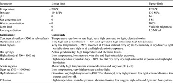
Cyanobacteria are photosynthetic microbes, which are found in extreme terrestrial habitats near the limits of life in cold deserts on the surface of the Earth (Friedmann Reference Friedmann1982; Overmann et al. Reference Overmann, Sandmann, Hall and Northcote1993; Siebert et al. Reference Siebert, Hirsch, Hoffmann, Gliesche, Peissl and Jendrach1996; Vincent & James Reference Vincent and James1996). These cyanobacteria possess a remarkable suite of survival mechanisms that makes them prime candidates for the potential colonization of the surface of early Earth and analogous early Mars (Wynn Williams et al. Reference Wynn Williams, Edwards and Garcia Pichel1999). On both these planets, they would have had similar needs for ultraviolet protection, so that protective pigmentation is a key biomarker for surface life to be able to exploit solar radiation as the optimal source of energy for metabolism. It is interesting to note that the key to survival of these extremophilic colonies is exactly that predicted by Charles Darwin in his book, On the Origin of Species by Natural Selection, first published in 1859 wherein he postulates that their ability to adapt to their environment is most critically important. A summary of the environmental parameters constraining the survival of life in stressed terrestrial locations is provided in Table 1.
Environmental parameters constraining life processes

Cyanobacterial survival mechanisms for dealing with ultraviolet stress include a variety of biochemical and avoidance strategies that result in communities that are stratified along gradients of light, moisture and temperature. The spatial distribution of pigments in these communities is as important as their concentration. Analysis of spatial structure in situ is a key advantage of laser Raman spectroscopy over analyses which requires disruption of the communities and extraction of their pigments. On lakebeds in Antarctic oases that are analogous to paleolakes on Mars (Wharton et al. Reference Wharton, Crosby, McKay and Rice1995; Grin & Cabrol Reference Grin and Cabrol1997; Doran et al. Reference Doran, Wharton, Des Marais and McKay1998) stromatolites comprise organic mats sandwiched between thin layers of sediment (Wharton Reference Wharton, Bertrand-Sarfati and Monty1994, Table 2). On nearby exposed rock ridges, endolithic cyanobacteria or algae associated with fungi (often as lichens) are found in layered communities up to 10 mm inside the translucent rock. They are often hidden from the surface as cryptoendoliths (Friedmann Reference Friedmann1982; Friedmann et al. Reference Friedmann, Hua and Ocampo-Friedmann1988). As these communities depend in part on protection by mineral substrata (Clark Reference Clark1998), either by burial or as the outer crust of the endolithic habitat, they would have been protected from subsequent environmental stresses after their extinction.
If burial was combined with freezing in permafrost and subsequent further deposition of wind-borne debris, it is possible that their residual biomolecules and associated mineral trace fossils (Friedmann & Weed Reference Friedmann and Weed1987) would have been preserved. The re-distribution of minerals such as iron and calcium is readily demonstrated by Raman spectroscopy of the habitat profile (Edwards et al. Reference Edwards, Russell and Wynn-Williams1997). Porphyrins that are derivatives of chlorophyll from ancient photosynthetic organisms are preserved in oil-bearing shales (Huseby et al. Reference Huseby, Barth and Ocampo1996) and have a distinctive Raman spectrum (Czernuszewicz et al. Reference Czernuszewicz, Rankin and Lash1996). The search for evidence of life on Mars could therefore profitably focus on detecting preserved biomolecules from former microbial communities by Raman spectroscopy (Fig. 6). Current technology precludes deep drilling on Mars, so any anaerobic chemolithotrophs dependent on oxidation–reduction gradients for energy would be inaccessible and the residues of putative cyanobacterial analogues near the surface would therefore be a more feasible target.
Raman spectroscopy was first proposed as a potential technique for remote planetary analysis by two groups, Wang et al. (Reference Wang, Jolliff and Haskin1995) concerned with lunar robotic analyses and Wdowiak et al. (Reference Wdowiak, Agresti and Nfirov1995) concerned with its development for planetary landers. A series of workshops charted the course of the developing Raman systems with a primary emphasis on the analysis of minerals (Treado & Treiman Reference Treado, Treiman, Wdowiak and Agresti1996; Haskin et al. Reference Haskin, Wang, Rockow, Joliff, Korotev and Viskupic1997; Israel et al. Reference Israel, Arvidson, Wang, Pasteris and Jolliff1997; Wdowiak et al. Reference Wdowiak, Agresti, Nfirov, Kudryavtsev, Clifford, Treiman, Newsom and Farmer1997). However, to date a Raman spectrometer was never included in the instrumentation package of a Martian lander mission, but it has now been adopted by an European Space Agency (ESA)/National Aeronautics and Space Administration (NASA) consortium as a part of the Pasteur suite for the forthcoming ExoMars-C mission due for launch in 2018 specifically to detect life signatures on the surface and subsurface of Mars. An additional point of relevance is that the miniaturized Raman spectrometer on the ExoMars-C mission will be a first-pass instrument, which relies upon the proven ability of the technique to determine the key spectral signatures of biological and biogeological materials that are characteristic of extant or extinct life in the presence of their host mineral matrices without pre-treatment or chemical extraction of specimens; a particular advantage of the Raman spectroscopic technique here is the recording of spectral signatures over a wide range of wavenumbers from host mineral oxides, sulphates, sulphides, phosphates, carbonates and silicates through to the vibrational modes of chemically functional groups such as OH, NH, C = O, C = C and CH. The specificity of the molecular spectral signals in the Raman spectrum for important biochemicals such as carotenoids, proteins, aromatic and aliphatic compounds along with minerals often in complex admixture renders the technique invaluable for unambiguous materials characterization that is critically important for remote operation and adoption of database search engines.
The first application of Raman micro-spectroscopy to the non-destructive analysis of an Antarctic endolith (Edwards et al. Reference Edwards, Russell, Seaward, Wynn-Williams and Armstrong1995) illustrates the principles, which makes the technique so useful for this purpose. Hitherto, chemical information about the endolith community and its biogeological system depended on the physical extraction of material using a dental pick; this method destroyed the specimen for further study and also meant that information about the interaction between the biology and geology, the key to biological survival in stressed environments, was lost. There is now a significant literature on the Raman spectra of key biochemicals produced in response to extremophilic organism survival strategies in stressed terrestrial environments that are considered to be Mars analogue sites, such as hot and cold deserts (in the Arctic, Antarctic and Atacama Desert for example), which manifest extremes of desiccation, low wavelength high energy radiation insolation, extremes of pH and low temperatures. In this paper, we shall demonstrate the versatility of the Raman spectroscopic technique for space mission applications through examples of case studies of life signature detection in the following scenarios:
• detection of key protective pigmentation chemicals against ultraviolet-radiation in extremophilic epilithic lichens in the maritime Antarctic exposed to ozone atmospheric depletion,
• the identification of carotenoids in admixture in Arctic snow algae on a glacier surface,
• characterization of porphyrin and carotenoid pigments along with mineral components in a geological stromatolic niche,
• identification of a halotolerant biological colony as a subsurface inclusion in sulphate crystals from a meteoric impact crater, and
• the identification of a range of biological protectant molecules in the cyanobacterial colonization of a volcanic basalt vacuole from the high-Arctic region.
Raman spectroscopy
Salient features relating to extremophilic chemical detection
The Raman spectra reported in this paper were all recorded in the laboratory using specimens obtained from several terrestrial cold desert sites; the instrumentation necessarily involved a range of radiation illumination and collection conditions and laser excitation wavelengths through the visible and into the near infrared regions of the electromagnetic spectrum. This has provided some useful information that will inform the correct selection of operational procedures and instrument design for remote miniaturized Raman spectroscopic instrumentation installed on the future rover and lander vehicles on planetary surfaces. For example, it is realized that not one single wavelength of excitation will be universally acceptable for the Raman spectroscopic detection of the mineral and organic components associated with the biological colonization of geological host matrices by extremophilic organisms found terrestrially or putatively on our neighbouring planets and their moons. So, whereas Raman excitation in the green region of the visible spectrum (at 514 or 532 nm) is found to be eminently satisfactory for the detection of carotenoids, particularly the conjugated skeletal C = C and C–C vibrational modes, because of the electronic resonant enhancement of the Raman scattering intensity by several orders of magnitude, this advantage is lost when the excitation wavelength is changed to the near infrared at 785 and 1064 nm. Paradoxically, the illumination of biological material in the green region of the spectrum often results in the generation of a strongly fluorescent background emission that swamps the weaker Raman scattering – which can be exacerbated by the adoption of alternative laser excitation of lower incident intrinsic energy in the near infrared where theoretical considerations advise that the Raman scattering intensity is significantly weaker by a factor of the fourth power of the excitation wavelength. Hence, excitation of the Raman spectrum of a material using a near ultraviolet wavelength of 250 nm gives a Raman spectrum that is some 300 times stronger than its counterpart excited using 1000 nm radiation, all other instrumental factors being equal; however, we would anticipate that the fluorescence emission would also be much more likely to be generated by exposure of the material to the low wavelength radiation.
Secondly, the Raman scattering intensity produced from a range of materials is dependent on several factors, including the irradiance (power density in W m−2) of the incident laser beam at the sample, but a delicate balance must be achieved between a very high incident laser irradiance and the potential for sample damage through attendant thermal degradation which is especially relevant for sensitive biological specimens and where the use of microscope objectives to increase the laser irradiance into small specimen spatial footprints which may only be of the order of several μm3 in dimension. A major advantage of Raman spectroscopy over many other analytical techniques is the ability to record the unique spectral signatures of inorganic and organic compounds in the same spectrum, non-destructively and without the involvement of chemical or mechanical pre-treatment or separation processes. The presence of extremophilic colonization of geological matrices is evident from their adaptation of their host environment, with the production of protective chemicals and waste materials and the biogeological modification of the minerals concerned; the importance of the latter has only recently been recognized and will form a vital cognitive strategy in future database construction for the automatic recognition of the presence of life in extreme environments.
A third point of importance relates to the detection of life signature biochemicals when life itself has become extinct, i.e. the chemical traces of previous life are still recognizable in the now-dead mineral matrix; in this both organic biochemicals and the biogeological components that have been synthesized by the living colonies now assume utmost importance. However, two spectroscopic factors need to be appreciated here, namely, that not all Raman-active chemical functionalities have the same scattering effectiveness through their molecular scattering coefficients in demonstrating their presence in a spectrum to an observer and this will dictate a range of sensitivities for the technique which will be achievable practically. For example, the C = O bond, an important chemical functionality in the organic biochemistry is weak in the Raman effect, whereas the C = C is very strong; hence, any system that has the same molecular ‘concentration’ of both functionalities will show the presence of the C = C unsaturation at much lower levels of detection. Generally, minerals such as calcite, gypsum and cinnabar will have extremely strong Raman spectral features, which are normally much more prevalent than their biochemical counterparts. It must also be realized that biochemicals produced in extremophilic scenarios are themselves subject to degradation by extremes of radiation exposure, temperature, geological processes and desiccation, so making an assessment of the capability of the analytical techniques employed to detect changes in their composition vital.
Experimental instrumentation
Raman spectra reported here were recorded using the following laboratory-based laser excitation wavelengths: 488, 514, 532, 633, 785 and 1064 nm, extending from the blue to the near infrared regions of the electromagnetic spectrum. The first prototype miniaturized Raman spectrometer to be constructed by the ESA for its ExoMars mission under the AURORA programme, specifically to search for evidence of life signatures (extinct or extant) on Mars utilized a 660 nm red laser as a result of early recommendations. This has now been refined to a second prototype that has been constructed using 532 nm green excitation. This prototype RLS spectrometer that is now being evaluated and tested with synthetic and natural samples is being measured against the laboratory instrumentation and a range of biological and non-biological specimens, which have relevance to the geology of Mars as has been divined from the NASA probes to the planet's surface.
A novel piece of equipment on the ExoMars rover vehicle is the drill, which will penetrate the surface of Mars to a depth of approximately 2 m and will obtain samples from this depth for analysis at the surface in the Pasteur instrumentation suite, following grinding and pulverizing, of which the RLS spectrometer is a component part. The underlying idea is to circumvent the heavily oxidizing and chemically corrosive surface peroxides and perchlorates produced by the high energy radiation insolation at the Martian surface, which is expected to be terminally destructive to life signature chemicals there. Hence, it is also important to evaluate the RLS instrument in the analysis of powdered samples that may contain mixtures of organic chemicals of significance to life detection interspersed with mineral component particles and to eventually examine these materials after exposure in a terrestrial Mars chamber that simulates the atmospheric and surface geological composition of Mars.
Extremophile Scenarios for Raman Spectroscopic Evaluation
Antarctic cold deserts
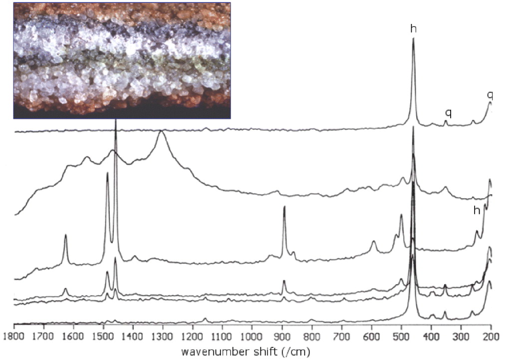
The specimen shown in Fig. 1 was collected by Dr David Wynn-Williams of the British Antarctic Survey, Cambridge, at Beacon Valley in the Dry Valleys in Antarctica, the coldest desert on Earth, during the summer season of 2002. The sandstone shows a normal orange-red colouration externally, but a depletion of the red mineral (haematite) is clearly visible in the vicinity of the endolithic organism, where green algal and black hyaline Chroococcidiopsis sp. cyanobacterial colonization layers are observed.
Endolithic colonization of Beacon Sandstone and Antarctica showing zonation. The lower interface of the algal zone dominated by cyanobacteria occurs 8 mm below the upper surface of the rock. The Raman spectra across this transect from outer to inner layers (spectra from top to bottom) reflects the predominance of the (h)aematite pigment at the surface and in the inner parts of the uncolonized rock matrix, which are replaced with organic signatures in the central zones due to the protective biochemicals that have been produced there by the cyanobacterial colonies.
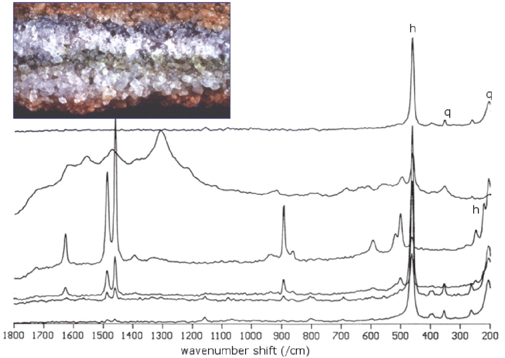
Quartz is the main component of the rock, with Raman spectral signatures at 128, 206, 355, 542, 696, 795, 807, 1064, 1081, 1161 and 1227 cm−1; the red colour is recognizable as haematite because of its characteristic Raman bands at 223, 291, 404, 495 and 609 cm−1. This is abundant below the organism stratum, but has been removed from the immediate vicinity, probably to permit photosynthetic-active radiation to reach endolithic community and as a possible reinforcement due to its ultraviolet radiation absorbing properties (Clark Reference Clark1998). The specificity of Raman spectroscopy permits one to distinguish the relatively weak signature of haematite in the crustal spectrum comprising mainly quartz; haematite has been proved to be a ultraviolet-screening compound and it is interesting that the organisms can maintain this mineral on the crust for the purpose of protecting against radiation that reaches the terrestrial surface (exacerbated by the depletion of Antarctic ozone). Also present are clear signatures of goethite, an iron (III) oxyhydroxide, which confers a distinctly yellowish colouration to the crustal zone; this particular crust colouration as a result of the modification by the underlying biological colony was used by the prospecting geologist as a visual clue to the presence of endolithic communities inside that particular rock. The major component detected spectroscopically in the intermediate zones was calcium oxalate monohydrate, with distinctive features at 1462 and 1494 cm−1 and supporting bands at 906 and 500 cm−1.
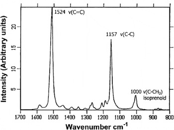
The green layer gives a strong fluorescence emission with 785 nm laser excitation and no spectral signatures are visible but the selection of alternative argon ion with 514 nm laser excitation in the green region provides bands from a carotenoid pigment which are clearly recognizable at 1524, 1157 and 1000 cm−1 (Fig. 2); these spectral signatures are consistent with an assignment to astaxanthin and represent the C = C, C–C stretching and C = CH bending modes, respectively. The same applies to the black zone in the endolith, but now the carotenoid present gives bands at 1516, 1154 and 1004 cm−1 indicative of its assignment to beta-carotene. Although no spectral signatures could be recorded for this system with the near-infrared laser at 1064 nm, differentiation between carotenoids with 514 nm excitation is possible in the green band and in the black band.
Fourier transform-Raman spectrum of a carotene (astaxanthin) found in an Antarctic Beacon sandstone endolith.

Arctic cold deserts
Firstly, a chasmolithic community living in a fracture inside a marble rock was collected during the AMASE (Arctic Mars Analogue Svalbard Expedition) in August 2004 to the Vest Spitsbergen Island in the Svalbard Archipelago, sited 80°N, inside the Arctic Circle. Although the temperatures are not as cold as those in Antarctica, at Spitsbergen Island, the winter temperature nevertheless reaches minus 30–35 °C and the biological colonization is considered extremophilic because of the low temperatures and the absence of sunlight for almost 6 months each year. The sample was found in a glacier moraine in Bockfjorden, at an altitude of 30 m above the sea level, and shows an epilithic community in the crustal zone which gives a greyish tonality to the white stone substrate, and a chasmolith present inside a crack where several coloured strata, namely pinkish, black, green and brown, are visible.
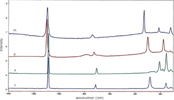
The Raman spectrum of the substratum presents bands at 1097 cm−1, which is seen as a shoulder on the stronger signatures at 1086 and 713 cm−1 with a shoulder at 725 and 283 cm−1 with a shoulder at 299 cm−1 and a doublet at 177 and 157 cm−1. This provides an excellent example of the ability of Raman spectroscopy to differentiate between isomorphic minerals in admixture; the bands at 1097, 725, 299 and 177 cm−1 are characteristic of dolomite, a calcium magnesium carbonate (CaMg(CO3)2), whereas calcite, the calcium carbonate isomorph, shows signatures at 1086, 713, 283 and 157 cm−1, very close in wavenumber, but readily distinguishable from the spectral signatures of the dolomite. Figure 3 gives a Raman spectral stackplot of magnesite, MgCO3, with Raman bands at 1094, 738, 326, 242 and 109 cm−1, dolomite, CaMg(CO3)2, with bands at 1097, 725, 299 and 177 cm−1, aragonite, CaCO3, with bands at 1086, 704, 208 and 154 cm−1, and calcite, CaCO3, with bands at 1086, 713, 283 and 157 cm−1.
Raman spectra of (m)agnesite, (d)olomite, (a)ragonite and (c)alcite – indicating the spectral discrimination between these carbonates.

On the surface crust, the grey tonality of the specimen is the result of cyanobacterial colonization, which gives the characteristic Raman spectral bands of scytonemin, which is exclusively produced by cyanobacteria under stress. This ultraviolet protective pigment is not found in the chasmolithic organisms inside the crack and has probably been produced as a strategic response to the increased radiation levels experienced at the rock surface in this location.
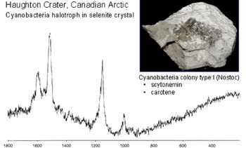
Secondly, the presence of salts in Shergotty–Nakhla–Chassigny (SNC) Martian meteorites and the acknowledgement of the extensive presence of sulphates in the Martian regolith, together with the recognition of the existence of lacustrine water sediments at or below the surface of Mars makes terrestrial halotolerant organisms important potential Martian analogues. Apart from the Raman spectroscopic identification of two different types of bacteria in a gypsum crystal (Nostoc and Gloeocapsa), it was also possible to detect organic signatures from bacterial colonies sited several millimetres below the surface in a transparent crystal of selenite, calcium sulphate dihydrate, from the surface breccia deposits in the 26 Mya Haughton meteoritic impact crater at Devon Island in the Canadian High Arctic. The Raman spectrum of the selenite crystal surface shows the strongest gypsum band at 1007 cm−1 and other weaker gypsum signatures; Raman spectra taken from a vertical transect through the crystal from the surface, using a confocal laser beam to obtain spatial information from within the crystal without mechanically cleaving it, provides evidence of additional features in which the Raman bands can be identified with the cyanobacterial signatures. Confocal laser probing of a dark-coloured inclusion within the selenite crystal reveals that it is a halotrophic cyanobacterial colony containing the radiation protectant scytonemin (Fig. 4).
Raman spectrum of halotrophic cyanobacterial colony inside selenite crystal, located approximately 5–8 mm below the upper surface. The Raman spectrum of the cyanobacterial inclusion obtained using confocal laser imaging shows the presence of scytonemin and a carotenoid assigned as beta-carotene.

Stromatolites
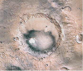
Shade adaptation is required by bottom-dwelling cyanobacteria in lakes while enabling them to avoid ultraviolet stress; during seasonal changes in the flow of melt-water, the thick cyanobacterial mats at the bottom of lakes becomes buried in silt. However, they permeate through the mineral layer to form a new mat each season, resulting in the production of stratified stromatolites. For example, the translucency of the 3 m thick ice cover on Lake Vanda in Antarctica permits the penetration of solar radiation through 70 m of water column to the bottom cyanobacterial mats where the receipt of photosynthetically active radiation (PAR) can be ∼120 μmol m−2 s−1. Even the PAR level of ∼15 μmol m−2 s−1 measured at the bottom of Lakes Hoare and Fryxell, which have thicker ice covers (about 4.5 m) is more than enough to sustain active photosynthesis by oxygenic mats. This has resulted in a wide diversity of cyanobacterial mat communities in many Antarctic Dry Valley lakes. Analogous habitats on Mars, such as Gusev and Noachis Craters, may provide suitable sources of fossil microbes and biomolecules such as porphyrins and cyanobacterial scytonemin. An example of such a potential scenario on Mars is Juventus Chasmae (Fig. 5). Modern stromatolites are considered to be analogues of Conophyton, which is a columnar stromatolite abundant in Precambrian rock when ultraviolet-stress would have been much greater.
Juventus Chasmae on Mars; the white area inside the crater is composed of magnesite and hydromagnesite rocks which have a counterpart terrestrially in Salda Golu Lake, Turkey.

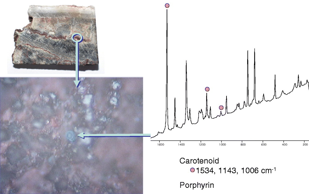
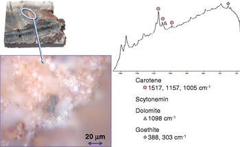
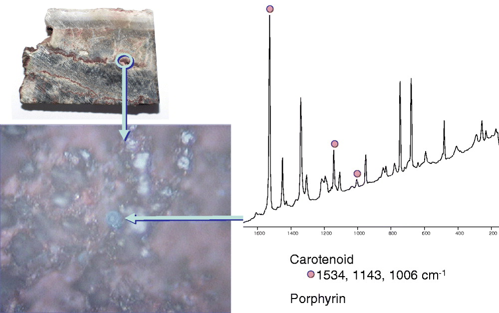
With climatic changes on a geological scale, the desiccation of lakes can occur and their cyanobacterial mats then become fossils. Their pigments, especially early porphyrins, which are chemical components related to chlorophyll are recognizable in oil-bearing shales. A primitive photosynthetic system capable of delivering an electric charge across a membrane bi-layer could have consisted of a porphyrin (pigment), a quinone (electron donor) and a carotene (electron acceptor) to provide a chain of conjugated bonds. Such molecules are valuable biomarkers on Earth and may fulfil a similar function for their recognition by remote life-detection systems on Mars. A recently recorded micro-Raman spectrum of the Trendall Pilbara stromatolite, from North Pole Dome in Western Australia, has identified a carotene and a porphyrin together in a fissure inside the rock (Fig. 6). This stromatolite is one of the oldest rocks found on Earth, with an age of 3600 Mya, dating from a very early period in the geological evolutionary history of our planet. However, while it would be facile to assume automatically that the bio-organic signatures that have been detected here are also of this age, this example does demonstrate very nicely the viability of Raman spectroscopy for the detection of organic signatures preserved in geological niches inside ancient rocks. In another sampling region of the same specimen, there is clear spectroscopic evidence for the presence of beta-carotene, scytonemin, dolomite and goethite (Fig. 7).
Trendall Pilbara stromatolite, Western Australia, 3600 Mya; Raman spectroscopic evidence for protective biochemicals, carotene and porphyrin produced in a protected geological niche.
Specimen of Trendall Pilbara stromatolite shown in figure 6, but different geological niche and a different chemical suite identified, namely, scytonemin, carotene, dolomite and goethite.

Hot deserts
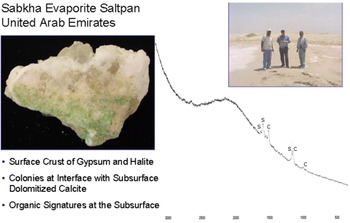
Dried terrestrial salt pans with their extremophilic halotrophic cyanobacterial colonization are good models for the study of Martian sites such as Gusev Crater. Here, an example from the Rhub-al-Khalil in the Arabian Desert shows a cyanobacterial colony in a dolomitized zone, several centimetres below the surface of a large gypsum crystal embedded in a halite matrix (Fig. 8). The Raman spectrum of the biological component clearly demonstrates the presence of photo-protective pigments such as scytonemin and carotenoids.
Sabkha surface crust with gypsum and halite crystals, Rhub-al-Khalil, Arabian desert; cyanobacterial colonization at interface with subsurface dolomitized calcite. The Raman spectrum shows the presence of scytonemin (s) and beta-carotene (b) in the biological zone.

Other hot desert halotrophic salterns are exemplified by those found in the Atacama Desert, Mojave Desert and the Negev Desert – all of which are accepted as Mars analogue terrestrial sites.
Mars meteorites
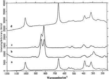
The specific information provided by Raman spectroscopic analysis of geological samples directly relevant to Mars can be illustrated here by the spectral stackplot of the Raman spectra recorded from the Nakhla SNC Martian meteorite, one of the currently accepted 35 Martian meteorites that have been collected on Earth, shown in Fig. 9. The rich diversity of chemical information that is provided by the micro-Raman spectra of this specimen exemplifies the geological applications of Raman spectroscopy to Martian materials.
Raman microscope spectral stackplot of the Nakhla meteorite, an SNC Martian meteorite, illustrating the discrimination offered between different regions; in each case a spectral footprint of about 2 μm is taken in the specimen and the geological heterogeneity should be noted. (a) Black particle, (b) white particle, (c) brown glassy particle, (d) clear glassy particle. Raman bands at 320, 350 386, 662 and 1009 cm−1 are characteristic of clinopyroxene, those at 815 and 841 cm−1 of olivine and those at 508 and 520 cm−1 of plagioclase. No bands arising from biomolecules can be identified in these spectra.

Conclusions
A detailed knowledge of the different chemical strategies adopted by terrestrial extremophiles, which are considered Martian analogues, is a prerequisite for planetary exploration. Geo- and bio-markers must be studied together to provide a better understanding of the adaptation abilities of the organisms. Geo- and bio-strategies are complementary and the survival of the organisms depends critically upon their being able to modify their microniche environments.
Raman spectroscopy is a useful analytical technique for planetary exploration, because it does not require sample manipulation, and macroscopic and microscopic analyses are possible when identifying components in the specimen. It is sensitive to organic and inorganic compounds and is not destructive to samples that are strictly limited in quantity or accessibility and so they can be used afterwards for other, perhaps more destructive, analyses. In this respect, the situation of a Raman spectrometer on a planetary rover or on a fixed lander, to which specimens are brought by a rover, are both acceptable space mission scenarios.
Laser excitation wavelength is an important parameter to consider, not only because fluorescence emission can swamp the weaker Raman scattering but also because of the different sensitivities shown towards compound identification by different excitation wavelengths. In our assessment 785 nm laser excitation gives a good balance between organic and inorganic compounds, but 514 nm excitation induces a resonance Raman effect for carotenes whose identification is reasonably straightforward, even in small concentrations, although the higher fluorescence emission from some systems can still mask several weaker Raman bands. The observation of more than two spectral biomarker bands is necessary normally for the unambiguous identification of the components of a specimen, particularly when there is a complex mixture of organics and inorganics present.
The ability to identify organic molecules and minerals present in admixture in a specimen at the same time is an advantage of the Raman analytical technique in astrobiology. Extremophiles are closely related to rock substrata, and biological and geological spectral signatures are shown together in the Raman spectrum; these are distinguishable and can be unambiguously assigned, even if some appear in a relatively low proportion. The Raman spectroscopic data presented in this paper from terrestrial Mars analogues and models will form an important part of the analytical scientific preparation for Mars exploration involving the search for life in the ExoMars mission.
Acknowledgements
The authors express their gratitude to colleagues in the European Space Agency ExoMars Raman RLS Science and Instrument Teams for interdisciplinary expertise in the achievement of acceptance of miniaturized instrumentation for the life-detection mission to Mars, and to the UK Science and Technology Facilities Research Council for the on-going funding support for Raman spectroscopic studies relevant to early evolution and the ExoMars mission.