Over the past decade, clinical indications for cardiac pacemaker systems in paediatric and CHD patients have been expanded significantly;Reference Walsh 1 however, due to a variety of safety concerns, the presence of an implantable cardiac device has been considered a contraindication for MRI.Reference Walsh 1 , Reference Martin, Coman, Shellock, Pulling, Fair and Jenkins 2 With the introduction of transvenous MRI-conditional pacemaker systems in 2008,Reference Sutton, Kanal and Wilkoff 3 MRI scanning became potentially accessible for pacemaker carriers with CHD. As cardiac MRI is regarded as the ‘gold standard’ for functional and anatomical imaging, particularly in the CHD population,Reference Bonello and Kilner 4 – Reference Kilner 6 this technique has a significant impact on clinical practice. Ventricular volumes and function can be assessed, complex anatomies visualised, blood flow measured, and postoperative results including myocardial scarring and fibrosis can be evaluated with high accuracy and reproducibility. Especially in CHD with impairment of the right ventricle, as, for example, in tetralogy of Fallot or systemic right ventricle due to d-transposition of the great arteries after atrial re-direction or congenitally corrected transposition of the great arteries, cardiac MRI is a valuable diagnostic tool to evaluate the progression of right ventricular dilatation and dysfunction. Here, MRI is used to detect patients who require re-intervention and to predict outcome.Reference Bonello and Kilner 4 As pacemaker leads and devices in the vicinity of the heart may cause artefacts,Reference Sasaki, Hansford and Zviman 7 and thus may seriously affect the validity of the investigation, we aimed to analyse safety, susceptibility artefacts, and image reading of cardiac MRI in CHD patients equipped with MRI-conditional pacemaker systems.
Materials and methods
Patient group
Patients with CHD and MRI-conditional pacemaker systems were enrolled if they had a clinical necessity for cardiac MRI. All MRI-conditional pacemaker systems had been implanted at least 6 weeks before the examination according to the manufacturer’s recommendations. Patients with epicardial or abandoned leads were excluded.
The study was approved by the institutional ethics committee following the ethical guidelines of the Declaration of Helsinki, and written informed consent was obtained from all participants.
Pacemaker function
Device interrogation to assess battery voltage, lead impedances and lead capture thresholds, sensing signal amplitudes, and percentage of stimulated beats for each lead was performed before, immediately after MRI, and at routine follow-up. Pacing mode was programmed to asynchronous MRI mode for the duration of the investigation, according to the manufacturer’s instructions. In patients with atrioventricular conduction block a DOO mode was chosen, and in patients with sinus node dysfunction but sustained atrioventricular conduction an AOO mode was chosen; stimulation frequency was set to at least 10–20 bpm higher than the patient’s resting frequency. After MRI, pacemaker and lead function were tested and the device was programmed to the original settings.
Cardiac MRI
Cardiac MRI scans were performed with a clinical whole-body 1.5-T MRI system (Philips Medical Systems, Best, The Netherlands) using a cardiac phased-array coil. Patients were continuously monitored with electrocardiographic telemetry, pulse oximetry, and blood pressure measurements. They were instructed to immediately inform the investigator of any unusual sensation such as heat or device movement during the MRI examination. A pacemaker specialist with an external defibrillator was present throughout the procedure.
The imaging protocol consisted of scout sequences for planning of standard cardiac views of the right ventricle – horizontal and vertical long axis, sagittal view – steady-state free precession cine transversal sequences for anatomical and functional analysis, and phase–contrast cine MRI for flow quantification in the great arteries. Additional pulse sequences were chosen according to the clinical indication for MRI – phase contrast cine MRI for right and left pulmonary artery, magnetic resonance angiography, single-phase three-dimensional steady-state free precession whole heart sequences, steady-state free precession cine short-axis, and coronal views. For all pulse sequences, the specific absorption rate was kept below 2.0 W/kg, as advised by the manufacturers of the MRI-conditional pacemaker systems.
Artefacts
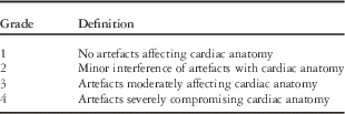
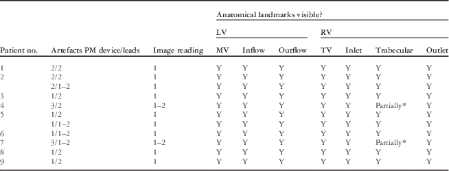
Each MRI examination was assessed by two experienced investigators in consensus based on a four-point grading scale, considering susceptibility artefacts caused by the metallic pacemaker device and leads (Table 1) according to Naehle et al.Reference Naehle, Kreuz and Strach 8
Grading scale for susceptibility artefacts caused by pacemaker device and leads.

Image reading
A four-point grading scale based on the scale for diagnostic value by Naehle et alReference Naehle, Kreuz and Strach 8 was applied to evaluate the certainty of diagnosis of all MRI scans (Table 2). For this purpose, anatomical landmarks of the left and right ventricle were identified. Mitral valve and inflow and outflow region/aortic valve were defined as landmarks for the left ventricle, and tricuspid valve, inlet, trabecular, and outlet region for the right ventricle. The pulmonary valve was not set as a landmark, as it is not always visible, even in cardiac MRI of uncompromised image quality, or might be absent, as for instance in patients with tetralogy of Fallot after valvectomy.
Grading scale for cardiac MRI image reading considering anatomical landmarks.

In brackets is the number of anatomical landmarks not represented in the MRI images.
Statistical analysis
All values are expressed as mean±standard deviation if not otherwise indicated. We used a Friedman non-parametric test to compare the lead parameters, such as pacing threshold, pacing impedance, and intrinsic signal amplitude, at three time points: before MRI, after MRI, and at last follow-up. The level of significance was set to 0.05.
Results
Patient group
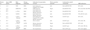
A total of 11 MRI examinations were performed in nine CHD patients with a pacemaker between October 2010 and April 2013. At MRI, the mean age of patients was 32.7±12.3 (19.5–61.3) years. Cardiac MRI was performed 130±261 (48–769) days after implantation of an MRI-conditional pacemaker system. Patients’ diagnoses were d-transposition of the great arteries after atrial re-direction (n=3), congenitally corrected transposition of the great arteries (n=2), tetralogy of Fallot (n=1), and double-outlet right ventricle with pulmonary stenosis (n=1) after surgical correction, as well as partial (n=1) and total anomalous pulmonary venous connection (n=1) after anatomical repair. An overview of patient characteristics is given in Table 3. Most patients had had previous corrective cardiac surgery (n=7) with a mean of two operations (range: 1–3). Leading indications for pacemaker implantation were sinus node dysfunction (n=6) and complete atrioventricular conduction block (n=3). Of the nine patients, three were pacemaker dependent. The majority (n=7, 78%) had been converted from pre-existing conventional to MRI-conditional pacemaker systems for various clinical reasons (Table 3).
Patient characteristics regarding main diagnosis, age, indication for pacing, pacemaker revision and MRI, and position of pacemaker device and leads.

cAVB=complete atrioventricular block; cc-TGA=congenitally corrected transposition of the great arteries; d-TGA=d-transposition of the great arteries; DORV/PS=double-outlet right ventricle with pulmonary stenosis; FW=free wall; IVS=interventricular septum; LAA=left atrial appendage; lat=lateral; LV=left ventricle; no.=number; PAPVC=partial anomalous pulmonary venous connection; Pas=pulmonary arteries; PPVI=percutaneous pulmonary valve implantation; PR=pulmonary regurgitation; PVs=pulmonary veins; RA=right atrium; RAA=right atrial appendage; RV=right ventricle; RVOT=right ventricular outflow tract; SND=sinus node dysfunction; TAPVC=total anomalous pulmonary venous connection; TOF=tetralogy of Fallot; TR=tricuspid regurgitation; vol/fx=volume/function.
Device and lead function/safety
Device interrogation immediately before MRI showed appropriate device function and functional lead parameters within the required limits for safe MRI scanning according to the manufacturer’s recommendations. Device testing directly after and at a mean routine clinical follow-up of 101±52 (64–210) days demonstrated no significant changes in lead impedance, capture threshold, sensing signal amplitude, or battery voltage (Table 4).
Comparison of pacemaker device and lead parameters before and after cardiac MRI and at follow-up (101±52 (64–210) days).

No significant changes related to the MRI examination were observed (p<0.05).
All MRI examinations were completed without adverse effects. Inhibition of pacing during MRI indicated by changes in heart rate or rhythm was not observed. None of the patients reported symptoms attributable to the pacemaker device.
Artefacts
In nine out of 11 MRI examinations, there was no or only minor interference of susceptibility artefacts with cardiac anatomy caused by the pacemaker device (grade 1 or 2). In the patient with total anomalous pulmonary venous connection and in one patient with d-transposition of the great arteries after atrial re-direction, artefacts moderately affected the underlying cardiac anatomy (grade 3). All examinations were rated grade 1 or 2 with regard to artefacts caused by pacemaker leads – that is, no or only minor artefacts in the cardiac anatomy were visible.
Image reading
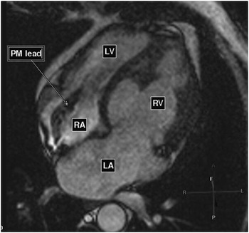
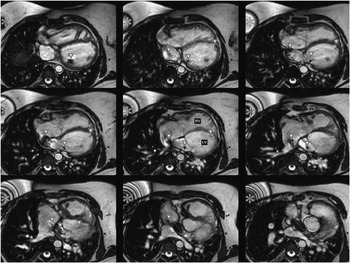
Both investigators independently evaluated all MRI examinations considering anatomical landmarks with high (grade 1) or sufficient-to-high certainty of diagnosis (grade 1–2; Table 5, Figs 1 and 2). In two patients, MRI scans were graded 1–2, as the trabecular region of the right ventricle was only partially visible. Nevertheless, clinical questions could be answered in all cases. The documented artefacts caused by the pacemaker leads did not affect image interpretation in any patient, regardless of the type of lead or image sequence.
MRI image in horizontal long axis (four-chamber view) of a 51-year-old patient with congenitally corrected transposition of the great arteries and MRI-conditional pacemaker system. LA=left atrium; LV=left ventricle (subpulmonary); PM lead=pacemaker lead; RA=right atrium; RV=right ventricle (subaortic).

MRI images in axial orientation of a 37-year-old patient with d-transposition of the great arteries after atrial re-direction and stenting of the superior caval vein baffle (dotted arrow). The star indicates minor artefacts caused by the right-sided pacemaker device, the arrow heads point at artefacts from pacemaker leads in the left atrial appendage and the apex of the left ventricle. LV=left ventricle (subpulmonary); RV=right ventricle (subaortic).

Grading of cardiac MRI considering susceptibility artefacts caused by pacemaker device and leads, as well as anatomical landmarks.

LV=left ventricle; MV=mitral valve; no.=number; PM=pacemaker; RV=right ventricle; TV=tricuspid valve; Y=yes.
* Trabecular RV region only partially visible.
Discussion
The primary aim of the present study was to analyse safety, susceptibility artefacts, and image reading of cardiac MRI in patients with CHD and MRI-conditional pacemaker systems. Our results suggest that cardiac MRI can be performed safely and with high certainty of diagnosis in this patient group if precautions and monitoring are carefully considered.
Device and lead function/safety
The function of the pacemaker device and leads remained stable in all patients immediately after MRI and at follow-up (101 days) with no significant changes in lead impedances, capture thresholds, sensing signal amplitudes, or battery voltage being detected compared with interrogation performed immediately before MRI. This is in accordance with the findings of other groups, suggesting that MRI of different anatomic regions may be performed safely in selected patients with pacemaker/implantable cardioverter defibrillator systems under well-controlled circumstances at 0.5,Reference Heatlie and Pennell 9 – Reference Sommer, Vahlhaus and Lauck 11 1.5,Reference Martin, Coman, Shellock, Pulling, Fair and Jenkins 2 , Reference Naehle, Kreuz and Strach 8 , Reference Gimbel, Bello and Schmitt 12 – Reference Wollmann, Thudt, Kaiser, Salomonowitz, Mayr and Globits 21 2,Reference Del Ojo, Moya and Villalba 22 and 3 T.Reference Gimbel 23 , Reference Naehle, Meyer and Thomas 24
Most data were collected for patients with conventional non-MRI-conditional cardiac devices.Reference Martin, Coman, Shellock, Pulling, Fair and Jenkins 2 , Reference Naehle, Kreuz and Strach 8 – Reference Sommer, Vahlhaus and Lauck 11 , Reference Cohen, Costa and Russo 14 , Reference Nazarian, Hansford and Roguin 15 , Reference Naehle, Strach and Thomas 17 – Reference Nazarian, Roguin and Zviman 20 , Reference Del Ojo, Moya and Villalba 22 – Reference Naehle, Meyer and Thomas 24 Gimbel et al stated the safety and efficacy of the Advisa MRI system (Medtronic; pulse generator: Advisa MRI, lead: CapSureFix 5086MRI) designed for safe whole-body MRI without positioning restrictions at 1.5 T. MRI-related complications occurred in none of the 148 patients who underwent MRI including chest scans, and minimal differences in pacing threshold values from pre-MRI to 1 month post-MRI were similar between the MRI and the control group.Reference Gimbel, Bello and Schmitt 12 Comparable results were described by Wollmann et alReference Wollmann, Steiner, Vock, Ndikung and Mayr 13 , Reference Wollmann, Thudt, Kaiser, Salomonowitz, Mayr and Globits 21 and Wilkoff et al,Reference Wilkoff, Bello and Taborsky 16 demonstrating no MRI-related adverse effects or clinically relevant changes in the function of MRI-compatible devices at 1.5 T, as shown in the present study.
All MRI examinations within the present study were performed safely and with high or sufficient-to-high certainty of diagnosis. Pulver et alReference Pulver, Puchalski and Bradley 18 reported on safe performance of cardiac and non-cardiac MRI at 1.5 T with good diagnostic quality in non-pacemaker-dependent paediatric and adult CHD patients, with pacemaker systems with predominantly epicardial pacemaker leads. The study was limited by the small patient number (cardiac MRI: n=9) and the short follow-up time (mean 5.5 months), and thust potential complications including long-term problems may have been missed. Moreover, pacemaker-dependent patients were not included in the study, and the exact specific absorption rate was not recorded.Reference Pulver, Puchalski and Bradley 18
Despite promising results, absolute safety of cardiac MRI in patients with cardiac devices cannot be guaranteed to date. Patient groups are often too small to allow statistically adequate safety evaluation, and, despite all precautions, the risk of heating of the lead tips with subsequent increases in pacing capture thresholds and serum troponin I levels indicating subclinical myocardial damage cannot always be fully eliminated.Reference Sommer, Naehle and Yang 19 , Reference Luechinger, Zeijlemaker and Pedersen 25 Thus, patients with cardiac devices should only be selected for cardiac MRI after detailed evaluation of risks potentially associated with this imaging modality and the clinical benefits of the acquired images.
Artefacts
In the present study, there was no or only minor interference of susceptibility artefacts by the pacemaker device and leads with cardiac anatomy, independent of the location of the pacemaker device. Evaluating the potential diagnostic limitations of cardiac MRI caused by susceptibility artefacts in patients with conventional pacemaker (n=15) and implantable cardioverter defibrillator systems (n=56), Sasaki et al described uncompromised interpretation of MRI images, regardless of the image sequence in all pacemaker patients. Artefact size was significantly greater in patients with implantable cardioverter defibrillators, especially with left-sided position, than in patients with pacemakers.Reference Sasaki, Hansford and Zviman 7 Naehle et al demonstrated good image quality and diagnostic value, allowing for final diagnosis mainly in patients with right-sided pacemaker devices (n=12). Remarkably, in that study, device-related artefacts leading to reduced image quality prohibited a reliable diagnosis in 65% of the cardiac MRI examinations in patients with a left-sided device, of which half (n=10) were implantable cardioverter defibrillators.Reference Naehle, Kreuz and Strach 8 These results indicate that the device-to-heart distance correlates with the occurrence of artefacts; however, the main factor appears to be the amount of ferromagnetic device components, which is naturally determined by the volume of the box being approximately one-third for pacemakers compared with implantable cardioverter defibrillators. In the present study, only patients with ‘low-volume’ pacemaker systems (12–14 cc) were investigated. The lesser content of ferromagnetic material due to their specific ‘MRI-conditional design’ may explain the better image quality even in cases of a left-sided device position. Thus, the MRI-conditional technology not only addresses safety issues but also serves to reduce susceptibility artefacts allowing improved image reading. These conclusions are also supported by a recent study of 150 non-CHD patients with MRI-conditional pacemaker systems (Advisa MRI, Medtronic), where 84% of the left ventricular images and 93% of the right ventricular images were of good or excellent image quality.Reference Schwitter, Kanal and Schmitt 26 Furthermore, the last study supports the results of the present study by showing that even in the lead-containing (subpulmonary) ventricle image artefacts are often of minor relevance. Again, the main point seems to be the content of metal, which for pacemaker leads is much less compared with the device.Reference Naehle, Kreuz and Strach 8
Image reading
Even more important than pure image quality is the possibility to generate state-of-the-art multi-modality MRI data in CHD patients with pacemakers. In the present study, this resulted in a high or at least sufficient certainty of diagnosis in all patients allowing for clinical decision-making. In addition, as previously described by Martin et al,Reference Martin, Coman, Shellock, Pulling, Fair and Jenkins 2 based on cardiac MRI, all clinical questions could invariably be answered in the present study. This can be exemplified by the assessment of the right ventricle for which cardiac MRI has become the imaging modality of choice.Reference Bonello and Kilner 4
MRI is an important tool for answering clinical questions related to right ventricular complications that are more common in CHD than in acquired heart disease. Thus, MRI is of particular benefit for patients with a systemic right ventricle in the context of d-transposition of the great arteries after atrial re-direction or (native) congenitally corrected transposition of the great arteries, allowing non-invasive evaluation of right ventricular function, volumes, and tricuspid regurgitation; however, increased prevalence of pacemaker devices in this groupReference Moons, Gewillig and Sluysmans 27 often excluded patients from cardiac MRI. The safe performance of cardiac MRI with high certainty of diagnosis in all patients with a systemic right ventricle (n=5) in the present study illustrates the potential benefit of MRI-conditional pacemaker systems in the CHD population. It is noteworthy to mention that in the group of patients with d-transposition of the great arteries after atrial re-direction, and congenitally corrected transposition of the great arteries, uncompromised evaluation in particular of the right ventricular region is facilitated by the fact that the ventricular pacemaker leads are placed in the subpulmonary morphological left ventricle, which is of minor interest for functional assessment.
Implications of MRI-conditional systems for CHD patients
Particularly in the CHD population, the widespread use of MRI-conditional pacemaker systems is currently restricted due to several limitations. To date only transvenous systems have been certified,Reference Ahmed, Morris, Allen, Khattar, Mamas and Zaidi 28 so that pacemaker patients whose size or anatomy do not allow venous access to the heart are excluded. As a consequence, epicardial leads are often used instead, resulting in lifelong exclusion from MRI for decades.
MRI-conditionality is a young and evolving technological field, where new developments may have a great impact on implant strategies in CHD. Besides the variety of lead types and sizes, there are other ‘general’ differences between MRI-conditional pacemaker systems, which physicians involved in CHD pacemaker therapy should be aware of.Reference Ahmed, Morris, Allen, Khattar, Mamas and Zaidi 28 In this context, it is important to mention that some MRI-conditional pacemaker systems exclude the thorax region.Reference Ahmed, Morris, Allen, Khattar, Mamas and Zaidi 28
Compared with adults with acquired heart disease, patients with CHD, especially children, are at increased risk of excessive cumulative levels of radiation dose, given that they often have to undergo multiple procedures over their lifetime.Reference Allen, Beekman and Garson 29 This results in the associated damaging biological effects of ionising radiation.Reference Andreassi, Ait-Ali, Botto, Manfredi, Mottola and Picano 30 Therefore, availability of cardiac MRI as a substitute for computed tomography or therapeutic catheterisation would be of great benefit for CHD patients with pacemakers, and it is important to discuss whether a more progressive strategy of MRI-conditional system upgrade should be considered for this group.
Study limitations
The study is limited by the relatively small size of the study population. As a result, all potential complications may not have been fully identified. All our patients had reached adulthood. The more unfavourable ratio of body size, and thus vascular and cardiac structures, to lead and device size in younger patients may enhance the impact of susceptibility artefacts, complicating the identification of anatomical landmarks, and in this way limit certainty of diagnosis. In addition, more abundant lead material in the form of lead loops intended to compensate for future length growth may give rise to an increase in lead-associated artefacts. Therefore, more data in larger and younger populations with CHD and pacemaker are needed to support our positive conclusion of MRI-compatible pacemaker implantation in patients with CHD.
Conclusions
The results of the present study suggest that cardiac MRI can be safely performed in patients with CHD and MRI-conditional pacemaker systems, provided that appropriate precautions such as limitation of specific absorption rate, adequate monitoring of patients during MRI examinations, and device interrogation before and after MRI are taken into account. Furthermore, susceptibility artefacts caused by pacemaker device and leads do not seem to limit certainty of diagnosis by MRI in this patient group, as anatomical landmarks can sufficiently be identified. All CHD patients eligible for transvenous lead access, including those with a clinically indicated revision of a pre-existing non-MRI-conditional system, should be considered for implantation of an MRI-conditional pacemaker system allowing full-body scanning, and thus paving the way for the usage of cardiac MRI for non-invasive monitoring of CHD.
Acknowledgements
The authors thank Alireza Khasheei (radiographer) for technical assistance, Julia Stein for performing statistical calculations on functional pacemaker measurements, and Anne Carney for editorial assistance.
Financial Support
This research received no specific grant from any funding agency, commercial, or not-for-profit sectors.
Conflicts of Interest
None.
Ethical Standards
The authors assert that all procedures contributing to this work comply with the ethical standards of the relevant national guidelines on human experimentation and with the Helsinki Declaration of 1975, as revised in 2008, and that the study has been approved by the institutional committee (ethics committee – Charité – Universitaetsmedizin Berlin).