INTRODUCTION
Entamoeba histolytica is a highly invasive parasite that is able to degrade extracellular matrix (ECM) components during invasion of basal lamina by penetrating the intestinal mucosa. This process is complex and requires a coordinated interaction of numerous molecules and the activation of specific signalling pathways (Meza, 2000). A large variety of cell surface adhesion molecules are necessary to communicate cells with their environment. These molecules lead the cell to adhere and displace. Specific adhesion of E. histolytica trophozoites to fibronectin (FN) substrate through at least 2 proteins, one of 37 kDa (Vázquez-Prado and Meza, 1992) and one of 140 kDa (Talamás-Rohana et al. 1992) followed by the formation of adhesion plates and FN degradation has been reported (Talamás-Rohana and Meza, 1988). The 140 kDa FN-binding molecule was further characterized by biochemical, antigenic, and functional criteria as a β1-integrin like molecule (EhFNR) that acts as a FN receptor (Talamás-Rohana et al. 1994, 1998). Additionally, Sengupta et al. (2001) reported a monoclonal antibody (3C10 mAb) against the amoebic FNR that inhibits cell adhesion to ECM components. Regarding trophozoite-host cell interaction, the main adhesion molecule is the Gal/Gal NAc lectin, a 260 kDa heterodimeric glycoprotein composed of a 170 kDa heavy subunit linked covalently by disulfide bonds to a 31–35 kDa light subunit. Additionally, a 150 kDa protein has been identified as part of this molecular complex (Petri et al. 2002; Mann, 2002). The 170 kDa heavy subunit (Hgl) shares an epitope with human integrins and shows sequence similarity with amino acid residues of the cytoplasmic tail of β2 and β7 integrins (Adams et al. 1993; Vines et al. 1998).
All these signalling molecules must be regulated during E. histolytica invasion and liver abscess formation. In order to have a better understanding about the mechanisms that involve EhFNR during the invasive process, we decided to study the regulation processes of this molecule during trophozoite-FN interaction and during liver abscess formation. A cDNA clone (5A) that codifies a fragment of the EhFNR was selected with the 3C10 mAb. This clone showed 99% identity with the Igl2 sequence and the protein showed cross-reactivity with EH3015 mAb raised against the Igl subunit. The 3C10 MAb was used as a probe to analyse the regulation of this amoebic receptor in trophozoites incubated on FN, and also in trophozoites recently recovered from a liver abscess that were stimulated by the micro-environment of the liver.
MATERIALS AND METHODS
Cell culture
Trophozoites of E. histolytica HM-1: IMSS (originally isolated and typified at Centro Médico Nacional, Instituto Mexicano del Seguro Social, México) were maintained as described (Diamond et al. 1978).
Isolation of trophozoites from hepatic lesions
Male hamsters (Meriones ungiculatus) from CINVESTAV-IPN animal facilities, complying with the international regulation, were used throughout the study. Animals (4/group) were infected intrahepatically with 1×10⁶ trophozoites of E. histolytica strain HM1: IMSS. At 1, 2, 3, 4, and 7 days post-infection, animals were anesthetized and killed by exsanguination. Livers were dissected and small portions were incubated into TYI-S-33 medium supplemented as described (Chávez-Munguía et al. 2004). After 48 h of incubation, liver sections were removed and the culture medium was changed. The number of parasites as well as the attainment of the logarithmic phase was variable according to the time of recovery.
Isolation of cDNA clones coding for the 140 kDa EhFNR molecule
A cDNA library (Jiménez-Delgadillo et al. 2004) was screened with the monoclonal antibody (3C10) specific for the 140 kDa EhFNR. After 3 rounds of screening, 1 clone (5A) was selected for further characterization. The cDNA insert of the λ ZAP II clone was subcloned in the pMOS blue vector and sequenced (GeneBank Accession number: AY842505) using an automatic sequencer ABI PRISM 310 (Applied Biosystems, Foster City, CA, USA).
Purification of the recombinant protein (GST-EhFNR)
The HindIII-EcoRI DNA fragment (1062 bp) from the clone 5A was subcloned in-frame in the expression vector pGEX-5X-3 (Invitrogen, Carlsbad, CA, USA) to obtain the plasmid pVA5s. Escherichia coli DH5α cells were transformed with the recombinant expression vector. Cells were harvested and the purification of the expressed protein was performed as described (Patra et al. 2000) with some modifications. Briefly, cells were centrifuged for 30 min at 40000 g. Pelleted cells were lysed by sonication and centrifuged 30 min at 8000 g. The pellet containing GST-EhFNR inclusion bodies was washed first with 5 mM EDTA, 2% deoxycolate in 50 mM Tris-HCl, pH 8·0, and then with distilled water and centrifuged as above. The purification process was verified by silver staining of a 10% SDS-PAGE.
Production of polyclonal antibodies against the GST-EhFNR
BALB/c mice were immunized intraperitoneally with 100 μg of recombinant protein (GST-EhFNR) with complete Freund's adjuvant (GIBCO, Carlsbad, CA, USA) the first time and incomplete Freund's adjuvant (GIBCO) twice more; each immunization was done 2 weeks apart (López-Monteon et al. 2003).
Fluorescence microscopy
Culture trophozoites plated on fibronectin (FN)-, collagen (Col)-, or bovine serum albumin (BSA)-coated cover-slips and axenized trophozoites (recovered from hepatic lesions and harvested during logarithmic phase) were incubated for 1 h at 37 °C, and fixed with 4% p-formaldehyde for 1 h. Fixed trophozoites were incubated overnight with the 3C10 mAb (1/1000). Cells were washed with PBS, incubated with FITC-labelled goat anti-mouse IgG (1/50). Preparations were observed in a Nikon E600 fluorescence microscope (Nikon, Melville, NY, USA). All photographs were taken under the same conditions (exposure time and magnification) with a JVCKY-F70B digital camera.
Immunoprecipitation and Western blot
Purified GST and GST-EhFNR were electrophoresed in a 10% SDS-PAGE and transferred to NCP. Recombinant GST-EhFNR protein was detected with the 3C10 mAb (1/1000), anti-GST-EhFNR (1/1000), anti-GST antibodies (1/17000), and with pre-immune sera (1/1000). Additionally, pure GST was reacted with anti-GST antibody (1/17000). In all cases, an alkaline phosphatase conjugated anti-mouse IgG was used as secondary antibody (1/5000). The immune reaction was developed with NBT and BCIP in alkaline phosphatase buffer. Plasma membrane (PM) and internal membranes (IM), representing vesicle-associated membranes, were isolated as described by Aley et al. (1980) from trophozoites in suspension (control), from trophozoites incubated for 5 h on FN-coated 150 ml vol. culture flasks, and from trophozoites incubated for 5 h on FN-coated 150 ml vol. culture flasks, that were immediately labelled with biotin. PM and IM were separated in a 7·5% SDS-PAGE, transferred to NCP, and incubated with the 3C10 mAb (1/1000) and a horseradish peroxidase (HRP)-conjugated secondary antibody (1/1000) or overlayed with streptavidin-peroxidase (St-HRP). Blots were developed by chemiluminescence (SuperSignal Pierce, Rockford, IL, USA).
For immunoprecipitation assays, IM were solubilized in lysis buffer (2% Triton X-100, 10 mM Tris-HCl, pH 7·5, 150 mM NaCl, containing the following proteinase inhibitors, 3 mM NEM, TLCK, and IA, 1 mM PMSF, and 5 μM aprotinin) with 90 strokes in a glass homogenizer and centrifuged at 10000 g for 30 min. Samples of solubilized IM (1 mg) were pre-cleared with 2% BSA-blocked protein G-agarose (50 μl/sample) (GIBCO). The pre-cleared supernatants were then incubated with the corresponding antibodies: 3C10 mAb (1/1000), anti-human β1 integrin (1/1000) (Upstate Biotechnology, Lake Placid, NY, USA), anti-EhFNR (1/5000) (Flores-Robles et al. 2003), and anti-GSTEhFNR (1/1000). As negative control, a polyclonal anti-GST antibody (1/17000) was used. After overnight incubation at 4 °C, 60 μl of protein G-Agarose were added to each sample and further incubated for 2 h at 4 °C. Recovering and treatment of the immune complexes were performed as described (Flores-Robles et al. 2003). For detection of immunoprecipitated EhFNR the following antibodies were used: 3C10 mAb, anti-EhFNR, and anti-GSTEhFNR. Additional assays were performed with the same method but using 3C10 (anti-140 kDa mAb; 1[ratio ]1000 for Ipp and 1[ratio ]2000 for Wb) and EH3015 (anti-Igl subunit mAb; 1[ratio ]100 for Ipp and 1[ratio ]500 for Wb) mAbs. Biotin-labelled PM were treated as above for immunoprecipitation and the pre-cleared supernatant was incubated with 3C10 mAb (1[ratio ]1000). Detection of the immunoprecipitated biotinylated EhFNR was achieved by overlay with St-HRP (1[ratio ]1000).
Quantification of EhFNR in biotinylated trophozoites
Trophozoites recovered from hepatic lesions (1, 2 and 7 days post-infection) were washed twice with sodium bicarbonate buffer (NaHCO3 0·1 M/NaCl 0·8%, pH 8·3), and incubated with biotin hydrazide (Sigma, Milwaukee, WI, USA) (1×10⁶ cells/10 μg biotin hydrazide) for 15 min at room temperature with occasional agitation. The reaction was stopped with washing buffer. To check membrane staining and cell viability, a sample of labelled cells was stained with streptavidin-FITC (1/100) and observed by immunofluorescence microscopy. Biotinylated cells were centrifuged and lysed with lysis buffer (10 mM Tris-HCl, pH 7·5, 50 mM NaCl and protease inhibitors). Cell lysate was centrifuged at 9000 g and the pellet containing the total membranes fraction was separated. The total membranes fraction (30 μg) was electrophoresed by triplicate in 7·5% SDS-PAGE and blotted to NCP. Blots were incubated separately as follows: St-HRP (1/1000), 3C10 mAb (1/1000), and anti-actin mAb (1/7000) (loading control) (Chemicon, Temecula, CA, USA). Afterwards, the last two were incubated with HP-conjugated secondary antibodies (1/1000), and all were developed by chemiluminescence. The relative amount of EhFNR protein on the cell surface with respect to total amoebic receptor in each sample was quantified from 2 separate experiments, using a Software SigmaGel (Jandel Scientifc, San Rafael, CA, USA).
Electron microscopy
PM and IM were fixed with 2·5% glutaraldehyde in 0·1 M sodium cacodylate buffer, pH 7·2, for 60 min and post-fixed with 1% osmium tetroxide in the same buffer for 60 min. After dehydration with increasing concentrations of ethanol and propylene oxide, samples were embedded in epoxy resin and polymerized at 60 °C for 24 h. Thin sections (60 nm) were contrasted with uranyl acetate and lead citrate. Sections were analysed in a Phillips Morgan 268-D electron microscope.
RESULTS
Isolation of a cDNA clone encoding the EhFNR molecule
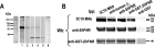
Screening of a cDNA expression library with the 3C10 mAb (Sengupta et al. 2001) revealed 1 highly positive clone, named 5A. This clone was purified through 3 rounds of immunoscreening and was further characterized. The unique open reading frame of the DNA insert (1062 bp) codifies for a 336 amino acid protein. The amino acid sequence (Fig. 1A) was compared with the sequences reported in the non-redundant GeneBank+EMBL+DDBJ+PDB databases using the BLASTN program (versión 2.2.10, Oct-19-2004). Results showed that the amino acid sequence of 5A, corresponds to the C-terminal region of the Igl2 subunit of the Gal/GalNac lectin and shows 96% identity with the Igl1 subunit (Cheng et al. 2001). Amino acid sequence alignment of these proteins showed the presence of the CXXC motif in 5A as described for Igl1 and Igl2 proteins (Fig. 1A, boxes). On the other hand, 5A showed conserved (D>E, R>K; double dot) and semi-conserved (P>T; dot) amino acid substitutions. The predicted amino acid sequence also revealed the presence of 2 CXC and 19 CXXC motifs in the C-terminal region of the molecule; no attachment site for GPI (big-PI Predictor GPI Modification Site Prediction, June-13-2002) and kinase domains were found in this sequence. This clone is not full length, however, it has a 21 amino acid sequence (from amino acid 316 to amino acid 336) showing characteristics of a transmembrane helix, usually present in transmembrane proteins. Antibodies against the fusion peptide produced by clone 5A, recognized the EhFNR in IM (data not shown). The identity of the 140 kDa protein (EhFNR) as Igl 150 kDa subunit was confirmed by cross-recognition of the 140–150 kDa protein immunoprecipitated with both 3C10 (anti-140 kDa antibody) and EH3015 (anti-Igl subunit antibody) mAbs and Western blotted with the 3C10 and the anti-Igl MAbs (Fig. 1B).

Fig. 1. (A) ClustalW alignment of 5A, Igl1, and Igl2 proteins. Identical amino acids are indicated by asterisks, conserved amino acids are indicated by double dots, and semi-conserved amino acids are indicated by a dot. The CXXC motif is boxed and the CXC motif is in bold. (B) Identity between EhFNR and Igl is shown by immunoprecipitation (Ipp) and Western blot (Wb) using the 3C10 mAb against EhFNR and the EH3015 mAb against the Igl subunit.
Characterization of the recombinant protein (GST-EhFNR)
To further characterize the function of the EhFNR, polyclonal antibodies against the recombinant protein (GST-EhFNR) were obtained. The DNA fragment (1062 bp) of the cDNA 5A clone, selected with the 3C10 mAb, was sublconed into the expression vector pGEX-5X-3 and after IPTG induction the recombinant protein was expressed as inclusion bodies, whose purification was achieved through extensive washes with detergent (Fig. 2A, lane 1, silver staining). The recombinant protein was recognized by the 3C10 mAb (Fig. 2A, lane 2), polyclonal anti-GST-EhFNR (Fig. 2A, lane 3), and anti-GST (Fig. 2A, lane 4) antibodies. To corroborate that the low molecular weight protein recognized by the anti-GST-EhFNR and the anti-GST polyclonal antibodies was GST, pure GST was reacted with anti-GST antibody (Fig. 2A, lane 5). Pre-immune serum was used as negative control (Fig. 2A, lane 6).

Fig. 2. (A) Identification and characterization of the recombinant protein GSTEhFNR. The relatedness between the fusion protein and the amoebic EhFNR is shown. (A) Purified GSTEhFNR (lanes 1–4, 6) was reacted with the 3C10 mAb (lane 2), polyclonal anti-GSTEhFNR (lane 3) and anti-GST (lane 4) antibodies, and pre-immune sera (lane 6). Purified GST was reacted with anti-GST polyclonal antibody (lane 5). Silver staining of purified GSTEhFNR is shown in lane 1. Molecular weight markers are shown at the left. (B) Western blot (Wb) analysis of the immunoprecipitation (Ipp) of the EhFNR complex by the 3C10 and anti-human β1 mAbs, polyclonal anti-EhFNR, anti-GSTEhFNR, and anti-GST antibodies. Electrophoretically separated proteins from immune complexes were transferred to NCP and reacted with the 3C10 mAb, polyclonal anti-EhFNR, and anti-GSTEhFNR antibodies. Molecular weights of proteins recognized by the antibodies are shown at the right.
The relatedness of the recombinant protein with the EhFNR was confirmed by immunoprecipitation assays with 3C10 and anti-human β1 integrin mAbs, polyclonal anti-EhFNR, and anti-GST-EhFNR antibodies (Fig. 2B). As previously reported by Sengupta et al. (2000), these antibodies immunoprecipitated 3 proteins that were differentially recognized by the antibodies used. The 3C10 mAb identified mainly the 150 and 140 kDa proteins, the anti-EhFNR antibody detected the 3 components (150, 140, and 130 kDa), and the anti-GST-EhFNR detected mainly the 140 kDa molecule. Anti-GST antibody was used as negative control (Fig. 2B).
Participation of the FN-binding receptor molecule during the invasion process
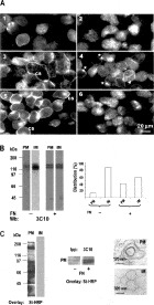
To study the role of the EhFNR during interaction of parasites with ECM components, cultured trophozoites were incubated on FN and the subcellular distribution of the EhFNR was analysed by immunofluorescence. Results clearly showed a redistribution of the EhFNR during interaction of trophozoites with FN, Col, and BSA (100 μg/ml) (Fig. 3A). After 1 h of incubation, the fluorescence pattern was diffuse, although some decorated vesicles were found (Fig. 3A2). Trophozoites incubated for 5 h showed a clear translocation of the EhFNR towards plasma membrane (43% of cells showed surface label) (Fig. 3A3). Longer times of interaction (6–9 h) induced a polarization of the label mainly in the cell-cell interface and still some receptors could be found on the cell surface (Fig. 3A4). Only 5–10% translocation to plasma membrane was observed when cells were incubated with doses of 1–10 μg/ml of FN (data not shown). The specificity of this mobilization was studied using other substrates. In trophozoites incubated with Col, staining of plasma membrane was found in 50% of cells (Fig. 3A5). However, cells incubated with BSA showed only 17% of translocation (Fig. 3A6). Glass was used as non-stimuli control (Fig. 3A1).

Fig. 3. Subcellular distribution of EhFNR in long-term cultured trophozoites incubated on FN. (A) Immunolocalization of EhFNR in long-term cultured trophozoites incubated on glass (A1), FN during 1 h (A2); 5 h (A3); 7 h (A4), or during 5 h on Col (A5) or BSA (C6). Trophozoites were fixed and reacted with the 3C10 mAb. Arrows, vesicle; asterisks, polarization of the EhFNR; CS, cell surface. (B) Subcellular distribution of EhFNR in trophozoites incubated on FN. Plasma membranes (PM) and internal membranes (IM) were prepared from trophozoites in suspension (−) or incubated 5 h on FN (+). Electroblotted proteins (20 μg) were analysed by WB with the 3C10 mAb. Densitometric analysis is representative of 1 experiment done in triplicate. (C) Left panel: PM and IM prepared from FN-incubated and biotinylated trophozoites. Biotin labelling was developed by overlay with St-HRP. Middle panel: EhFNR was immunoprecipitated from biotinylated PM of trophozoites incubated with (+) or without (−) FN using the 3C10 mAb; biotin labelling was developed as above. Right panel: Electron microscopy images of PM and IM, prepared by the procedure described by Aley et al. (1980), are shown for morphological comparison.
To corroborate these findings, the distribution of the molecule was analysed in PM and IM by semi-quantitative Western blot. As shown in Fig. 3B, a drastic change in the allocation of the receptor was found in trophozoites incubated on FN (+) when compared with trophozoites in suspension (−). In the absence of FN, 10% of the protein was associated with PM, whereas 41% of the label was associated with this fraction in the FN condition. This result clearly confirms that the EhFNR molecule is redistributed towards PM as a consequence of FN binding. To demonstrate that the EhFNR molecule in fact was moving from one membrane compartment (IM) to another (PM) and was not the result of cross-contamination of fractions during the purification procedure, FN-incubated and biotinylated trophozoites (+) were subjected to the same procedure. Biotin label was identified by overlay with streptavidin-peroxidase only in the PM fraction (Fig. 3C). None of the biotin label was associated with IM even after 30 min exposure of the blot. Additionally, the increased presence of the EhFNR in the biotin-labelled PM was shown by immunoprecipitation with the 3C10 mAb. This result confirmed that the EhFNR is mostly associated to PM in trophozoites incubated in the presence of FN (Fig. 3C, middle panel). Electron microscopy analysis of PM and IM fractions showed a clear morphological difference between the two fractions, as has been described previously (Aley et al. 1980).
Most of the time trophozoites used to perform biochemical and molecular studies are long-term cultured cells. Therefore, the stimuli that these cells obtain from the environment in culture media are different compared to those obtained by trophozoites in the host-parasite interface during abscess development.
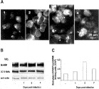
To explore the modulation of the amoebic receptor, as a consequence of the interaction of trophozoites with host cells in an animal model, we analysed the distribution of the EhFNR by immunofluorescence with the 3C10 mAb, in trophozoites recently recovered from a hepatic lesion. In trophozoites recovered from lesions with 24 h of progression the fluorescence labelling was found decorating the cell surface (Fig. 4A2). In contrast, vesicles of different size and clearly defined by the 3C10 mAb were evident after 48 h of host-parasite interaction (Fig. 4A3). These structures were arranged throughout the cytoplasm or were located at one pole of the cell. At 7 days, the 3C10 label was heterogeneously distributed throughout the cells and the appearance of labelled trophozoites at this time of cell recovery was very similar to that of long-term cultured cells (Fig. 4A4 and 4A1 respectively). Even though the cells were not synchronized and the time in culture was variable, the distribution of the EhFNR was reproducible according to the host-parasite interaction time. Since immunofluorescence results suggested redistribution of the EhFNR, the translocation of this protein towards PM after the host-parasite interaction was confirmed through analysis of the level of this molecule in the cell surface (Fig. 4B). The amount of biotinylated EhFNR increased in a time-dependent manner, with a maximum peak shown at 2 days post-infection (Fig. 4C). The total amount of the protein determined with the 3C10 mAb did not show changes (Fig. 4B, 3C10). Anti-actin antibody was used as a control of protein loading (Fig. 4B, anti-actin).

Fig. 4. Subcellular distribution of EhFNR in trophozoites recovered from hepatic lesions. (A) Immunolocalization of the EhFNR. Trophozoites recovered at different times post-infection were adhered to cover-slips, fixed, and reacted with the 3C10 mAb. (A1) Long-term cultured cells; (A2), 1 day; (A3), 2 days; (A4), 7 days. Arrows, vesicles; asterisks, polarization of the EhFNR; CS, cell surface. (B) Translocation of EhFNR. Trophozoites recovered in the same conditions were biotinylated, and total membranes were separated by 7·5% SDS-PAGE, transferred to NCP, and incubated with streptavidin-HRP (St-HRP). Replicates were also incubated with 3C10 mAb, and with anti-actin mAb as loading control. (C) Densitometric analysis of B represents the mean of 2 separate experiments.
Data presented above were obtained with trophozoites recovered from lesions and kept in culture at least 1 week before they were subcultured. During this time in culture, cells could have modified the expression and distribution of the receptor. Therefore, in order to avoid this technical bias, trophozoites recovered at different times post-infection and cultured for different periods up to 48 h without subculturing, were used to repeat some experiments. Under these conditions the same results were obtained (data not shown).
DISCUSSION
Entamoeba histolytica trophozoites can invade the mucosa of the host large intestine, degrade ECM proteins in lamina propria, and reach other organs such as the liver, where abscess development occurs. Participation of amoebic receptor molecules in this process is crucial and the host-parasite interface would lead to activation of several signalling pathways and differential gene expression (Guillén, 1993; Meza, 2000).
Several lines of investigation have suggested that the Gal/GalNAc lectin functions as part of a signalling complex (Vines et al. 1998; Mann, 2002; Petri et al. 2002). A 150 kDa protein that co-purifies with the Gal/GalNAc lectin (Igl) has been described as part of this molecular complex (Cheng et al. 2001). This protein lacks sugar binding domains (Mann, 2002; Petri et al. 2002) and although, its function remains unclear, passive immunization with the EH3015 monoclonal antibody or active immunization with purified 150 kDa antigen, induced protection of hamsters from amoebic liver abscess formation, suggesting an important role for this molecule in the invasive process (Cheng et al. 1999; Cheng and Tachibana, 2001). Comparison of the 5A cDNA insert sequence with the E. histolytica genome (Loftus et al. 2005) showed 99% and 96% identity with the genes that codify for the intermediate Igl2 and Igl1 subunits, respectively. The use of mAb EH3015 (Cheng et al. 2001) against the Igl of the Gal/GalNAc lectin in immunoprecipitation assays confirmed the identity of the EhFNR as the Igl subunit.
The predicted amino acid sequence of the 5A clone revealed the presence of 2 CXC and 19 CXXC motifs in the C terminal region of the molecule similar to those found in the intermediate subunit (Igl) of the Gal/GalNAc lectin (Cheng et al. 2001), G. lamblia variant-specific surface proteins (VSPs) (Nash, 2002), and laminin LE domain (Tunggal et al. 2000). These CXXC-rich proteins fall into the group of transmembrane receptor kinase proteins (TMK) with highly variable extracellular domains and with cytoplasmic kinase domains (Loftus et al. 2005; Beck et al. 2005). Beck et al. (2005) have suggested that the motif CXXC present in the extracellular domain of TMK may form a rod-like structure, as it occurs in laminin, to facilitate trophozoite interaction with host cells. Therefore, the presence of CXXC motif in the amino acid sequence of the 5A clone can be an important element in the adhesion process during host-parasite interaction.
Entamoeba histolytica migration is essential for the development of amoebiasis, a human disease characterized by invasion, destruction of tissues, and hepatic abscess formation (Meza, 2000). The notion that liver abscess formation requires the regulation and concerted action of a variety of amoebic proteins is supported by the fact that a diverse set of genes is specifically up-regulated. These genes codify for proteins associated with stress response, signal transduction, regulation of transcription, and vesicular trafficking (Bruchhaus et al. 2002).
The continuous presence of the trophozoite in the liver requires a strong adaptive response involving specific regulation of many amoebic proteins. To analyse the regulation of the EhFNR after trophozoite interaction with FN and after trophozoite recovery from liver lesions, a cDNA clone coding for a partial sequence of this receptor and the 3C10 mAb were used. The recombinant protein of the selected clone was purified and polyclonal antibodies against this recombinant protein confirmed its identity with the EhFNR. Using the 3C10 mAb to trace the EhFNR, we found that 40% of the receptor protein was allocated to PM when trophozoites were incubated on FN. Using an experimental approach in which trophozoites are newly recovered from liver lesions (Chávez-Munguía et al. 2004), we show in this work that the EhFNR is specifically regulated during abscess development. This specific regulation did not depend on an increase of mRNA (using the 5A cDNA insert as probe) or protein levels (using the 3C10 mAb). On the contrary, EhFNR increased 4-fold on total membranes of cells recovered 2 days post-infection.
The EhFNR behaves functionally and antigenically as an integrin-like molecule (Talamás-Rohana et al. 1994, 1998). According to studies on integrin dynamics in migratory cells, integrins are either ripped off from the membrane and left behind, or collected into vesicles that are transported along the cell body as the cell migrates (Kawakami et al. 2001; Laukaitis et al. 2001; Weeb et al. 2002). In neutrophils the β2 integrin is held in intracellular vesicles and then mobilized to the cell surface after stimulation (Bainton et al. 1987). This mechanism provides means of rapid modulation of integrin function, occurring within minutes.
Entamoeba histolytica is known for its extraordinarily high content of intracellular vesicles, which contain molecules that have been described as participants in virulence or pathogenesis. In this work, using the 3C10 mAb, we observed a differential distribution of the EhFNR molecule associated with these vesicles and/or plasmatic membrane in cells recovered from hepatic lesions. Staining of vesicles was clearly observed at early times while in cells recovered a long time post-infection, the surface membrane was also stained. The size of vesicles was variable and intensity of the staining was higher in trophozoites recovered from hepatic lesions than in long-term cultured cells.
In addition, our results show that some of the vesicles are in an apparent heterotypic fusion process, mainly in cells recovered from hepatic lesions. Rab proteins modulate the vesicular traffic controlling the aggregation and fusion of vesicles (Novick and Zerial, 1997; Pfeffer, 2001; Whyte and Munro, 2002). In E. histolytica the presence of Rab-like proteins has been reported (Temesvari et al. 1999; Welter et al. 2002) and a gene with homology to Rab 7 during the formation of hepatic abscess has been identified (Bruchhaus et al. 2002). More recently, it has been shown that Rab 7 co-localizes with Rab 5 forming pre-phagosomal vacuoles before fusion with phagosomes (Saito-Nakano et al. 2004). We also suggest that the distribution pattern of the EhFNR can reflect its translocation in a route of vesicular traffic dependent on Rab-7 protein, similar to the process described in eukaryotic cells (Bucci et al. 2000). Further experiments are necessary to confirm this hypothesis.
We propose that during the interaction of trophozoites with the micro-environment in the liver, a process of mobilization and/or translocation of the EhFNR may ocurr; moreover, a portion of the cell surface containing this receptor could be endocytosed and recycled during adhesion of trophozoites to host tissue. The redistribution of the EhFNR (Igl) in response to host cells and/or ECM components (i.e. FN) strongly suggests an important role for this molecule during the invasive process.
This work was supported in part by Grant 28077M from Conacyt, México. We thank Dr Bibiana Chávez from the Departamento de Patología Experimental at CINVESTAV for the preparation of electron microscopy photographs from membranes. Also we thank Belem de Luna and Enrique Martínez for their technical assistance and María Guadalupe Aguilar González from the Departamento de Genética y Biología Molecular at CINVESTAV for DNA sequencing. Polyclonal anti-GST antibody was a gift from Dr López-Monteon and monoclonal antibody EH3015 was a generous gift from Dr Hiroshi Thachibana. Department of Infectious Diseases, Tokai University School of Medicine, Isehara, Kanagawa, Tokyo, Japan.