Introduction
Alfalfa weevil, Hypera postica (Gyllenhal) (Coleoptera: Curculionidae), is one of the most economically important insect pests of alfalfa (Medicago sativa Linnaeus; Fabaceae) in North America. It is univoltine in Canada (Bereza Reference Bereza1970) and the northern Great Plains states of the United States of America (Anonymous 1954). Adults overwinter in or near alfalfa fields, emerge and mate in spring; egg production is high, averaging about 600–800 per female, with a recorded maximum of 2414 eggs per female (Yakhontov Reference Yakhontov1934). Eggs, which are laid in ground litter and in old and new alfalfa stems, hatch in about two weeks, and after feeding on alfalfa plants and developing through four instars, larvae reach maturity in three to five weeks. Adults emerge after pupation in plant litter, feed on the plants for a short period, then seek sheltered sites to hibernate until the following spring (Hagan Reference Hagan1918; Hobbs et al. Reference Hobbs, Nummi and Virostek1959; Harcourt et al. Reference Harcourt, Guppy and Binns1977). In western Canada, peak larval feeding activity extends from mid-June to early July, at early to mid-bloom stages of alfalfa (Schaber et al. Reference Schaber, Lysyk and Lactin1994).
Eurasian in origin, the alfalfa weevil is thought to have had three successful introductions into the United States of America: near Salt Lake City, Utah, in 1902 (Titus Reference Titus1911), near Yuma, Arizona, in 1939 (Wehrle Reference Wehrle1940), and in Maryland in 1951 (Poos and Bissell Reference Poos and Bissell1953). The alfalfa weevil was believed to have first entered Canada from Montana, United States of America, with the first Canadian records from southeastern Alberta and southwestern Saskatchewan in 1954 (Hobbs Reference Hobbs1954; Hobbs et al. Reference Hobbs, Nummi and Virostek1959; Beirne Reference Beirne1971). It was accidentally introduced into southern Ontario (Beirne Reference Beirne1971) and southwestern Québec in the 1950s–1960s (Loan Reference Loan1971), likely from the eastern United States of America, and is now found across Canada.
Numerous biological control agents of alfalfa weevil were introduced into North America from multiple European countries in the last century (Day Reference Day and Papavizas1981; Kingsley et al. Reference Kingsley, Bryan, Day, Burger, Dysart and Schwalbe1993; Radcliffe and Flanders Reference Radcliffe and Flanders1998; Kuhar et al. Reference Kuhar, Youngman and Laub1999). The most effective parasitoids introduced to help control alfalfa weevil belong to the genus Bathyplectes Förster (Hymenoptera: Ichneumonidae). At least three Bathyplectes species were introduced to Ontario, Canada from Europe in 1970–1971 (Harcourt and Guppy Reference Harcourt, Guppy, Kelliher and Hulme1984): Bathyplectes anurus (Thomson), B. curculionis (Thomson), and B. stenostigma (Thomson). A subsequent morphological revision of European Bathyplectes (Horstmann Reference Horstmann1974) found that the species B. curculionis is very difficult to distinguish from the species B. exiguus (Gravenhorst), which is also present in North America (Carlson Reference Carlson, Krombein, Hurd, Smith and Burks1979). In addition, Horstmann (Reference Horstmann1974) found morphological variation within B. curculionis and hypothesised that B. curculionis might be a complex of several cryptic species. In a more recent molecular-based study in Virginia, United States of America, Moore (Reference Moore2014) found two distinct haplotypes of the DNA barcoding region of the mitochondrial gene cytochrome oxidase 1 and hypothesised that it may be indicative of incipient speciation. In addition to Bathyplectes species, another parasitoid of alfalfa weevil larvae, Oomyzus incertus Ratzberg (Hymenoptera: Eulophidae), as well as two parasitoids of alfalfa weevil adults, Microctonus aethiops Nees and M. colesi Drea (Hymenoptera: Braconidae), also were released in Ontario in 1970–1971 (Harcourt and Guppy Reference Harcourt, Guppy, Kelliher and Hulme1984). These species, along with the egg parasitoid Anaphes luna (Girault) (Hymenoptera: Mymaridae), which spread naturally from the United States of America, became established in eastern Canada (Harcourt and Guppy Reference Harcourt, Guppy, Kelliher and Hulme1984). Along with the fungus Zoophthora phytonomi (Arthur) Batko (Zygomycetes: Entomophthoraceae) (Harcourt et al. Reference Harcourt, Guppy, MacLeod and Tyrrell1974), this parasitoid complex provides generally effective control of the alfalfa weevil in eastern North America. Insecticidal control is seldom required in areas where three or more of the parasitoid species are established (Flanders and Radcliffe Reference Flanders, Radcliffe, Radcliffe, Hutchison and Cancelado1996; Radcliffe and Flanders Reference Radcliffe and Flanders1998).
Biological control of the alfalfa weevil in western North America has not been as successful as in the east, possibly because of differences in the establishment success of control agents, climate or other environmental factors, the presence of hyperparasitoids, or physiological differences among strains of the alfalfa weevil (Maund and Hsiao Reference Maund and Hsiao1991; Hsiao Reference Hsiao, Symondson and Liddel1996; Achata Böttger et al. Reference Achata Böttger, Bundy, Oesterle and Hanson2013). Bathyplectes curculionis (in one or more of its forms, see Discussion and Key) was introduced into Utah, United States of America from Europe in 1911. It is the only parasitoid of alfalfa weevil that is well established in western North America (Hagen Reference Hagen1990; Brewer et al. Reference Brewer, Donahue and Morrison1997; Rand Reference Rand2013). It was commonly found in southwestern Alberta, Canada, in 1954 along with the first findings of alfalfa weevil (Hobbs Reference Hobbs1954; MacNay Reference MacNay1957; Hobbs et al. Reference Hobbs, Nummi and Virostek1959). In rearing studies conducted in 1956, parasitism rates by B. curculionis as high as 17% were recorded from alfalfa weevil larvae from southwestern Saskatchewan, Canada, the only area of the province where B. curculionis was found (C.H. Craig, unpublished data on file at Saskatoon Research and Development Centre, Saskatoon, Saskatchewan, Canada). Despite the Carlson (Reference Carlson, Krombein, Hurd, Smith and Burks1979) report that the Canadian distribution of the wasp was from southern Québec west to southern Alberta, the distribution of B. curculionis in western Canada is not well known.
The objectives of this investigation were to determine the distribution and population size of alfalfa weevil in Saskatchewan, to assess population trends and parasitism by Bathyplectes and other biological control agents in selected alfalfa production regions in Alberta, Saskatchewan, and Manitoba, Canada, and to provide diagnoses, both morphological and molecular, for the described Bathyplectes species in North America.
Materials and methods
Alfalfa weevil populations in Saskatchewan 2001–2006
In conjunction with a project to detect the presence and spread of alfalfa blotch leafminer (Agromyza frontella (Rondani); Diptera: Agromyzidae) across Saskatchewan, alfalfa fields were inspected for the presence of and injury by alfalfa weevils in surveys conducted in 2001–2006, with one site in southwestern Manitoba sampled from 2001 to 2005. Sampling sites were selected on a semi-random basis, initially in the southern and eastern regions of the province where occurrence of alfalfa blotch leafminer was expected. Selected fields were aligned with major highways, usually more than 40–50 km from the previous field, and were hay or seed fields with alfalfa density greater than 50% of the stand. On occasion, when no alfalfa fields were found at the required distance, alfalfa in roadside ditches was sampled if the cover was more than 100 m in length and greater than 50% alfalfa. Fields were visited once, usually in June or early July, and sampled by sweeping and/or stem collecting of alfalfa in a preordained pattern depending on the configuration of the field; care was taken to sample representative portions of the field and not field edges. When sweeping, 10 180° walking sweeps were taken at each of 10 stops in the field that were 25–50 m apart, and the sweep net emptied into separate plastic bags for each stop. At each of the 10 stops, three undisturbed stems from nearby alfalfa were collected and placed in paper bags, with one bag for each field. Sweep and plant samples were later examined under a stereomicroscope at the Saskatoon Research and Development Centre of Agriculture and Agri-Food Canada (Saskatoon, Saskatchewan, Canada), and alfalfa weevil adults and larvae were identified by morphological features (Whiteford and Quisenberry Reference Whiteford and Quisenberry1990).
Alfalfa weevil populations across Saskatchewan 2010–2012
Because of concerns about increasing alfalfa weevil infestations in Saskatchewan (Hartley Reference Hartley2008), a formal survey of alfalfa hay fields across the province was conducted in 2010–2012, with the assistance of forage crop specialists from the Saskatchewan Ministry of Agriculture, to monitor alfalfa weevil distribution and to determine the identity and prevalence of other insect pests in the crop. In the latter half of June and first part of July, crop specialists sampled alfalfa hay fields in all nine agricultural districts of the province by sweep net and visual inspection of stems as described previously, with sampling of two to eight fields per crop district, and one visit per field. Efforts were made to survey all areas of the province wherever alfalfa hay was grown. The insects and alfalfa stems that were collected were examined at Saskatoon Research and Development Centre at Saskatoon, where the percentage of stems damaged by alfalfa weevil was determined and insects were identified and/or categorised as alfalfa pest, beneficial (known biological control agents) or benign (nonharmful) insects. The distribution and numbers of alfalfa weevils were determined from these samples.
Alfalfa weevil populations at selected sites across Alberta, Saskatchewan, and Manitoba 2013–2014
In conjunction with a study to examine accuracy of growing-degree-day models for the prediction of alfalfa weevil population development (Soroka et al. Reference Soroka, Grenkow, Cárcamo, Meers, Barkley and Gavloski2019), in 2013–2014 five alfalfa hay fields in Alberta, five in Saskatchewan, and three in Manitoba were sampled weekly by sweeping and stem sampling from the beginning of adult alfalfa weevil discovery in May until after larval populations peaked in late June to early July. The methodology was similar to that of previous surveys, with stem samples examined in situ and insect samples examined at Saskatoon Research and Development Centre, Lethbridge Research and Development Centre, Lethbridge, Alberta, or Alberta Agriculture and Forestry, Crop Diversification Centre South, Brooks, Alberta. Voucher specimens of alfalfa weevils collected from 2010 to 2014 are deposited at the Saskatoon Research and Development Centre, Saskatoon, Saskatchewan, Canada, and the E.H. Strickland Entomological Museum, University of Alberta, Edmonton, Alberta, Canada.
Bathyplectes and other alfalfa weevil biological control agents
Contents of the sweep samples that were sent to the Saskatoon laboratory in the sampling during 2010–2012 and 2013–2014 above were examined for the presence of adult Bathyplectes parasitoid wasps. Further, in early July of 2013 and 2014, when most alfalfa weevil larvae were in or approaching fourth instar, bulk sweep collections were made at sites at or near the sampling sites described in the 2013–2014 sampling and the alfalfa weevil larvae transported to Saskatoon, where they were placed, in groups of up to 1000, into 1-m3 screened emergence cages (BugDorm, Taichung, Taiwan) containing mature stands of the alfalfa cultivars Grimm (during 2013) or AC Longview (during 2014) that had previously been sprayed with a solution of Malathion 500 insecticide at a rate of 3 L/ha to eradicate insects. Insects in the collecting containers at the top of the cages were retrieved weekly until the advent of killing frosts in October. After the first snowfall, the cages were removed from the field and the alfalfa left under natural overwintering conditions. The cages were returned after snow melt in the following spring but before any overwintering insects became active, and the insects emerging from the overwintered alfalfa were collected until the end of June. Bathyplectes specimens were identified by A.M.R.B., and other reared hymenopteran specimens of interest were submitted for identification by other taxonomists of the Canadian National Collection of Insects, Arachnids, and Nematodes, Ottawa, Ontario, Canada. Bathyplectes species were identified using keys modified from the revision of Horstmann (Reference Horstmann1974). Voucher specimens are deposited in the Canadian National Collection of Insects, Arachnids, and Nematodes (unique identifiers are listed in Table 5).
Molecular sequencing and sequence analysis
Molecular sequences from the cytochrome oxidase I gene of the mitochondrion (the DNA barcode region) were obtained from selected specimens of Bathyplectes collected in the current study, and from historical specimens in the Canadian National Collection of Insects, Arachnids, and Nematodes, by submission of tissue samples to the Canadian Centre for DNA Barcoding, Guelph, Ontario, Canada. Methods for DNA extraction, polymerase chain reaction protocols, and DNA sequencing followed established Canadian Centre for DNA Barcoding protocols (http://ccdb.ca/resources). Specifically, glass fibre plate DNA extraction was used with 96 well plates using insect lysis buffer as detailed in Ivanova et al. (Reference Ivanova, deWaard and Hebert2006). Amplification of DNA by polymerase chain reaction used the following master mix per 12.5 μL reaction: 6.25 μL 10% trehalose, 2 μL distilled H20, 1.25 μL 10× buffer, 0.625 μL 50 mM MgCl2, 0.125 μL of each primer (10 μM), 0.0625 μL 10 mM dNTPs, 0.06 μL of Platinum Taq polymerase (Invitrogen, Thermo Fisher Scientific, Waltham, Massachusetts, United States of America), and 2 μL of DNA template. Depending on the specimen, the following primer sets were used (see Table 4) individually or in combination: LCO1490 and HCO2198 (Folmer et al. Reference Folmer, Black, Hoeh, Lutz and Vrijenhoek1994); LepF1 and LepR1 (Hebert et al. Reference Hebert, Penton, Burns, Janzen and Hallwachs2004); C_LepFoIF and C_LepFolR (Hernández-Triana et al. Reference Hernández-Triana, Prosser, Rodríguez-Perez, Chaverri, Hebert and Gregory2014); and C_ANTMR1D (Smith and Fisher Reference Smith and Fisher2009). Thermocycle programmes for polymerase chain reaction followed the conditions outlined in the primer publications listed above. Sequencing was performed on an Applied Biosystems 3730xl DNA analyser (Thermo Fisher Scientific). In addition, some published DNA barcode sequences were downloaded from GenBank (Benson et al. Reference Benson, Cavanaugh, Clark, Karsch-Mizrachi, Lipman, Ostell and Sayers2017). Sequences were aligned and translated to amino acids to confirm the lack of stop codons or indels in Geneious 10.0.7 (Kearse et al. Reference Kearse, Moir, Wilson, Stones-Havas, Cheung and Sturrock2012). Sequence lengths ranged from 275 to 658 base pairs (see Table 4), and analyses of relationships were performed using the entire 658 base pair alignment with gaps treated as missing data. Sequence divergences were calculated using DNADist version 3.5c (Felsenstein Reference Felsenstein1993) implemented through BioEdit (Hall Reference Hall1999). Barcode index numbers (BINs) based on 2% or greater sequence divergence of DNA barcodes were calculated using the Barcode of Life Data Systems version 4 (BOLD) (Ratnasingham and Hebert Reference Ratnasingham and Hebert2013). Relationships of specimens were analysed using a parsimony approach implemented in Tree Analysis Using New Technology (TNT) (Goloboff et al. Reference Goloboff, Farris and Nixon2003), with outgroup DNA barcode sequences obtained from two other campoplegine genera: Lemophagus Townes and the putatively closely related genus Rhimphoctona Förster (Hymenoptera: Ichneumonidae) (Wahl Reference Wahl1991). Optimal tree length was found 20 times using the default settings of “xmult” plus 10 cycles of tree drifting. The optimal trees found were then imported into Winclada 1.00.08 (Nixon Reference Nixon2002) for tree viewing. In addition, relationships of specimens were analysed using maximum likelihood with the rapid hill-climbing algorithm and a single starting tree, implemented in RaxML 8.0 (Stamatakis Reference Stamatakis2014). The data were partitioned by codon position and the model used was GTR + Gamma, as determined by PartitionFinder2 (Lanfear et al. Reference Lanfear, Frandsen, Wright, Senfeld and Calcott2017). Branch support was calculated by conducting 100 bootstrap replicates in RaxML using the same model parameters. All sequences are deposited in GenBank (www.ncbi.nlm.nih.gov/genbank) (MN729629–MN729686; Table 5).
Results
Alfalfa weevil distribution
In total, 330 data points from 177 sites were obtained in 11 years of investigation, 2001–2006 and 2010–2012 in Saskatchewan, and 2013–2014 in Alberta, Saskatchewan, and Manitoba (Supplementary Table 1). By alfalfa type, 280 were hay fields, 36 were seed fields, 10 were roadside ditch sites, and four were wildlife habitat or research sites.
Alfalfa weevil populations in Saskatchewan 2001–2006
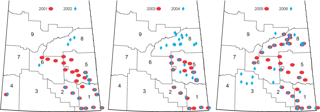
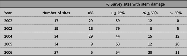
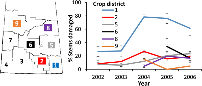
Alfalfa weevil presence was light in early surveys and was initially concentrated in the southeastern corner of the province, from which came the greatest producer concern. In a survey of 11 alfalfa fields in southeastern Saskatchewan and one southwestern Manitoba site in early July of 2001 (Fig. 1; Supplementary Table 1), only one site near Estevan had appreciable alfalfa weevil populations at 14.5 ± 9.1 alfalfa weevil larvae and adults per sweep, while counts at the other 10 sites were two alfalfa weevils/sweep or lower. Because alfalfa weevil had been found in the southwestern corner of the province since 1954, general surveying for the alfalfa weevil was not carried out in this area. However, a hay field near Maple Creek had an alfalfa weevil population of 0.9 ± 0.3 alfalfa weevils per sweep in 2001, lower than numbers found in the area in the 1990s (J.S., unpublished data). In a September 2001 survey of 13 additional fields in east central Saskatchewan, foliar damage typical of alfalfa weevil larval or adult feeding as diagrammed by Johansen and Retan (Reference Johansen and Retan1971) was found at low levels at six sites (Supplementary Table 1). Because alfalfa weevil numbers were generally low in early surveys and difficult to determine by sweeping, in the following five years damage symptoms to alfalfa stems and leaves (Johansen and Retan Reference Johansen and Retan1971) were used as alfalfa weevil population indicators. The number of sites surveyed and the survey area were expanded over 2002–2006 to include sites across the entire eastern half of the province (Fig. 1). The number of survey sites with no evidence of alfalfa weevil feeding decreased over the time of the survey, while those with levels of feeding of over 50% of the stems examined increased (Table 1). We initially categorised our findings by ecozone and ecoregion (Ecological Stratification Working Group 1995), but found no pattern of alfalfa weevil distribution by ecoregions, which run diagonally from southeast to northwest across the province, during this time period. Rather, alfalfa weevil distribution appeared to be related to provincial crop district boundaries. Crop district 1, in the southeastern corner of the province (including the one Manitoba location), had the highest proportion of stems damaged in all years surveyed (Fig. 2), well above the nominal economic threshold for alfalfa hay (20–30 larvae per 90° sweep if 12% hay loss is acceptable; Aasen and Bjorge Reference Aasen and Bjorge2009) or seed production (20–25 larvae per 90° sweep or 35–50% of foliage tips damaged; Aasen and Biorge Reference Aasen and Bjorge2009; Saskatchewan Ministry of Agriculture 2019) in the last three years of the survey. All other crop districts had variable, but much lower, levels of damage from alfalfa weevils through the sampling period. This included five alfalfa fields in the northwestern portion of crop district 3, north and east of Maple Creek that was surveyed only in 2006 (Fig. 1), which had an average of 33.3 ± 7.8% stems with alfalfa weevil damage.
Crop districts 1–9 and sites sampled for alfalfa weevil injury in alfalfa across Saskatchewan, 2001–2006 (n = 169).

Percentage of survey sites with varying levels of alfalfa stems exhibiting alfalfa weevil feeding symptoms in Saskatchewan and one Manitoba site, 2002–2006 (number stems examined per site = 30).

Mean per cent alfalfa stems damaged (± standard error) by alfalfa weevils in six crop districts across eastern Saskatchewan 2002–2006. Economic threshold for alfalfa seed – 20–30 larvae per 90° sweep or 35–50% of foliage tips with damage (Saskatchewan Ministry of Agriculture 2019).

Alfalfa weevil populations across Saskatchewan 2010–2012
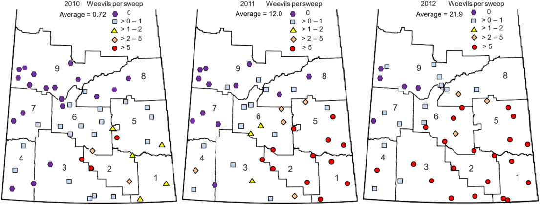
The 2010–2012 survey encompassed the entire agricultural area of the province. In total, 135 fields were sampled, 48 in 2010, 45 in 2011, and 42 in 2012, with both sweep and plant samples taken. The initial pattern of alfalfa weevil population distribution in 2010 was similar to that seen in the earlier survey, with numbers low across Saskatchewan, averaging 0.72 ± 0.21 larvae per sweep, but highest in the southeastern quadrat of the province (Fig. 3). Alfalfa weevils expanded rapidly in number and population distribution across the province in 2011 and 2012, with average number of alfalfa weevils per sweep increasing to 12.0 ± 4.07 in 2011 and 21.9 ± 4.97 in 2012. By the end of the survey, only 12% of the sites monitored were free of alfalfa weevil (Table 2); these sites were in crop districts 7 and 9, in the north and western regions of the province (Fig. 3). The greatest number of alfalfa weevils collected in the three year survey, 135 ± 50 alfalfa weevil larvae and adults per sweep from Churchbridge in southeastern Saskatchewan, on 29 June 2011, was well above the 20 per sweep nominal threshold (Saskatchewan Ministry of Agriculture 2019). The survey also illustrated how quickly alfalfa weevil became a dominant pest of the crop in the province, with 10, 27, and 55% of the fields surveyed having alfalfa weevil as the primary insect pest in 2010–2012, respectively. On a per site basis, 0, 16, and 33% of fields surveyed in 2010–2012, respectively, had alfalfa weevil numbers greater than nominal thresholds.
Densities of alfalfa weevils and sites where they were collected in nine crop districts of Saskatchewan, 2010–2012. Total number of sites surveyed in 2010 = 48, in 2011 = 45, and in 2012 = 42.

Percentage of survey sites with varying densities of alfalfa weevils in Saskatchewan 2010–2012.

Alfalfa weevil populations at selected sites across the prairies 2013–2014
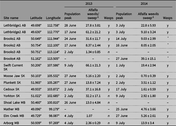
Eleven of the 13 sites surveyed in each year were sampled in both years. In 2013, peak numbers were generally high, with six of the sites having population peaks greater than 20 alfalfa weevils per sweep (Table 3); the greatest numbers of alfalfa weevil adults and larvae were found in the southwest near Lethbridge, Alberta and Swift Current, Saskatchewan. The range in date of occurrence of peak populations was from 17 June to 9 July. Maximum numbers of alfalfa weevils were much lower in 2014 than in 2013, with only two sites in southern Alberta having peak alfalfa weevil populations over 20 per sweep. The dates of occurrence of peak populations, from 16 June to 17 July, were later in 2014 than in 2013, with nine of the 13 sites sampled in 2014 having their greatest numbers in July (Table 3). In a comparison of monthly mean, minimum, and maximum temperatures, and monthly precipitation accumulation from January of 2012 until July of 2014 (Environment and Climate Change Canada 2019a) with comparable 30-year long-term normal values from 1981 to 2010 (Environment and Climate Change Canada 2019b), the most consistent weather factor among sites was a trend in low temperature across the prairies in the winter of 2013–2014. Mean December 2013 temperatures were lower than normal, and February 2014 temperatures were much colder than normal, averaging −17.4 °C over the 11 sites, 7.2 °C colder than the average February long-term normal temperature of −9.9 °C.
Sampling sites for prairie-wide alfalfa weevil survey, the date of alfalfa weevil population peak, the number of alfalfa weevil adults and larvae per sweep at the peak, and the presence (y) or absence (n) of Bathyplectes wasps at each site over the sampling period, 2013–2014.

AB, Alberta; MB, Manitoba; SK, Saskatchewan.
* Not determined.
Bathyplectes curculionis and other biological control agents
All female specimens of Bathyplectes collected in the current study were identified morphologically as B. curculionis. Males of B. curculionis and B. exiguus are indistinguishable; hence, males were identified simply as Bathyplectes species in Table 4 and Figs. 4–5. With respect to female B. exiguus, Horstmann (Reference Horstmann1974) cited the longer ovipositor sheath length as the only character that could distinguish female B. exiguus from B. curculionis. We often found this character difficult to use because of differences in the position of the ovipositor relative to the apex of the metasoma that affected its apparent length, but none of the females collected in the current study appeared to have the ovipositor sheaths longer than the metatibia (i.e., B. exiguus). See the key and subsequent notes at the end of the results for more discussion of the distinction between B. curculionis and B. exiguus.
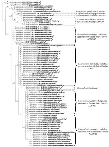
Strict consensus cladogram from parsimony analysis of DNA barcoding region of COI sequence for Bathyplectes specimens and outgroups. Number of equally parsimonious cladograms = 103, tree length = 780, consistency index = 0.62, and retention index = 0.88. Numbers above branches are synapomorphies supporting nodes or terminals. See Table 5 for range of genetic distances between barcode index numbers. bp, base pairs. †Denotes historically collected specimens from the Canadian National Collection of Insects, Arachnids, and Nematodes. *Denotes specimens collected in the current study.

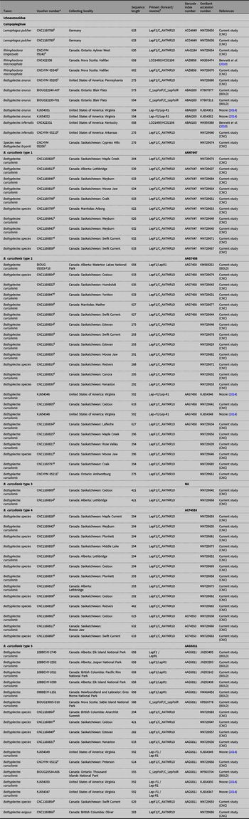
In terms of DNA barcodes, sequence was analysed from a total of 77 ichneumonid specimens (Table 4; Figs. 4–5). Of these, sequences were obtained from 47 specimens of Bathyplectes curculionis collected in the current study from Alberta, Saskatchewan, and Manitoba. In addition, four historically collected Bathyplectes specimens from the Canadian National Collections of Insects, Arachnids, and Nematodes were also sequenced, three that keyed out as B. curculionis, and one (CNC1160866) with ovipositor sheaths longer than the metatibia that was identified as B. exiguus (see Notes on North American species). Thirteen B. curculionis sequences were also downloaded from GenBank, five from the study of Moore (Reference Moore2014), and eight from sequences submitted by the Centre for Biodiversity Genomics. The total number of specimens of the B. curculionis–B. exiguus species complex was 64. For comparison, DNA barcodes were obtained from eight specimens of authoritatively identified specimens of other Bathyplectes species from the Canadian National Collections of Insects, Arachnids, and Nematodes and GenBank (Table 4). Finally, five outgroup specimens were sequenced (Table 4; Figs. 4–5).
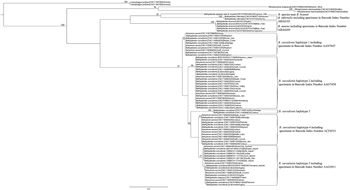
Maximum likelihood analysis of DNA barcoding region of COI for Bathyplectes specimens and outgroups. Bootstrap values above 50% are given for major branches, based on 100 replicates.

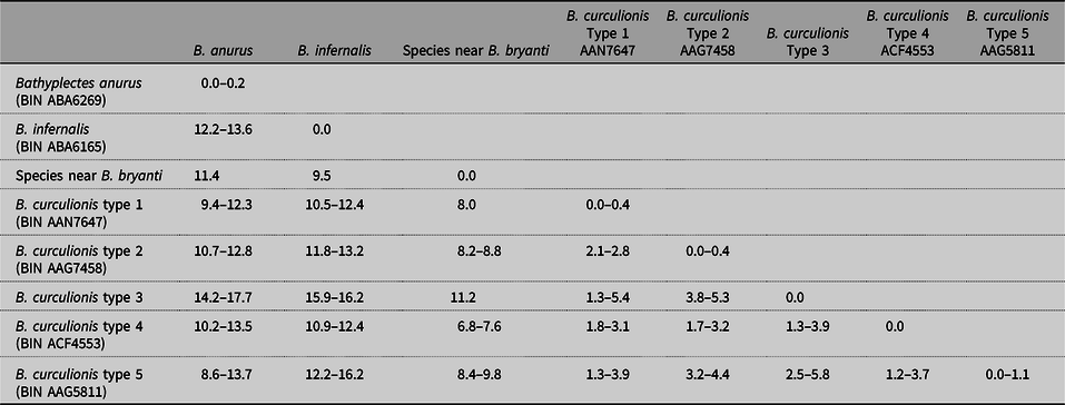
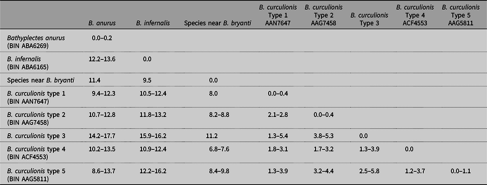
The DNA barcode region of both Bathyplectes anurus and B. infernalis was clearly differentiated from specimens identified morphologically as B. curculionis, with sequence divergences ranging from 8.6–17.7% (B. anurus to B. curculionis) and 10.5–16.2% (B. infernalis to B. curculionis) (Table 5). In addition, a single historical specimen from the Canadian National Collections of Insects, Arachnids, and Nematodes previously identified as B. bryanti (CNCHYM052206) was molecularly distinct compared to all other specimens (8.0–11.2% divergence from specimens of B. curculionis). Morphological comparison of this specimen with the holotype of B. bryanti revealed significant differences; specifically, the areolet of the fore wing is not petiolate in the sequenced specimen but is in the holotype (see Notes on North American species), and the carinae of the propodeum differed. Consequently, this specimen is thought to be a different species than B. bryanti (referred to as “Species near Bathyplectes bryanti” in Tables 4–5; Figs. 4–5). Note that apart from the 77 specimens from which DNA was obtained, additional specimens of Bathyplectes were also submitted to BOLD, including several specimens of B. stenostigma. Unfortunately, no sequence was obtained from any of the specimens of B. stenostigma.
The DNA barcode of B. curculionis was more variable than B. anurus and B. infernalis. Whereas the DNA barcode region of the latter two species was very conserved (0.5% sequence divergence within B. anurus and 0.2% within B. infernalis for specimens in BOLD), the DNA barcodes for B. curculionis diverged by up to 5.8% in our study (Table 5). Accordingly, on the BOLD website, specimens of B. curculionis are placed in four BINs (Figs. 4–5; Tables 4–5). In addition, two specimens (CNC1160809 and CNC1160814) with sequences below 500 base pairs (the threshold for being placed in a BIN) were molecularly distinct from these four BINs in both the parsimony and likelihood analyses (Figs. 4–5). In Table 5, the genetic differences between the five different haplotypes of B. curculionis are considered separately.
The parsimony analysis found 103 equally parsimonious trees (consistency index = 0.62). The overall topology of the tree was consistent among all these trees, and all ambiguous relationships were merely differences between particular specimens of B. curculionis. The strict consensus of the 103 trees is shown in Figure 4. Lemophagus pulcher (Szepligeti) (Hymenoptera: Ichneumonidae) was used to root the cladogram, and Rhimphoctona was the closest genus to Bathyplectes. Within Bathyplectes, there was a grouping consisting of Bathyplectes anurus + (species near B. bryanti + B. infernalis). This grouping was sister group to Bathyplectes curculionis. The DNA barcodes confirmed the morphological identifications. All 47 of the sequenced Bathyplectes specimens collected in this survey clustered within the B. curculionis clade, including all of the sequenced males, which confirmed their species identification. There was moderate sequence divergence among the 47 collected specimens in that some specimens were found in each of the five haplotypes (Fig. 4). Attempts were made to find diagnostic morphological characters that supported each of these haplotypes, but none were found. In fact, the character that Horstmann (Reference Horstmann1974) used to distinguish varieties in this group (the length of the ovipositor sheath) did not appear to be diagnostic between our haplotypes. For example, the one historically collected Canadian National Collections of Insects, Arachnids, and Nematodes specimen that was identified as B. exiguus (CNC1180866) clustered within BIN AAG5811 with an identical DNA barcode to a B. curculionis specimen (CNCHYM05212) from Peterson, Saskatchewan, which had the ovipositor sheaths shorter than the metatibia. The maximum likelihood analysis (Fig. 5) found the same relationships as the parsimony analysis (Fig. 4), although there was weak bootstrap support (below 50) for several nodes. Rhimphoctona always clustered with Bathyplectes, but some bootstraps found that Bathyplectes was paraphyletic with respect to Rhimphoctona. In addition, the sister group to B. curculionis was equivocal – either (B. anurus + (species near B. bryanti + B. infernalis)), which is the same as the parsimony analysis (species near B. bryanti + B. infernalis), or B. anurus. Finally, some of the groupings within the Bathyplectes curculionis complex were equivocal (see nodes in Fig. 5 for which no bootstrap values are provided; i.e., bootstraps below 50).
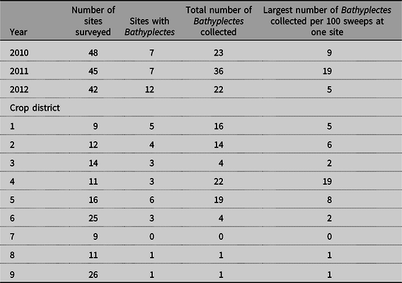
With respect to its distribution, Bathyplectes adults were swept from alfalfa fields from eight of the nine crop districts in Saskatchewan in the 2010–2012 survey (Table 6). Distribution of Bathyplectes was correlated with alfalfa weevil distribution, with the greatest number of sites containing Bathyplectes found in crop districts 1, 2, and 5 (Table 6), in the south and eastern parts of the province where alfalfa weevil populations were the highest (Fig. 3). Conversely, crop districts 7, 8, and 9, in northern and northwestern Saskatchewan, had the fewest number of Bathyplectes (Table 6) along with the lowest levels of alfalfa weevils in the survey (Fig. 3). The most Bathyplectes specimens per site in the three years of sampling were found near Maple Creek in crop district 4 on 12 July 2011, when 19 adult wasps were collected in 100 sweeps of the field.
Specimen data for Bathyplectes and related taxa sequenced for the DNA barcoding region of COI.
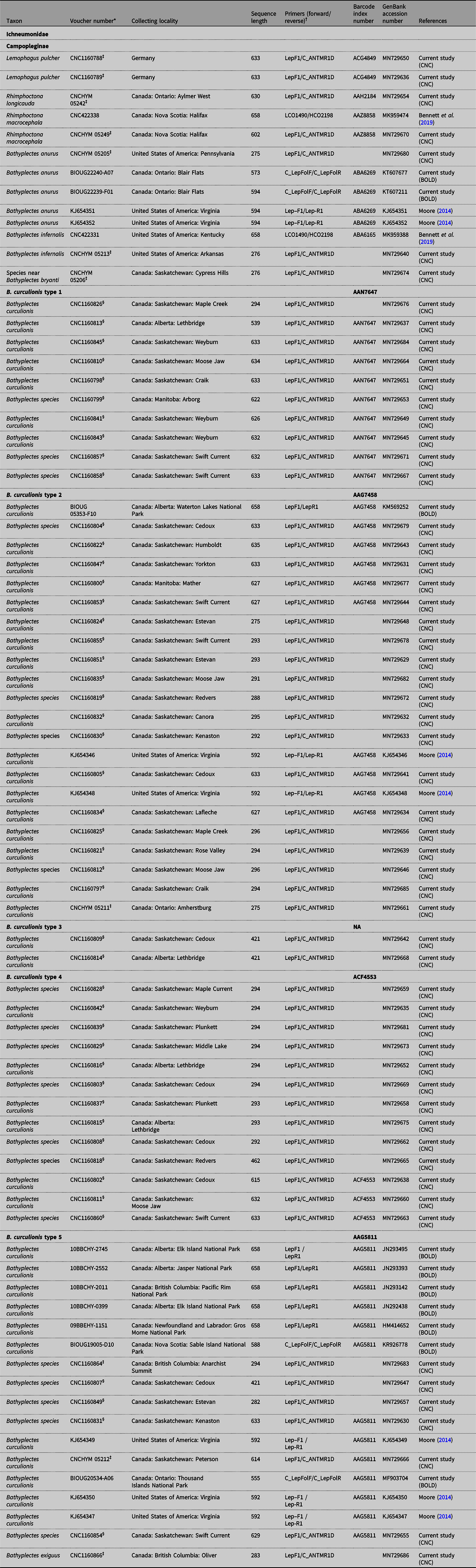
* All specimens with Canadian National Collection of Insects, Arachnids, and Nematodes unique identifiers (CNC prefix) are deposited in Canadian National Collection of Insects, Arachnids, and Nematodes, all with BIOUG or 10BBCHY are deposited at the Centre for Biodiversity Genomics, University of Guelph (Guelph, Ontario, Canada). Sequences from specimens with a KJ prefix in their voucher number were obtained from GenBank from the study of Moore (Reference Moore2014).
† See Methods section for references describing the primers used in polymerase chain reactions.
‡ Denotes historical Canadian National Collection of Insects, Arachnids, and Nematodes specimen sequenced in the current study.
§ Denotes fresh specimen collected in the current study. Specimens identified only as Bathyplectes species are males that could not be morphologically identified to species.
Sequence similarity matrix (% divergence) for DNA barcoding region of COI (n = 77). Missing data were not included in distance measurement calculations. Bathyplectes curculionis based on morphological identification of at least one female in each haplotype or barcode index number, as calculated by BOLD (2% or less sequence divergence within each barcode index number for sequences 500 or more base pairs). Divergences between barcode index numbers that are less than 2.0% are from comparisons of sequences less than 500 base pairs (see Table 4 for sequence lengths).
Number of Bathyplectes collected in 100 sweeps of alfalfa fields in Saskatchewan in late June or early July, 2010–2012, classified by year and crop district.

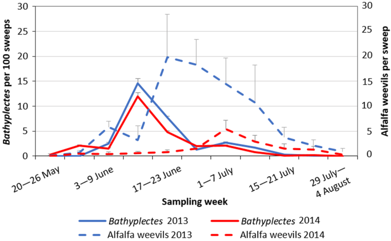
Monitoring of a select number of sites across the three prairie provinces during 2013–2014 facilitated determination of seasonal B. curculionis population development. Bathyplectes wasps in varying numbers were found at eight sites in Alberta and Saskatchewan in 2013 and 10 sites in all three provinces in 2014 (Table 3). One Bathyplectes curculionis male (CNC1160799) was found in 100 sweeps from Arborg, Manitoba, on 9 July 2014, and one B. curculionis female (CNC1160800) was collected in 100 sweeps from the site near Mather, Manitoba, on 24 July 2014. These specimens are the first confirmed records of B. curculionis in Manitoba. Despite the lower number of alfalfa weevils found in 2014 than in 2013, the population curves of B. curculionis are similar for the two years in shape and number (Fig. 6); the total numbers of Bathyplectes collected in the two years were 250 and 266 for 2013 and 2014, respectively. Peak Bathyplectes numbers were found in mid-June, two to three weeks before peak alfalfa weevil larval numbers (Fig. 6). The greatest numbers of Bathyplectes collected per site were 66 ± 40 per 100 sweeps along with 61 ± 21 alfalfa weevil larvae and adults per sweep on 17 June at Lethbridge2 in 2013 and 74 ± 40 per 100 sweeps with 1.3 ± 0.5 alfalfa weevils per sweep at Swift Current on 11 June 2014.
Number of Bathyplectes adults per 100 sweeps and mean number alfalfa weevils per sweep from eight alfalfa fields in 2013 and 10 in 2014 across the three prairie provinces.

Inspection of emergence cages with 1000 alfalfa weevil larvae added from each of 12 sites on 3–11 July 2013 (no live alfalfa weevils were retrieved from the Brooks3 site) resulted in collection of 659 alfalfa weevil adults in total, or 5% emergence, from July to October, with low numbers possibly due to prolonged drought of the alfalfa over the summer. No Bathyplectes wasps were collected from the cages over that summer or the spring of 2014. Of the few insects that were collected, one Eulophidae (Hymenoptera), recovered on 23 August 2013, from a cage stocked with alfalfa weevil larvae collected from Elm Creek, Manitoba, on 3 July, was identified by Canadian National Collections of Insects, Arachnids, and Nematodes systematist John Huber as the introduced alfalfa weevil parasitoid Oomyzus incertus (Ratzeburg) (Hymenoptera: Eulophidae). One wasp collected on 27 June 2014 from an emergence cage that was initially placed on alfalfa on 3 July 2013 and stocked with alfalfa weevils from Lethbridge, Alberta, was identified by Canadian National Collections of Insects, Arachnids, and Nematodes systematist Henri Goulet as the introduced alfalfa weevil braconid parasitoid Microctonus colesi Drea (Hymenoptera: Braconidae).
In 2014, the alfalfa caged for infestation of larvae in July was more luxurious and vigorous than that used in 2013. The total number of alfalfa weevil adults retrieved from some of the 12 cages (no live larvae were collected from Brooks4) was greater by the end of the summer than the number of alfalfa weevil larvae originally placed in them. Further, sentinel cages that had not been infested with larvae from our monitoring sites had small numbers of alfalfa weevil adults, indicating that our initial insecticide treatment had not totally eradicated the background endemic Curculionidae population. In light of these developments, the origin of the Bathyplectes wasps that emerged from the cages could not be guaranteed. In sum, 53 Bathyplectes adults were collected from all cages from their placement in early July until 1 October 2014, while 93 Bathyplectes wasps were collected from the cages from 11 May until 22 June 2015, with a peak of 35 ± 2.6 Bathyplectes adults collected on 25 May.
Other alfalfa weevil-specialist parasitoids collected from emergence cages included 120 specimens of O. incertus retrieved on 15 August 2014 from an emergence cage that was originally stocked with alfalfa weevils collected from Moose Jaw, Saskatchewan, in July. Twenty-five specimens (range 1–8, averaging 3.1 ± 2.4 specimens per site) of O. incertus were collected on 15 and 22 August 2014, from emergence cages earlier stocked with alfalfa weevils from Lethbridge, Rosemary, and Millicent, Alberta; Swift Current, Yorkton, Plunkett, and Cedoux, Saskatchewan; and Woodside, Manitoba. In the spring of 2015, three O. incertus were found in the cage previously stocked with alfalfa weevils from Moose Jaw, two from Millicent, and one each from cages with alfalfa weevils from Plunkett and Woodside. No O. incertus specimens were found in the sentinel cages in any year. No M. colesi was identified from any of the emergence cages stocked in 2014.
Identification guide to the described species of North American Bathyplectes
The purpose of this identification guide is to aid North American biological control workers who are involved in programmes to help control Hypera Germar species. No attempt has been made to describe and include undescribed North American species in this work, and therefore, some specimens, especially those not reared from pest hosts, may not key out correctly. Nevertheless, the importance of Bathyplectes to biological control and the lack of a current illustrated key to all described North American species justify its publication.
Generic diagnosis
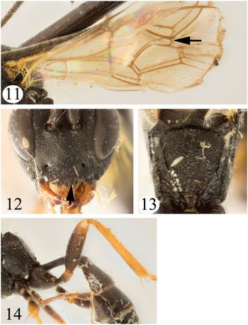
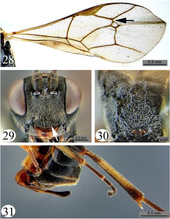
Bathyplectes can be distinguished from all other genera of Campopleginae by combination of the following characters: (1) glymma (lateral depression anterior to spiracle) of the first metasomal tergite absent; (2) areolet of fore wing closed, often pentagonal (Figs. 7, 15, 23), but quadrangular (strongly petiolate anteriorly) in some species (Figs. 11, 28); (3) suture separating tergite and sternite of first metasomal segment far ventral to middle; (4) hind wing vein 1/Cu&cu-a (= nervellus) angulate (Fig. 7), intercepted by vein 2/Cu (= discoidella), although 2/Cu usually unpigmented; (5) eye margins not indented medially near antennae (Fig. 8), their surface bare or only sparsely setose, lacking long, dense setae between ommatidia; (6) protibia with a strong to weak tooth anteriorly at apex; (7) ovipositor length ranging from nearly as long as apical depth of metasoma (i.e., the “height” of the apex of the metasoma in lateral view) (see Figs. 10, 31) to slightly shorter than length of metasoma (Fig. 27); and (8) mandibles lacking a ventral flange. The unusual characters for Campopleginae are the complete lack of a glymma and, in most Nearctic species (except B. bryanti, B. infernalis, and some specimens of B. stenostigma), the pentagonal areolet of the fore wing. Combination of these two characters will diagnose most specimens. The tooth on the protibia (if it is visible) is also uncommon in the subfamily.
Described species
Thirty species worldwide, including seven in North America (not including B. glacialis Jussila from Greenland) (Yu et al. Reference Yu, Achterberg and Horstmann2016).
Distribution
Holarctic and oriental
Hosts
All substantiated host records are from weevils (Coleoptera: Curculionidae) of the genus Hypera (Carlson Reference Carlson, Krombein, Hurd, Smith and Burks1979), but most species have no host records.
Taxonomic literature
Horstmann (Reference Horstmann1974) revised the European species including all but two of the described Nearctic species: B. bryanti (Viereck) and B. sessilis (Provancher). Carlson (Reference Carlson, Krombein, Hurd, Smith and Burks1979) provided nomenclature for the North American species as well as distributional information.
Key to described species of Bathyplectes of North America

Notes on North American species
Bathyplectes anurus (Thomson, 1887) (Figs. 7–10). This species was introduced from Europe and has successfully established across much of North America, including California, Florida, and the northeastern United States of America and Ontario (Dysart and Day Reference Dysart and Day1976; Carlson Reference Carlson, Krombein, Hurd, Smith and Burks1979). So far it has not been recorded in any other provinces in Canada. The cocoons are dark brown with a raised, white equatorial band and have the unusual ability to jump when disturbed (Day Reference Day1970).
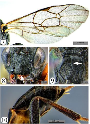
Bathyplectes anurus. 7, Fore wing; 8, head, anterior view (arrow points to impuncate region at apex of clypeus); 9, propodeum, dorsal view (arrow points to indentation of medial, longitudinal carina near junction of areolar and petiolar areas; 10, posterior metasoma and metathoracic leg, lateral view.

Bathyplectes bryanti (Viereck, 1925) (Figs. 11–14). This species is only known from the female holotype from Alberta (deposited in the Canadian National Collection of Insects, Arachnids, and Nematodes). The host, male, and cocoon of B. bryanti are unknown.
Bathyplectes bryanti female holotype. 11, Fore wing; arrow points to strongly petiolate areolet of fore wing; 12, head, anterior view; 13, propodeum, dorsal view; 14, metasoma and metathoracic leg, lateral view.

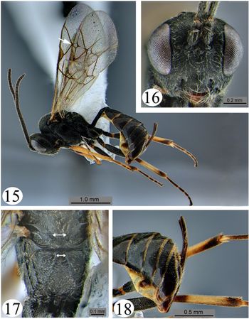
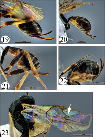
Bathyplectes curculionis (Thomson, 1887) (Figs. 15–23). This species is now recorded in all Canadian provinces from British Columbia to Québec. The cocoon is brown with a white, flattened (not raised) equatorial band.
Bathyplectes curculionis (haplotype 2). 15, Lateral habitus (arrow points to narrowly sessile (pentagonal) areolet of fore wing); 16, head, anterior; 17, propodeum, dorsal view (double-headed arrows indicate width of apex of scutellum compared to width of space between medial longitudinal carinae of propodeum at anterior of propodeum); 18, metasoma and metathoracic leg, lateral view.

Bathyplectes curculionis. 19, Haplotype 5 (CNCHYM 05212), metasoma and metathoracic leg, lateral view; 20, haplotype 4 (CNC1160802), metasoma and metathoracic leg, lateral view; 21, haplotype 1 (CNC1160847), metasoma and metathoracic leg, lateral view; 22, haplotype 3 (CNC1160809), metasoma and metathoracic leg, lateral view; 23, fore wing, haplotype 5 (CNCHYM 05212) (arrow points to widely sessile (pentagonal) areolet of fore wing, contrast with Fig. 15).

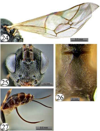
Bathyplectes exiguus (Gravenhorst, 1829) (Figs. 24–27). Carlson (Reference Carlson, Krombein, Hurd, Smith and Burks1979) hypothesised that B. exiguus is adventive in North America because species that later became junior synonyms of B. exiguus were collected in the late 19th century and early 20th century prior to introductions of Bathyplectes for biological control. This includes Campoplex (Bathyplectes) phytonomi Viereck known from Ontario, Québec, and British Columbia. The holotype of C. (B.) phytonomi is a male (Canadian National Collection of Insects, Arachnids, and Nematodes), and therefore of questionable identity (it could be B. curculionis), but female paratypes clearly have the ovipositor sheath longer than the metatibia and the ovipositor strongly upcurved, similar to Figure 27. Specimens of this species in the Canadian National Collection of Insects, Arachnids, and Nematodes are also known from St. John’s Newfoundland and Sable Island, Nova Scotia (both new provincial records).
Bathyplectes exiguus. 24, Fore wing; 25, head, anterior view; 26, propodeum, dorsal view; 27, metasoma and metathoracic leg, lateral view.

Bathyplectes infernalis (Gravenhorst, 1820) (Figs. 28–31). This species was introduced to North America from its native Europe and southwest Asia (Carlson Reference Carlson, Krombein, Hurd, Smith and Burks1979). There are no published records of it from Canada, but it is established in various states in the United States of America including California, Michigan, Mississippi, and New York (Carlson Reference Carlson, Krombein, Hurd, Smith and Burks1979).
Bathyplectes infernalis. 28, Fore wing (arrow points to strongly petiolate (quadrangular) areolet of fore wing); 29, head, anterior view (arrow points to punctation of clypeus that extends to apical edge); 30, propodeum, dorsal view; 31, metasoma and metathoracic leg, lateral view.

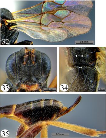
Bathyplectes sessilis (Provancher, 1875) (Figs. 32–35). This species is known only from the female holotype from Québec in the Provancher Collection (University of Laval, Ville de Québec). Townes (Reference Townes1945) synonymised B. sessilis with B. exiguus, but Carlson (Reference Carlson, Krombein, Hurd, Smith and Burks1979) resurrected it because he believed that it had been incorrectly synonymised. Carlson (Reference Carlson, Krombein, Hurd, Smith and Burks1979) did not elaborate on the reason for this decision. The original description (Provancher Reference Provancher1875) states that the ovipositor is almost as long as the abdomen which for described North American species resembles only B. exiguus; therefore, the Carlson (Reference Carlson, Krombein, Hurd, Smith and Burks1979) decision must have been based on something other than the ovipositor length. The host, male, and cocoon are unknown.
Bathyplectes stenostigma. 32, Fore wing; 33, head, anterior view; 34, propodeum, dorsal view (double-headed arrows indicate width of apex of scutellum compared to width of space between medial longitudinal carinae of propodeum at anterior of propodeum); 35, metasoma and metathoracic leg, lateral view.

Bathyplectes stenostigma (Thomson, 1887). Bathyplectes stenostigma was introduced to the eastern United States of America from Europe starting in 1964 (Radcliffe and Flanders Reference Radcliffe and Flanders1998). It had become established by 1970 and has spread from the east to as far west as Wyoming (Brewer et al. Reference Brewer, Donahue and Morrison1997). It was also released and established in Ontario (Carlson Reference Carlson, Krombein, Hurd, Smith and Burks1979) (the only Canadian province from which it is currently recorded). As noted in the key, its cocoon is covered by a brown, wizened bag that does not have a white, equatorial band.
Discussion
The data collected from the current investigation document the pattern of spread of alfalfa weevil from 2001 to 2012 from several ecoregions across the southern portion of Saskatchewan to the eastern half of the province, and then north and west to include most of the agricultural areas of the province. This rapid rate and area of expansion contrast with the previous distribution pattern of the alfalfa weevil in the province. The first Canadian discovery of alfalfa weevil was in southern Alberta and near Maple Creek in southwestern Saskatchewan in 1954 (Hobbs Reference Hobbs1954; Hobbs et al. Reference Hobbs, Nummi and Virostek1959). Although small numbers of the alfalfa weevil were found as far east as Weyburn, Saskatchewan, about 100 km west of the Manitoba border, in 1955 (J.C. Arrand, unpublished observations, data on file at Saskatoon Research and Development Centre, Saskatoon, Saskatchewan, Canada), its traditional occurrence was in an area of about 116 000 ha below 51°N (Craig Reference Craig, Davis and Hower1972), stretching from Regina, Saskatchewan, to southwestern Alberta; this distribution remained static for about 45 years (Soroka and Goerzen Reference Soroka and Goerzen2002). Alfalfa weevil was slow to gain pest status, with the first record of significant alfalfa weevil injury to alfalfa in Saskatchewan seen in irrigated alfalfa fields near Maple Creek in 1984 (Craig Reference Craig1984), 30 years after its first discovery in the province. Damage, sporadic in occurrence, remained confined to this area for the next 15 years. However, in 2000 the alfalfa weevil was reported for the first time as a significant pest in alfalfa and clover (Trifolium Linnaeus; Fabaceae) fields in the Estevan area in southeastern Saskatchewan (Hartley Reference Hartley2000). As distribution has advanced across the province, so has economic impact of alfalfa weevil (Hartley Reference Hartley2006, Reference Hartley2008).
First reports of noticeable alfalfa weevil damage in Manitoba were from the southwestern area of the province in 2001 (Gavloski Reference Gavloski and Jones2001), suggesting that it had expanded its range into Manitoba from populations in southeastern Saskatchewan. By 2005, damage was reported across southern Manitoba and north into the Interlake region (Gavloski Reference Gavloski and Beres2005), and by Reference Gavloski2008, it was found across the Interlake into northern and northwestern areas abutting the Saskatchewan border (Gavloski Reference Gavloski2008). Infestations of alfalfa weevil now occur across Manitoba (Gavloski Reference Gavloski2017).
Traditionally an economic problem only below 51°N in Alberta as in Saskatchewan, alfalfa weevil has been moving north in Alberta. In a 2014–2016 survey of alfalfa insects in Alberta, Sim and Meers (Reference Sim and Meers2016) found a notable expansion of range as far north as the Parkland Ecoregion north of Edmonton, 53.5°N, considerably farther north than typically observed (Sim and Meers Reference Sim and Meers2016). In the Alberta survey, several of the new sites in the Alberta Aspen Parkland where alfalfa weevil was found were close to the Saskatchewan border, with a discontinuous distribution from southern Alberta, and may represent a more westerly than northerly expansion pattern (see Sim and Meers Reference Sim and Meers2016, Fig. 8).
The possible explanation for this distribution spread from the first collections of the alfalfa weevil in the southwestern prairie provinces to the east, then northwards, and then westwards may be related to variation in geographical climate changes. Temperature is the most critical environmental factor to affect alfalfa weevil natural history, and in northern latitudes the overwintering temperatures near the soil surface, where alfalfa weevils overwinter, play a vital role in their survival and spread (Cook Reference Cook1925; Essig and Michelbacher Reference Essig and Michelbacher1933; Yakhontov Reference Yakhontov1934; Flanders et al. Reference Flanders, Radcliffe and Krueger1994; Saeidi and Moharramipour Reference Saeidi and Moharramipour2017). Soil temperatures near the soil surface can differ tremendously from ambient air temperatures, depending on the type and depth of insulation such as snow or leaf litter (Peterson Reference Peterson1960; Larsen et al. Reference Larsen, Brun and Enz1983; Sharratt et al. Reference Sharratt, Baker, Wall, Skaggs and Ruschy1992; Cárcamo et al. Reference Cárcamo, Herle, Otani and McGinn2009). A comparison of long-term weather data from southwestern (Maple Creek, Swift Current) and southeastern Saskatchewan (Estevan) weather stations revealed that snow cover levels did not decrease in either region in the 1960–1990 and 1981–2010 time periods. Instead, soil temperatures over the winter increased at Estevan compared to southwestern areas of the province (Environment and Climate Change Canada 2019b), which may have been a critical factor in alfalfa weevil range expansion and population growth eastward in that time period. An in-depth correlation of weather parameters and alfalfa weevil distribution is necessary to define the relationships between such parameters and alfalfa weevil survival and spread across the Canadian Prairies.
Regardless of the underlying reasons, the pattern of alfalfa weevil distribution observed in Saskatchewan is similar to the west to east distribution pattern of other curculionid pests in the province, including cabbage seedpod weevil Ceutorhynchus obstrictus Marsham (Dosdall et al. Reference Dosdall, Weiss, Olfert and Cárcamo2002; Olfert and Weiss Reference Olfert and Weiss2006) and pea leaf weevil Sitona lineatus Linnaeus (Coleoptera: Curculionidae) (Vankosky et al. Reference Vankosky, Dosdall and Cárcamo2009; Olfert et al. Reference Olfert, Weiss, Cárcamo and Meers2012). A better understanding of the factors underlying alfalfa weevil spread on the prairies could aid in determining the ultimate distribution of these and other potential Curculionidae pests.
DNA barcoding was used to elucidate the identity of the Bathyplectes species collected because of difficulties in identifying Bathyplectes specimens, especially males, morphologically. In terms of relationships indicated by our phylogenetic analyses, the maximum likelihood bootstrap trees, which found Rhimphoctona as paraphyletic to Bathyplectes, were not expected. The natural history of Rhimphoctona (parasitoids of Coleoptera deep in tree trunks) and the many associated adaptations to this lifestyle such as the very long ovipositor (Wahl Reference Wahl1991) do not support the hypothesis that these two genera are paraphyletic. We prefer to accept the sister–group relationship proposed by the parsimony analysis (Fig. 4). In terms of relationships within Bathyplectes, all of the species were well supported by 26 or more derived substitutions (Fig. 4) and bootstrap values of 87 or higher (Fig. 5). The congruence of the parsimony and likelihood topologies implies that either model is appropriate for analysing these data. The lack of support for some nodes within Bathyplectes in the likelihood analysis (Fig. 5) suggests that additional genes may be required to examine relationships within Bathyplectes further.
The relatively moderate sequence divergence (5.8%) within specimens identified morphologically as B. curculionis was not expected, but was similar to the results of Moore (Reference Moore2014) in Virginia who found two separate haplotypes. Using the guidelines suggested by BOLD, wherein 2% sequence divergences of the DNA barcode region are indicative of species differences (Ratnasingham and Hebert Reference Ratnasingham and Hebert2013), the current results could be used to justify the division of B. curculionis (including B. exiguus – see below) into as many as five species. The argument against this action is that no consistent morphological characters were found that correlated with the five BINs. In addition, whereas 5.8% divergence in the DNA barcoding region may be indicative of species differences in many taxa (see Ratnasingham and Hebert Reference Ratnasingham and Hebert2013 for data on Aves, Teleostei, Lepidoptera, and Hymenoptera), the interspecific differences between the DNA barcodes of other Bathyplectes species imply that the 2% divergence criterion may too low for species within this genus, as exemplified by our 8.6–17.7% divergence between B. anurus and B. curculionis, 10.5–16.2% between B. infernalis and B. curculionis, and 12.2–13.6% between B. anurus and B. infernalis. Similar high interspecific rates of divergence have been observed in other genera of ichneumonids. For example, Bennett and Barnes (Reference Bennett and Barnes2014) found interspecific differences in DNA barcodes up to 15.0% among species of the tryphonine genus Chiloplatys Townes. On the basis of these arguments, we conclude that the moderate genetic differences observed among the collected specimens in the current study are likely intraspecific and not indicative of cryptic species. This can be explained by the relatively broad geographic range from which B. curculionis was collected within Europe for release in North America. Known introductions of B. curculionis originated from geographically distant countries that included France, Sweden, the former Union of Soviet Socialist Republics, and “the Near East” (Radcliffe and Flanders Reference Radcliffe and Flanders1998); therefore, it would be expected that North American populations are genetically diverse.
The DNA barcode analysis also raised questions regarding the status of B. exiguus relative to B. curculionis. The revision of Horstmann (Reference Horstmann1974), based on morphological examination, including the primary types of the two species, concluded that they were distinct but very closely related species. Our DNA barcode results did not separate these two species in that a specimen with ovipositor sheath longer than the metatibia (Fig. 27) had an identical 283 base pair fragment of the DNA barcode to one with the sheath shorter than the metatibia (Fig. 19). On this basis, it could be argued that B. exiguus is a synonym of B. curculionis, but the rather striking morphological differences in the ovipositor length and shape argues against this synonymy. The ineffectiveness of DNA barcodes to differentiate some campoplegine ichneumonids has been found previously. For example, no consistent differences in three gene regions were found between two Campoplex species with different hosts and ecology (Jenner et al. Reference Jenner, Jenner, Kuhlmann, Bennett, Cossentine, Mason and Gillespie2013). In the absence of further evidence such as breeding experiments, both B. curculionis and B. exiguus are considered valid species.
Based on our conclusion that only B. curculionis was collected in this survey (albeit in five haplotypes), it is possible to discuss the distribution of B. curculionis in a similar manner as alfalfa weevil. Relative to eastern North America, less is known about its parasitism of alfalfa weevils in the west. Bathyplectes curculionis became established in Utah shortly after its first introduction from Europe in 1911 (Chamberlain Reference Chamberlain1926) and is now the predominant parasitoid of alfalfa weevil in the western United States of America (Brewer et al. Reference Brewer, Donahue and Morrison1997; Rand Reference Rand2013). It was found with alfalfa weevil in Alberta in the first record of the pest in southeastern Alberta/southwestern Saskatchewan, likely entering from Montana, in 1954 (Hobbs Reference Hobbs1954), and was reared from alfalfa weevil larvae collected in several sites in south western Saskatchewan in 1956 (C.H. Craig, unpublished data, on file at Saskatoon Research and Development Centre, Saskatoon, Saskatchewan, Canada). However, its numbers there did not match the rapid increase in numbers of the alfalfa weevil seen in the area (McLeod Reference McLeod1962), and although Carlson (Reference Carlson, Krombein, Hurd, Smith and Burks1979) listed the distribution of B. curculionis in Canada as southern Québec west to southern Alberta, there have been few records of the ichneumonid in western Canada outside of southeastern Alberta/southwestern Saskatchewan. Our data indicate a fairly general distribution of B. curculionis across Saskatchewan, and considering the discovery of the parasitoid in Manitoba in this study and in records by Ostermann (Gavloski and Ostermann Reference Gavloski and Osterman2018) in that province, it is likely that B. curculionis now occurs in most areas where alfalfa weevil is found on the Canadian Prairies.
Bathyplectes curculionis is a solitary endoparasitoid, ovipositing most frequently in younger instar alfalfa weevil larvae. It has one complete generation in spring followed by a partial or complete second generation in southerly areas of the continent (Essig and Michelbacher Reference Essig and Michelbacher1933; Radcliffe and Flanders Reference Radcliffe and Flanders1998, and references therein). Our Bathyplectes population curves showed univoltinism to be the case across the three prairie provinces, with adult populations peaking in mid-June. This information has important implications for alfalfa weevil management in terms of avoiding the use of insecticides when the adult parasitoids are most active. Increased parasitism could help delay alfalfa weevil resistance to insecticides, which presently may be developing in southern Alberta (Meers Reference Meers2016).
Considering other parasitoids of alfalfa weevil, the multivoltine, gregarious, eulophid endoparasitoid O. incertus was originally introduced from France into North America for alfalfa weevil control, with establishment in eastern United States of America by 1960 (Streams and Fuester Reference Streams and Fuester1967). Although multiple early releases of O. incertus in western United States of America did not result in establishment there, it recently has been found in the border states of Montana and North Dakota (Rand Reference Rand2013). According to the distribution list of Burks (Reference Burks, Krombein, Hurd, Smith and Burks1979), in Canada this species was not found west of Québec. Our recovery of a specimen of the eulophid from alfalfa weevils collected near Elm Creek, Manitoba in 2013 extends that distribution westward. Although we cannot say with absolute certainty that release cages in 2014 were not contaminated by alfalfa weevil larvae and O. incertus was not already present, the high numbers of adult O. incertus recovered from the cage stocked with alfalfa weevil larvae from Moose Jaw, Saskatchewan, and the absence of Oomyzus Rondani from our sentinel cages leads credence to our assertion that O. incertus is present in that area and may be present at many locations across the three prairie provinces. We can say with certainty that it is now present at Saskatoon, Saskatchewan. In a recent assessment of parasitism of alfalfa weevil at four sites in Manitoba, Ostermann (2017) found O. incertus at all four sites surveyed, with parasitism levels ranging from 1.4–10.0%. Parasitism by B. curculionis also occurred at all four sites, at levels from 7.1–39.4% (Ostermann 2017). Unlike those of B. curculionis, O. incertus females prefer to parasitise older instar alfalfa weevils (Miller Reference Miller1970), and if numbers of the eulophid increase in western Canada, the wasp may complement B. curculionis in biological control of alfalfa weevil.
Microctonus colesi is a thelytokous endoparasitoid of alfalfa weevil larvae whose origins are unclear (Drea et al. Reference Drea, Dysart, Coles and Loan1972; Radcliffe and Flanders Reference Radcliffe and Flanders1998). First found in Pennsylvania in 1962 (Coles and Puttler Reference Coles and Puttler1963), it soon became established in the northeastern United States of America (Day et al. Reference Day, Coles, Stewart and Fuester1971) and, subsequently, southern Ontario (Harcourt and Guppy Reference Harcourt, Guppy, Kelliher and Hulme1984). The species is locally important in controlling alfalfa weevil in the central, northern states of the United States of America (Radcliffe and Flanders Reference Radcliffe and Flanders1998), but, despite introductions (Coulson Reference Coulson1992), it was not found in surveys of the northern states of North Dakota, South Dakota, Wyoming, and Montana (Rand Reference Rand2013; Rand et al. Reference Rand, Pellisier, Jabbour, Lundgren and Waters2018). Apart from two specimens from western Alberta, the most westerly occurrence of M. colesi as recorded in the distribution map of Pucci (Reference Pucci2013) is in the state of Wisconsin. Our finding of a single specimen from Lethbridge despite considerable collecting effort across the Canadian Prairies confirms that M. colesi is present in Alberta, but we could not confirm its presence in Saskatchewan and Manitoba.
Biological control of alfalfa weevil has met with spectacular success in some areas of North America (Day Reference Day and Papavizas1981; White et al. Reference White, Allen, Moffitt and Kingsley1995), modest success in others (Kingsley et al. Reference Kingsley, Bryan, Day, Burger, Dysart and Schwalbe1993, Long et al. Reference Long, Getts, Goding, Stevens, Clark, Putnam and Mostafa2017), and limited success in still others (Pitcairn and Gutierrez Reference Pitcairn and Gutierrez1989). Generally, alfalfa weevil is seldom of economic importance in regions where three or more biological control agents have become established (Flanders and Radcliffe Reference Flanders, Radcliffe, Radcliffe, Hutchison and Cancelado1996). Since the spread of alfalfa weevil across western Canada is a relatively recent phenomenon, as this long-term study confirms, and since it can take five years or more for parasitoids to build up to effective populations (Day Reference Day and Papavizas1981), some time may be required before pest–parasitoid interactions stabilise to impact alfalfa weevil populations in the region. Similar to the surveys of Rand et al. (Reference Rand, Pellisier, Jabbour, Lundgren and Waters2018) in Wyoming, South Dakota, and Montana, our investigation did not find any M. aethiops on the prairies, nor did we collect specimens of B. anurus, two species that are instrumental in control of alfalfa weevil in eastern North America (Radcliffe and Flanders Reference Radcliffe and Flanders1998). Small numbers of both B. anurus and M. aethiops were introduced into the Maple Creek area of Saskatchewan from Ontario in 1985 (C.H. Craig, unpublished data, on file at Saskatoon Research and Development Centre, Saskatoon, Saskatchewan, Canada), and the reasons for their lack of establishment were never determined. As alfalfa weevil becomes a chronic economic problem across western Canada, the re-introduction of these two species should be considered, as was suggested for Montana (Jaronski et al. Reference Jaronski, Rand and Reddy2017). The presence of B. curculionis and other biological control agents encountered in the current investigation, along with the establishment of B. anurus and M. aethiops, holds promise for natural suppression of alfalfa weevil populations below economic thresholds, contributing to sustainable production of alfalfa on the Canadian Prairies.
Acknowledgements
Funding from the Pesticide Risk Reduction Programme of the Pest Management Centre, Agriculture and Agri-Food Canada, for part of this research is gratefully acknowledged. We thank the Saskatchewan Ministry of Agriculture’s Terry Kowalchuk and forage specialists Glenn Barkley, Bill Bilegetu, Al Foster, John Hauer, Lyndon Hicks, Todd Jorgenson, Lorne Klein, Trevor Lennox, Nadia Mori, the late Don Perrault, Ian Schemenauer, Sara Sommerfeld, Charlotte Ward, and especially André Bonneau, for resources and sampling. Hector Cárcamo, Carolyn Herle, and staff of Agriculture and Agri-Food Canada, Lethbridge; Scott Meers, Shelley Barkley, and staff of Alberta Agriculture, Brooks; and John Gavloski and staff of Manitoba Agriculture carried out sampling and insect identification. Larry Grenkow and Brian Galka of Agriculture and Agri-Food Canada, Saskatoon, volunteer Wesley Wizniuk, as well as many summer students assisted in collection and analysis. David Giffen and Jon Williams, Agriculture and Agri-Food Canada Saskatoon, generated maps and graphs. We thank John Huber, Canadian Forest Services, Ottawa, and Henri Goulet, Ottawa Research and Development Centre, Agriculture and Agri-Food Canada, Ottawa, for their identification of parasitoids; and Boyd Mori, Saskatoon Research and Development Centre, Agriculture and Agri-Food Canada, Saskatoon, for alfalfa weevil strain identification. The generous expertise of Gary Gibson and Peter Mason of the Ottawa Research and Development Centre, Agriculture and Agri-Food Canada, Ottawa, is gratefully acknowledged. Finally, Diana Barnes, Ottawa Research and Development Centre, Agriculture and Agri-Food Canada, Ottawa, is thanked for taking images of Bathyplectes species.
Supplementary material
To view supplementary material for this article, please visit https://doi.org/10.4039/tce.2020.35.