Introduction
One complete cycle of folliculogenesis in the cow is estimated to take between 160 and 180 days (Russe, Reference Russe1983; Lussier et al., Reference Lussier, Matton and Dufour1987). Of these, approximately 140 days are spent between activation of the primordial follicle and the secondary stage, that is during preantral development. A close examination of preantral follicle development revealed characteristics that defined each stage (Fair et al., Reference Fair, Hulshof, Hyttel, Greve and Boland1997a, Reference Fair, Hulshof, Hyttel, Greve and Boland1997b; Fair, Reference Fair2010). Activation of follicle growth marks the transition from primordial to primary follicles that is accompanied by morphological changes in the surrounding granulosa cells and in the oocyte’s cytoplasmic organelles to face the increasing energy demand of growth (Fair et al., Reference Fair, Hulshof, Hyttel, Greve and Boland1997b). The oocytes of primordial and primary follicles are transcriptionally inactive; transcription begins during the secondary stage of development and ceases completely in the early antral stage when the oocyte reaches 120 µm and the follicle, 2–3 mm diameter (Fair et al., Reference Fair, Hulshof, Hyttel, Greve and Boland1997a, Reference Fair, Hulshof, Hyttel, Greve and Boland1997b; De La Fuente and Eppig, Reference De La Fuente and Eppig2001). The secondary stage of development is also when acquisition of communication between the oocyte and surrounding granulosa cells through gap junctions occurs, and cytoplasmic cortical granules and the zona pellucida become apparent (Fair et al., Reference Fair, Hulshof, Hyttel, Greve and Boland1997a, Reference Fair, Hulshof, Hyttel, Greve and Boland1997b; Fair, Reference Fair2010).
Development of methods to efficiently grow preantral follicles after their isolation from the ovarian tissue has tremendous advantages, from treating human infertility to accelerating livestock genetic improvement and preserving endangered species germplasm. With the growing interest in developing methods to efficiently culture isolated preantral follicles in vitro (Kreeger et al., Reference Kreeger, Fernandes, Woodruff and Shea2005; Xu et al., Reference Xu, West, Shea and Woodruff2006; McLaughlin and Telfer, Reference McLaughlin and Telfer2010; Jorssen et al., Reference Jorssen, Langbeen, Marei, Fransen, De porte, Leroy and Bols2015; Xiao et al., Reference Xiao, Zhang, Romero, Smith, Shea and Woodruff2015; Laronda et al., Reference Laronda, Rutz, Xiao, Whelan, Duncan, Roth, Woodruff and Shah2017), it has become imperative that the developmental stages are morphologically well defined in isolated follicles, so that the most appropriate culture conditions can be utilized. This is particularly relevant in species in which preantral follicular development is a lengthy process and follicles require specific conditions for optimal growth at different stages, such as cows and other large mammals including humans (Xu et al., Reference Xu, West, Shea and Woodruff2006; McLaughlin and Telfer, Reference McLaughlin and Telfer2010; Hornick et al., Reference Hornick, Duncan, Shea and Woodruff2012; Araujo et al., Reference Araujo, Gastal, Figueiredo and Gastal2014; Xiao et al., Reference Xiao, Zhang, Romero, Smith, Shea and Woodruff2015; Apolloni et al., Reference Apolloni, Bruno, Alves, Ferreira, Paes, Moreno, de Aguiar, Brandao, Smitz, Apgar and de Figueiredo2016; Rossetto et al., Reference Rossetto, Saraiva, Bernuci, Silva, Brito, Alves, Magalhaes-Padilha, Bao, Campello, Rodrigues and Figueiredo2016).
The classification scheme used for staging growing preantral follicles (i.e. not primordial) – namely primary, early secondary and secondary – is mostly based on histological analysis (Gougeon and Chainy, Reference Gougeon and Chainy1987; van Wezel and Rodgers, Reference van Wezel and Rodgers1996; Braw-Tal and Yossefi, Reference Braw-Tal and Yossefi1997), with limited reports on isolated follicle (Langbeen et al., Reference Langbeen, Jorssen, Fransen, Rodriguez, Garcia, Leroy and Bols2015). Extrapolation of this scheme to classify follicles isolated from the ovaries, however, creates room for variation of interpretation of what each follicular stage looks like in its three-dimensional form. Histological analysis only provides information at a two-dimensional level, therefore cell number, for example, can only be estimated. The goals of this study were to characterize the developmental stages of isolated preantral follicles more comprehensively by morphological appearance, evaluate whether morphological classification could predict follicular cell number and diameter, and to evaluate the agreement between the present results and previously published data that used histological analysis and isolated follicles.
Materials and methods
Preantral follicle isolation
Bovine ovaries were sourced from a local abattoir and transported to the laboratory in warm phosphate-buffered saline solution (PBS) within 4 h of harvest. Preantral follicles were mechanically isolated from the ovaries using a homogenizer (Omni Macro Homogenizer, Omni International, Kennesaw, GA, USA) and subjected to sequential filtration steps as described below. Following wash in warm PBS containing 100 UI penicillin/100 μg/ml streptomycin and removal of adjacent tissue, ovaries were halved and homogenized for 1 min at 6000 rpm. The homogenate was initially filtered through a cloth to remove large debris. Next, the filtrate was strained through a 200 µm followed by through a 40-µm cell strainer. Material retained in the 40-µm strainer was rinsed with warm wash medium (TCM199, containing 3 mg/ml BSA, 25 mM HEPES, 100 UI penicillin/100 μg/ml streptomycin, 1 mM sodium pyruvate, 100 nM NEAA) into a Petri dish and follicles were searched under a stereomicroscope (Olympus SZX16, Tokyo, Japan). Preantral follicles with a visible oocyte, intact basement membrane and absence of antrum were individually selected and placed in 10-µl drops of warm wash medium under mineral oil. Next, follicles were incubated in 0.02% (v/v) trypan blue (Life Technologies, Carlsbad, CA, USA) in PBS for 1 min to assess viability. After three washes in PBS containing 0.1% (w/v) polyvinylpyrrolidone (PVP), unstained follicles were considered viable and immediately fixed in 4% paraformaldehyde for 15 min at room temperature. Follicles that retained trypan blue stain were considered unviable and discarded.
Preantral follicle stage classification by morphological assessment, cell count, and diameter
Isolated preantral follicles were visualized under an inverted microscope (Revolve, Echo Laboratories, San Diego, CA, USA) at ×20 magnification and classified as follows: primary follicles had one layer of cuboidal granulosa cells; early secondary, two to three, and secondary follicles had more than three layers of granulosa cells. Follicle diameter was determined by averaging perpendicular measurements of the two greatest diameters of each follicle utilizing the measurement function available in the Revolve software. For cell counting, follicles were stained with 1 µg/ml Hoechst 33342 for 10 min, mounted onto a glass slide containing Prolong Gold anti-fade (Invitrogen, Carlsbad, CA, USA), and coverslipped. Stained nuclei were counted with ImageJ software (National Institutes of Health, Rockville, MD, USA) using the cell count function. In total, 256 follicles (76 primary, 126 early secondary, 54 secondary) were analyzed in 13 replicates.
Statistical analysis
Data were analyzed using analysis of variance with the generalized linear model procedure using cell number and diameter as dependent variables and developmental stage as an independent variable. Results are reported as least square means ± standard error of the mean. The relationship between follicular diameter and cell number with all follicles combined was analyzed by the Pearson correlation coefficient. Finally, we calculated cell density in each follicle as a ratio of cell number to follicle diameter to obtain number of cells per micrometre, and analyzed the correlation of this cell density with size and cell number in each follicular stage using the Spearman correlation coefficient. All analyses were performed in SAS software version 9.4. A significance level of 0.05 was utilized in all statistical analyses.
Results
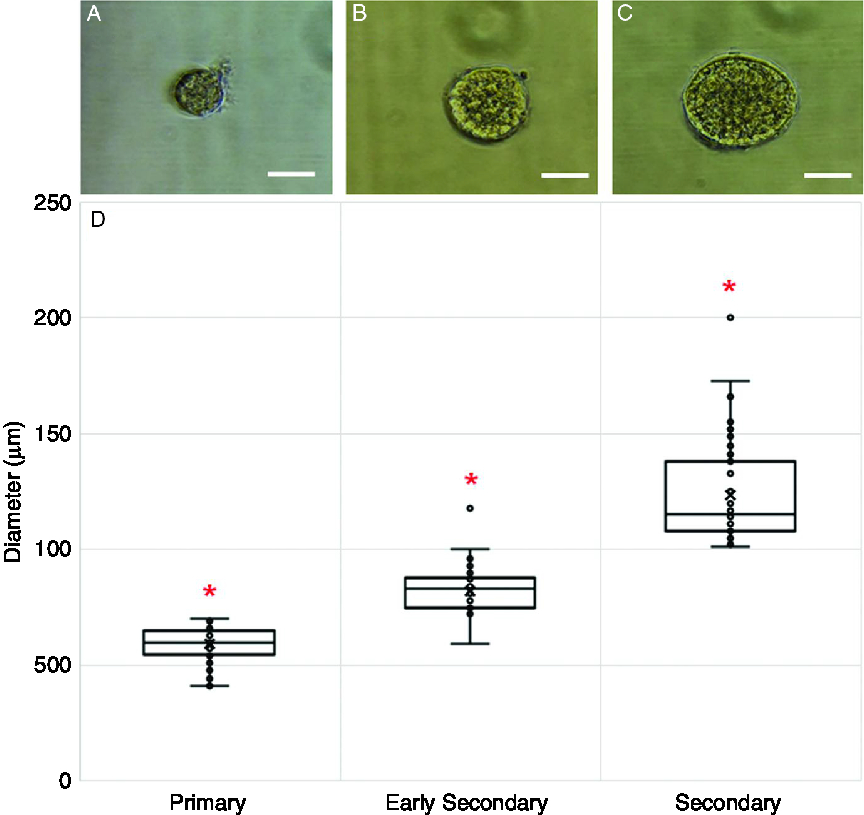
In total, 256 follicles (76 primary, 126 early secondary, 54 secondary) were morphologically classified upon isolation, measured and stained for cell count. Figure 1(A–C) shows representative images of follicles classified by morphology into each of the three stages. The average diameter for follicles of each stage increased from primary to early secondary stage and to secondary stage (P < 0.01), as shown in Fig. 1(D).
Representative images of freshly isolated bovine follicles classified as primary (A), early secondary (B) and secondary (C). (D) Mean diameter was different for each developmental stage (P < 0.01) and was 59.30 ± 1.45 µm (range: 41–70 µm) for primary follicles (n = 76), 82.29 ± 1.14 µm (range: 59–118 µm) for early secondary follicles (n = 126), and 122.51 ± 1.73 µm (range: 101–200 µm) for secondary follicles (n = 54). Results are the least square means ± standard error of the mean. Scale bars (A–C) 50 µm.

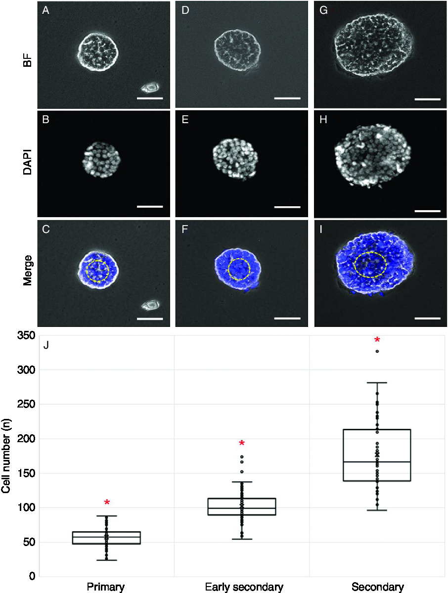
Representative images of follicles of each stage stained with Hoechst 33342 for cell count are depicted in Fig. 2(A–I). Cell number increased between primary and secondary follicles (P < 0.001), and there was a difference in cell number for each stage evaluated (Fig. 2J).
Representative images of freshly isolated bovine follicles classified as primary (A–C), early secondary (D–F) and secondary (G–I). (A, D, G) Each follicle as seen in bright field (BF). (B, E, H) The same follicles following DNA staining with Hoechst 33342 and imaged using the 4′,6-diamidino-2-phenylindole (DAPI) channel for cell counting. (C, F, I) Merged images of transmitted light and DAPI channel. (J) Mean cell number was different for each developmental stage (P < 0.01); primary follicles (n = 76) had a mean of 57.3 ± 3.3 cells (range: 24–88 cells), early secondary follicles (n = 126) 102.3 ± 2.5 cells (range: 54–174 cells), and secondary follicles (n = 54) had mean 175.3 ± 3.9 cells (range: 96–327 cells). Results are least square means ± standard error of the mean. Scale bars (A–I) 50 µm.

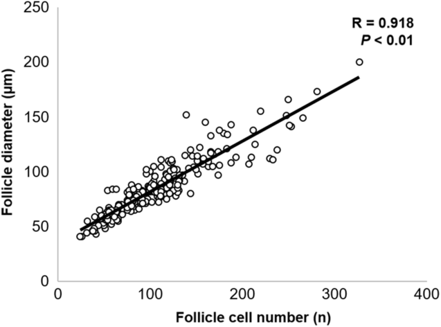
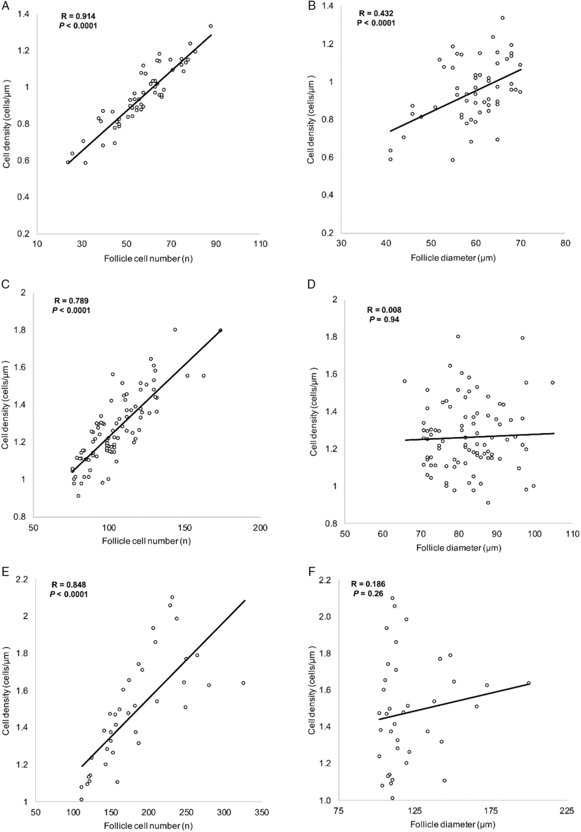
Correlation coefficient analysis showed a strong positive correlation between diameter and cell number (P < 0.01; Fig. 3). Examination of the correlation between cell density, follicle diameter and cell number for each follicular stage revealed a positive relationship between cell density and cell number in all follicle stages evaluated (P < 0.001; Fig. 4A, C, E). The relationship between cell density and follicle size, however, was only significant in primary follicles (Fig. 4B, D, F).
Positive correlation between cell number (x-axis) and follicle diameter (y-axis). Pearson correlation coefficient R = 0.918 (P < 0.01).

Spearman correlation between granulosa cell density (cells/µm), cell number and follicle diameter for primary, early secondary and secondary preantral follicle stages. In primary follicles (A, B) there was a significant positive correlation between cell density of a follicle and its diameter and cell number. In early secondary (C, D) and secondary follicles (E, F), however, a significant correlation with cell density was only found for cell number, but not for follicle diameter, demonstrating that, as preantral follicles develop, cell proliferation rate exceeds diameter growth rate.

We evaluated the diameter and cell number results in comparison with the publication by Braw-Tal and Yossefi (Reference Braw-Tal and Yossefi1997), a publication frequently cited as the source of bovine preantral follicle classification in which the authors utilized histological sections of bovine ovarian cortex to categorize preantral follicles. We found that our measurements (Figs 1D and 2J) agreed well with the ranges described previously for primary (10–40 cells, 40–80 µm diameter), with some discrepancy in the early secondary (41–100 cells, 81–130 µm diameter) and secondary follicles (101–250 cells, 131–250 µm diameter). A second comparison was performed with the study by Langbeen et al. (Reference Langbeen, Jorssen, Fransen, Rodriguez, Garcia, Leroy and Bols2015) that evaluated the diameter of freshly isolated preantral follicles from bovine, ovine and caprine ovaries. The diameter of bovine primary follicles in that study (47.4 ± 5.5 µm) was in agreement and within the range found in the current study; follicles classified as secondary (65.0 ± 12.5 µm) by Langbeen et al. (Reference Langbeen, Jorssen, Fransen, Rodriguez, Garcia, Leroy and Bols2015) were within the diameter range of early secondary follicles in the present study.
Discussion
Consistent and accurate classification of follicles is the first step in studying preantral stages of folliculogenesis. The present work expands on previously published methods for the classification of bovine preantral follicles based on histological sections (Braw-Tal and Yossefi, Reference Braw-Tal and Yossefi1997) and freshly isolated follicles (Jorssen et al., Reference Jorssen, Langbeen, Marei, Fransen, De porte, Leroy and Bols2015; Langbeen et al., Reference Langbeen, Jorssen, Fransen, Rodriguez, Garcia, Leroy and Bols2015). Improvements made here are the use of a wider range of follicle sizes that allows for a more comprehensive capture of preantral follicle stages and, more importantly, individual cell count. As the pivotal early work with isolation of fetal (Hulshof et al., Reference Hulshof, Figueiredo, Beckers, Bevers and van den Hurk1994), postnatal and adult (Figueiredo et al., Reference Figueiredo, Hulshof, Van den Hurk, Ectors, Fontes, Nusgens, Bevers and Beckers1993) bovine preantral follicles, there has been a knowledge gap on detailed classification of follicles once they are isolated from the ovary. An accurate classification system becomes even more important as many research groups studying preantral follicle development are using the bovine as an animal model of human folliculogenesis, and the use of isolated follicles is becoming increasingly common as we search for efficient ways to promote folliculogenesis in vitro. In the present study, bovine preantral follicles between 40 and 200 µm were isolated from adult ovaries using a similar method to the one described by Langbeen and colleagues (Langbeen et al., Reference Langbeen, Jorssen, Fransen, Rodriguez, Garcia, Leroy and Bols2015). As expected, follicle diameter and number of cells increased as stage of development progressed. More importantly, the mean number of cells and diameter for each stage were significantly different from each other, confirming their distinctiveness. The ranges for follicle diameter and cell number in the present study were mostly in agreement with the ones reported by Braw-Tal and Yossefi (Reference Braw-Tal and Yossefi1997) utilizing histological specimens. In a previous study that categorized isolated bovine follicles by morphology and diameter, Langbeen et al. (Reference Langbeen, Jorssen, Fransen, Rodriguez, Garcia, Leroy and Bols2015) reported that secondary follicles had an average diameter of 65.0 ± 12.5 µm. In that study, follicle size was limited by filtration to 100 µm, from which we conclude that the follicles classified as secondary in that study corresponded to our early secondary stage.
To our knowledge, this study was the first to approach counting of granulosa cells as a means of characterizing stage of development of freshly isolated follicles. We found this was a more precise method, because it used the entire follicle instead of estimation models based on histological sectioning or solely counting cells seen in one cross-section of a follicle. For example, Lundy and colleagues (Reference Lundy, Smith, O’Connell, Hudson and McNatty1999) reported that sheep primary follicles contained, on average, 19 cells in the cross-section showing the oocyte. However, using an estimation model, the total number of granulosa cells in the follicle ranged from 30 to 520 (Lundy et al., Reference Lundy, Smith, O’Connell, Hudson and McNatty1999). Likewise, quantification of DNA content in follicles has also led to a large range in granulosa cell number such as primary follicles containing 72–233 and 90–410 cells depending on the equation used (Wang et al., Reference Wang, Roy and Greenwald1991). By counting cells in whole mount follicles we found that the range and variability in the number of cells increased as follicles progressed from the primary to the secondary stage, but to a much less degree. We also noted that, within follicles of each stage, greater numbers of granulosa cells were associated with increased cell density at all stages, but increase in follicle diameter was only associated with increased cell density in primary follicles. This finding suggests that there may be variability in granulosa cell proliferation rate within follicles of similar size. Gap junction formation between oocytes and granulosa cells is variable at the secondary stage and this may explain the differences in proliferation rate based on the ability of the oocyte to communicate with granulosa cells and signal for cell division (Kacinskis et al., Reference Kacinskis, Lucci, Luque and Bao2005). It has been previously shown that once the second layer of granulosa cells forms in early secondary follicles, the cells begin to divide inwards with their axes perpendicular to the basal lamina, and that proliferation of cells occurs more in the inner layers compared with cells lining the basal lamina (Da Silva-Buttkus et al., Reference Da Silva-Buttkus, Jayasooriya, Mora, Mobberley, Ryder, Baithun, Stark, Franks and Hardy2008). This finding supports the hypothesis that granulosa cell–oocyte contact may be important for cell proliferation in early stages and corroborates our finding that cell density was only associated with follicle size in primary follicles, that is in follicles containing only one layer of granulosa cells. The relationship between pattern and rate of granulosa cell proliferation and developmental potential of preantral follicles remains to be explored and could provide an efficient method to predict successful follicular development.
In conclusion, this study using isolated bovine follicles provides insight into the relationships between morphology, diameter and cell number of growing preantral follicles between the primary and secondary stage of development. Overall agreement with previous bovine preantral follicle classification studies confirmed the accuracy of our measurements and validated our classification method. These data give a foundation for preantral follicle classification, which is becoming ever more important as research is increasingly focusing on preantral folliculogenesis.
Acknowledgements
The authors thank all personnel involved in collection and transportation of ovaries from the slaughterhouse.
Financial support
This work was supported by the USDA Regional Research Project: W 3112, Reproductive Performance in Ruminants; and by the Henry A. Jastro Shields Graduate Research Award (UC Davis).
Conflicts of interest
The authors have no conflict of interest to declare.
Ethical approval
The experiments described here used bovine ovaries purchased from a local slaughterhouse. No animal use protocol was required as there were no live animals involved in the research.