Since its introduction by Jatene et al in 1976,Reference Jatene, Fontes and Paulista 1 the arterial switch operation remains the surgical mainstay for transposition of the great arteries. Aortic root dilatation, aortic regurgitation, and pulmonary artery stenosis are possible late complications of the arterial switch operation. We report aortic root dilatation, severe aortic regurgitation, and pulmonary stenosis that were accidentally diagnosed 23 years after the arterial switch operation for transposition of the great arteries in situs inversus. The modified Bentall procedure and pulmonary artery reconstruction were successfully performed.
Case report
A 1-day-old infant with transposition of the great arteries in situs inversus exhibiting a single coronary artery, atrial septal defect, and patent ductus arteriosus underwent balloon atrial septostomy. At 39 months of age, the patient underwent palliative surgery of a right modified Blalock–Taussig shunt and pulmonary artery banding. After 2 months, the arterial switch operation with the Lecompte manoeuvre, atrial septal defect closure, left pulmonary artery patch plasty with a glutaraldehyde-preserved equine pericardial xenograft (Xenomedica AG, Lucerne, Switzerland), and pulmonary artery band removal were performed at another hospital.
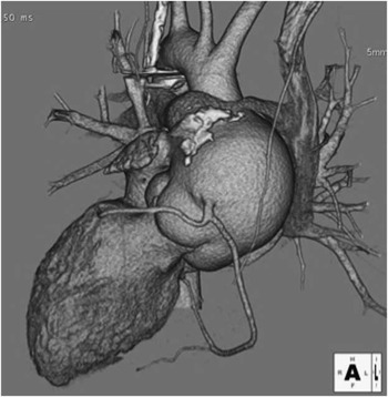
At 26 years of age, the patient presented to our hospital with spontaneous left pneumothorax. Further evaluation revealed aortic root dilatation, severe aortic regurgitation, and pulmonary stenosis. Multi-slice computed tomography with three-dimensional reconstruction revealed anterior displacement of the pulmonary trunk and left pulmonary artery due to aortic root dilatation. The diameters of the aortic valve annulus, the aortic sinus, and the previous site of aortic anastomosis were 29, 60, and 24 mm, respectively (Fig 1). Cardiac catheterisation revealed a right ventricular systolic pressure of 84 mmHg and a main pulmonary artery pressure of 44 mmHg. The patient was asymptomatic after conservative treatment for pneumothorax via chest drainage, and surgical repair was planned. Under general anaesthesia, the previous median sternotomy was revised. After careful dissection of adhesions, the heart and great arteries were exposed. Cardiopulmonary bypass was established with femoral arterial perfusion and right atrial drainage. The aorta was transected following aortic cross-clamping, and cardioplegia was introduced into the single coronary artery. The redundant aortic leaflets were mildly thinned and resected. The modified Bentall procedure was performed, and the single coronary artery was reconstructed using the Carrel patch technique. Next a vertical incision was made in the pulmonary artery. The pulmonary valve had no stenotic lesion, and there was no right ventricular outflow tract stenosis. Owing to the fact that the Xenomedica patch was severely calcified and protruded internally, we excised it and reconstructed the pulmonary trunk using a cut-open prosthetic graft (HEMASHIELD® Platinum™ Woven Double Velour 12-mm vascular graft; Boston Scientific, Natick, Massachusetts, United States of America). The left pulmonary artery was reconstructed with another graft. The post-operative course was uneventful. Post-operative multi-slice computed tomography with three-dimensional reconstruction showed no stenotic lesions in the reconstructed pulmonary artery. Cardiac catheterisation performed 1 year after the operation revealed a right ventricular systolic pressure of 37 mmHg and a main pulmonary artery pressure of 38 mmHg.
Pre-operative multi-slice computed tomography with three-dimensional reconstruction revealed aortic root dilatation and resultant anterior displacement of the pulmonary artery.

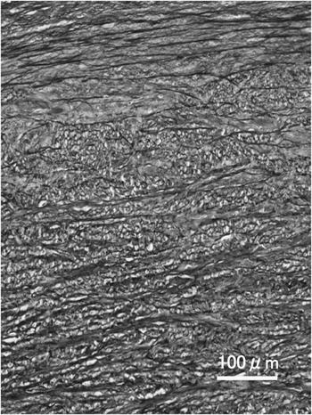
Pathological examination of the dilated aortic wall revealed intimal atherosclerosis, linear necrosis of the tunica media characterised by fragmentation of the elastic lamina and decrease in its thickness, and acid mucopolysaccharide deposition (Fig 2). Pathological examination of the aortic valve revealed severe mucoid degeneration, moderate fibrosis, and hyalinosis.
Pathological examination of the dilated aortic wall. Linear necrosis of the tunica media characterised by fragmentation of the elastic lamina (Elastic van Gieson; ×100).

Discussion
Aortic root dilatation, aortic regurgitation, and pulmonary stenosis are major complications after the arterial switch operation, and their development is considered to be time dependent.Reference Bové, De Meulder and Vandenplas 2 Prior pulmonary artery banding and single coronary were considered to be risk factors for developing such complications.Reference Mavroudis, Stewart and Backer 3 , Reference Schwartz, Gauvreau and Nido 4 Our patient last visited a hospital at 15 years of age, and pre-operative computed tomography at 26 years of age revealed significant aortic root dilatation. Pathological examination of the dilated aortic root showed intimal atherosclerosis, linear necrosis of tunica media, and acid mucopolysaccharide deposition.
In the reconstruction of the pulmonary artery, there is some controversy in the use of the Hemashield graft in terms of graft obstruction due to neointimal hyperplasia.Reference Kobayashi, Backer and Zale 5 In our case, we used the Hemashield graft to reconstruct the pulmonary artery because of the following reasons. First, most of such failure occurred in younger patients. Second, endothelialisation on the inner surface of the Hemashield graft should prevent infectious complications of the graft and thrombus formation during the longer period.
The elastic tissue of the normal adult pulmonary artery comprises 50–60% of that in the aorta.Reference Plank, James and Wagenvoort 6 The configuration of the elastic lamina changes over time. In the foetal pulmonary artery, the configuration of the elastic lamina resembles the aorta, a parallel arrangement with little fragmentation. After birth, the elastic fibrils remain parallel, but some fragmentation exists. At 2 years of age, the adult elastic fibril configuration, which is irregular, sparse, and fragmented, is established in the pulmonary artery.Reference Heath, Wood, DuShane and Edwards 7
Histological features of the pulmonary artery suggest an inherent vulnerability to systemic circulation. In our case, aortic root dilatation was probably caused by this intrinsic vulnerability and possible atherosclerosis. The pathological findings in our case and the histological features of the pulmonary artery suggest that aortic root dilatation results from variation in the histological structure, causing a difference in the arterial wall pressure resistance between the aorta and the pulmonary artery under high-pressure systemic circulation, possibly compounded by atherosclerosis.
Our patient had abnormal coronary anatomy. Some authorsReference McMahon, Ravekes and Smith 8 , Reference Losay, Touchot and Capderou 9 reported that coronary artery anatomy per se was not clearly associated with the development of neoaortic regurgitation; however, an unusual pattern of translocation of the coronary artery can cause the distortion of the neoaortic root. In addition, in our case, pulmonary artery banding was performed at 39 months and caused high pressure to overload the pulmonary root. It is possible that pulmonary artery banding accelerated changes in the configuration of the elastic lamina, and resulted in aortic root dilatation. Considering these findings, aortic root dilatation might be inevitable. Owing to the fact that this type of complication is sometimes asymptomatic, long-term follow-up is essential after the arterial switch operation.
Acknowledgement
The authors thank Sinichiro Shimizu, MD, PhD for pathological diagnosis.