Fetal echocardiography has improved the prenatal diagnosis of cardiovascular diseases and, in some cases, made intra-uterine treatment possible.Reference Zielinsky, Costa, Oliveira, Bonow, Silva and Hagemann1, Reference Hagemann and Zielinsky2 Doppler echocardiography is a suitable tool for noninvasive evaluation of fetal cardiac diastolic function.Reference Danford, Huhta and Murphy3–5 The advent of pulsed wave Doppler has now enabled the assessment of flow through the fetal heart.Reference Rizzo, Arduini, Romanini and Mancuso6 In this respect, the assessment of diastolic function in the fetus has been restricted to analysis of diastolic flows across the mitral and tricuspid valves,Reference Allan, Chita, Al-Ghazali, Crawford and Tynan7 but the value of such studies have been constrained by the high rate of the fetal heart, and the dependence of this parameter on conditions of loading.
Tissue Doppler now represents one of the most recent echocardiographic approaches with which to analyze fetal cardiac diastolic function. The method was first used in the 1980’s, but came to the fore with the studies of Sutherland and Fleming, and their colleagues.Reference Sutherland, Stewart and Groundstroen8, Reference Fleming, Mc Dicken, Sutherland and Hoskins9 This type of assessment allows a direct evaluation of myocardial velocities during the whole cardiac cycle, and avoids the limitations of high heart rate and conditions of loading associated with analysis of atrioventricular diastolic flow.Reference Allan, Chita, Al-Ghazali, Crawford and Tynan7, Reference Mc Dicken, Sutherland, Moran and Gordon10, Reference Miyatake, Yamagishi and Tanaka11 Our group has now studied several parameters relating to fetal diastolic function, which have confirmed the expected presence of abnormalities in fetuses of diabetic mothers.Reference Zielinsky, Costa, Oliveira, Bonow, Silva and Hagemann1, Reference Hagemann and Zielinsky2, Reference Zielinsky12–Reference Firpo and Zielinsky18 It was suggested that these abnormalities may precede myocardial hypertrophy in these fetuses as an early manifestation of mild myocardial dysfunction.Reference Firpo and Zielinsky18–Reference Nagueh, Kopelen and Lim20 The purpose of this study, therefore, was to assess fetal diastolic function, using the tissue Doppler approach, in fetuses of diabetic mothers with or without myocardial hypertrophy, and to compare the findings to those obtained in fetuses of nondiabetic mothers.
Methods
We designed a contemporary transverse study to assess diastolic function by tissue Doppler, in 62 fetuses with a gestational age between 25 weeks to term. Of the fetuses, 47 were from mothers with previous or gestational diabetes, and 15 from nondiabetic mothers. The fetuses were included in the study sequentially and non-intentionally, from April, 2002, to June, 2003. We excluded 3 fetuses from the study, because of poor echocardiographic windows.
Gestational diabetes was diagnosed following the guidelines of the American Diabetes Association, and the World Health Organization, as well as the Brazilian Diabetes Society.21–24 We used a Siemens Aspen echocardiography system, a Phillips HP Sonos 5500 system, or a General Eletric Vivid III system, all with phased array transducers and native tissue Doppler capabilities. Fetal cardiac anatomy was analyzed during the ultrasound evaluation, prior to the assessment of cardiac function, to detect congenital malformations and to confirm the gestational age.Reference Sabbagha and Hughey25 All fetal echocardiographic examinations were comprehensive, using the segmental sequential approach, according to previously published techniques.Reference Zielinsky, Haertel and Lucchese26 We diagnosed myocardial hypertrophy whenever the septal thickness at end-diastole as measured in M-mode and cross-sectional mode was two standard deviations above the mean, according to gestational age.Reference Allan, Allan, Horsnberger and Sharl27, Reference Devereux, Alonso and Lutas28
Right and left ventricular myocardial velocities were assessed in the four chamber apical view, using the base of the right ventricular wall for measurements of the tricuspid annulus, the area of the membranous septum and fibrous continuity between the aortic and mitral valves for aortic mitral annulus, and at the left ventricular wall for the mural mitral annulus. The size and width of the sample-volume were approximately 1 millimetre. The filter used and gain were set low, to exclude signs of high frequency. The Nyquist limit was adjusted to 15 to 20 centimetres per second.
In addition to fetal heart rate, we assessed peak diastolic inflow velocities, the “E” and “A” waves, at the tips of the leaflets of the tricuspid and mitral valves, permitting establishment of the ratio of these waves for the mitral and tricuspid valves, the peak myocardial velocities, again the “E” and “A” waves, at the tricuspid annulus in the right ventricle, and the aortic and mural components of the mitral annulus in the left ventricle, again permitting calculations of the ratios of the “E” and “A” waves, the ratio between E and E′ for the tricuspid and mitral valves, and the left ventricular diastolic and systolic diameters, along with the septal and mural thicknesses of the left ventricle.
We included for measurement only those examinations in which we obtained images of good quality, with an adequate angle of insonation, and without “fused signals”. From the total initial sample, we excluded 6 fetuses because of these limitations. All examinations were recorded on VHS tapes and the parameters were measured off line using the software of the echocardiography equipments. All mothers taking part in the study signed an informed consent approved by the Hospital Ethical Committee. For statistical analysis, we used the SPSS 11 software. Quantitative data were described by mean and standard deviation. Velocities measured by tissue Doppler, and peak inflow diastolic velocities, were registered as a mean of five measurements taken in fetal apneoa. Comparison of several echocardiographic parameters among the three study groups was performed by analysis of variance, and post hoc analysis with the Student Neumenn-Keuls test. We used analysis of co-variance to control maternal and gestational ages. P-values of less than 0.05 were considered significant. We did not assess intra-observer and inter-observer variation in this study, since such variation has previously been described for all tissue Doppler variables.Reference Fernandéz, Zamorano and Azevedo29
Results
Our sample was made up of 62 fetuses, 47 from mothers with previous or gestational diabetes (75.8%), and 15 fetuses from nondiabetic mothers (24.2%). We divided the sample into 3 groups: 13 fetuses of diabetic mothers with myocardial hypertrophy, 34 fetuses of diabetic mothers without myocardial hypertrophy, and 15 fetuses of normal mothers, without diabetes. Mean maternal age in the three groups was of 31.03 years, ranging from 25.93 to 33.62 years, and mean gestational age was of 32 weeks, ranging from 25.93 weeks to 33.62 weeks.
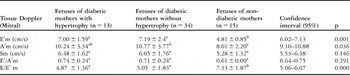
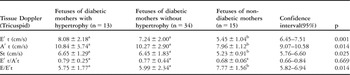
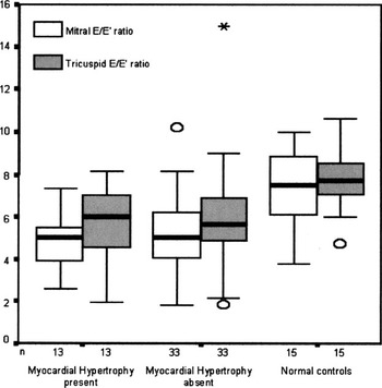
Gestational age varied among the groups when assessed by ANOVA, although this was not confirmed when using the co-variance test. When maternal age was analyzed in the three groups with ANOVA and co-variance analyses, the observed difference among the groups was statistically significant. Table 1 show the means and standard deviations of the diastolic myocardial velocities obtained at the post-lateral basal segments of the left ventricle, in other words the mural annulus of the mitral valve. Significant alterations of the E′ and A′ waves at this level were observed when comparing fetuses of diabetic mothers to fetuses of normoglycemic mothers. We also found a significant difference in diastolic velocities, the E′ and A′ waves, when measured for the aortic annulus of the mitral valve for same sample (Table 2). In Table 3, we show the diastolic velocities, the E′ and A′ waves, for the tricuspid valvar annulus at the lateral wall of the right ventricle. The peak flow velocities at mitral and tricuspid levels in fetuses of diabetic mothers with or without of left ventricular hypertrophy, and in fetuses of normal pregnant women, are shown in Tables 4 and 5, respectively. In Figure 1, we show the median of the ratio between mitral and tricuspid inflow E wave and the atrioventricular annular E′ wave as shown by tissue Doppler, giving the ratio between E and E′. In fetuses of diabetic mothers, with or without myocardial hypertrophy, this ratio was significantly lower than for control fetuses (p = 0.00 and p = 0.014), respectively.
Means and standard deviations of the myocardial diastolic velocities obtained at the basal post-lateral segment of the left ventricle (the mural annulus of the mitral valve).

a ≠ b – different characters show statistical significant differences between the groups.
Means and standard deviations of the myocardial diastolic velocities at the basal septal segment (anterior mitral annulus).

a ≠ b – different characters show statistical significant differences between the groups.
Means and standard deviations of the myocardial diastolic velocities obtained at the lateral wall of the right ventricle (tricuspid annulus).

a ≠ b – different characters show statistical significant differences between the groups.
Mitral inflow velocities in fetuses of diabetic mothers with or without left ventricular hypertrophy and fetuses of normoglycemic mothers.

a ≠ b – different characters show statistical significant differences between the groups.
Tricuspid inflow velocities in fetuses of diabetic mothers, with or without left ventricular hypertrophy, and fetuses of normoglycemic mothers.

a ≠ b – different characters show statistical significant differences between the groups.
Median comparison between myocardial velocities by pulsed Doppler, E wave, and by Tissue Doppler, E′ wave, at the mitral and tricuspid annulus.

Discussion
Tissue Doppler is a useful non-invasive technique with which to assess the velocities of contraction and expansion of the myocardium in systole and diastole.Reference Garcia, Thomas and Klein30–Reference Pai and Gill33 When analyzing fetal cardiac diastolic function, account must be taken of both ventricles, making tissue Doppler an excellent parameter for non-invasive analysis.Reference Isaaz34 Harada et al.,Reference Harada, Tsuda, Orino, Tanaka and Takada35 using this technique, established the normal values and changes in myocardial velocities of the left and right ventricular walls, as well as velocities for the septum, as related to gestational age in normal fetuses, with the A′ wave proving to be higher than the E′ wave. As gestational age advances, there is a decrease in the ratio between the waves, in agreement with the alterations observed in the patterns of velocity of the atrioventricular valves obtained using conventional Doppler.
Gestational diabetes mellitus has been used as a model of fetal diastolic dysfunction due to myocardial hypertrophy and the consequent reduction of ventricular compliance.Reference Zielinsky, Satler and Luchese36 In this respect, previous studies from our groupReference Zielinsky, Costa, Oliveira, Bonow, Silva and Hagemann1, Reference Hagemann and Zielinsky2, Reference Zielinsky13, Reference Zielinsky, Hagemann, Daudt and Behle15, Reference Firpo and Zielinsky18 have shown that, as a result of an increased myocardial mass and left ventricular hypertrophy, fetuses of diabetic mothers may have a higher preload than normal controls. We assessed these changes using the excursion of the primary atrial septum, along with flows through the pulmonary veins, the venous duct, and the oval foramen, and by using left atrial shortening.Reference Zielinsky, Piccoli and Gus37–Reference Zielinsky, Sallum and Satler40 We are currently evaluating the myocardial performance index, along with the deceleration times of the atrioventricular annuluses and the ventricular septum.
In this study, we have shown that diastolic myocardial velocities at the level of the aortic and mural annuluses of the mitral valve, as well as at the level of the tricuspid annulus, are significantly higher in fetuses of diabetic mothers than in fetuses of normal mothers. Further post-hoc analysis has shown that the observed alterations in diastolic function in the fetuses of the diabetic mothers do not depend on the presence of fetal ventricular hypertrophy. We also found that the velocities of the E′ at the level of the left and right ventricular walls, as well as at the ventricular septum, are almost always lower than those of the A′ wave, and that myocardial velocities taken at the right and left ventricular walls are almost always higher than the ones observed at the level of the ventricular septum, at both early and late diastole, confirming the findings of Harada et al.Reference Harada, Tsuda, Orino, Tanaka and Takada35 for fetuses of normal mothers. These differences in velocities of flow are observed, therefore, in fetuses of diabetic mothers, but this does depend on the site of the tissue Doppler sample. This finding may be related to the physiology of the longitudinal myocyte.
Significantly lower ratios between E and E′ were observed in both atrioventricular valves in fetuses of diabetic mothers when compared to control fetuses. This is the result of higher myocardial velocities at the mitral and tricuspid annuluses, rather than because of changes in early atrioventricular diastolic flows. In adults with aortic stenosis, the ratio between E and E′ for the mitral valve has been correlated with increased pressures of left ventricular filling, with progressively lower E′ velocities, and higher ratios between E and E′.Reference Bruch, Stypmann, Grude, Gradaus, Breithardt and Wichter41 On the other hand, others have shown significantly lower ratios between E and E′ for the mitral valve in patients with ischaemic disease and more compromised left ventricular function.Reference Baykan, Yilmaz, Celik, Orem, Kaplan and Erdol42 In the fetus, the impaired ventricular diastolic function as a result of decreased relaxation and compliance occurring in maternal diabetes seems to prompt higher myocardial velocities in the atrioventricular annuluses, in order to counter the limited ventricular distensibility in early diastole.
The data we have obtained strengthens the notion that tissue Doppler is more specific than conventional Doppler for the diagnosis of fetal diastolic dysfunction, since many of these patients have not presented alterations in trans-mitral or trans-tricuspid flow as assessed by pulsed Doppler.
Thus, we conclude that maternal diabetes mellitus is associated with alterations in left ventricular diastolic function in the fetus, and not only as a result of myocardial hypertrophy. These changes may still be present in the immediate neonatal period, prompting subtle or overt heart failure, which are characteristically transient. It has already been demonstrated that septal thickness, which has increased during fetal life, is usually normal at 2 to 3 months of age, this corresponding with normalization of levels of insulin in the blood.Reference Zielinsky, Costa, Oliveira, Bonow, Silva and Hagemann1 Postnatal assessment of diastolic dysfunction secondary to maternal diabetes, therefore, would best be accomplished by analyzing the behaviour of a multiparameter score, a topic already under investigation by our group.