Introduction
Oocyte development takes place in ovarian follicles. During folliculogenesis, oocytes grow and reach the metaphase stage of meiosis II (MII). To be developmentally competent, it is very important that the cytoplasm matures at the same time as oocyte meiosis resumes. This includes accumulation of proteins and energy substrates, organelle reallocation and changes in the structure of chromatin. If meiosis resumed precociously before cytoplasmic maturation, the developmental competence of the oocyte, e.g. the ability to be fertilized and the ability to develop to blastocyst, will be negatively affected.
The importance of cytoplasmic maturation was confirmed in a study reported by Cheng et al. (Reference Cheng, Fan, Wen, Tong, Zhu, Lei, Sun and Chen2003). When the cytoplast at germinal vesicle (GV) stage was used as the recipient and the karyoplast at metaphase in meiosis I (MI) or at MII as donor, the constructed oocyte extruded a polar body after electrofusion and culture. While both the cytoplasm and the polar body had a metaphase spindle in the MI–GV pair, only a clutch of condensed chromatin was observed in the cytoplasm and the polar body of the MII–GV pair. When the MI cytoplast was used as the recipient and the GV or MII karyoplast as donor, the reconstructed oocyte also extruded a polar body. Each had one spindle and a group of metaphase chromosomes in the cytoplasm and the polar body, respectively. When the MII cytoplast was used as the recipient and the GV or MI karyoplast as the donor, the reconstructed oocytes were activated, became parthenogenetic embryos and even developed to hatching blastocysts after electrofusion. Immunoblotting showed that mitogen-activated protein kinase activity was high in MI and MII cytoplasts, but not detected in the GV cytoplast. These results demonstrated that the synchronized maturation of the cytoplasm and nucleus is very important.
Synchronization of cytoplasmic and nuclear maturation is regulated by CNP-cyclic guanosine monophosphate (cGMP) signalling. CNP is encoded by a natriuretic peptide precursor C (Nppc) gene expressed mainly in mural granulosa cells. This protein stimulates the natriuretic peptide receptor B (NPRB) on the membrane of cumulus cells to produce cGMP. cGMP of cumulus origin diffuses into oocytes to suppress phosphodiesterase 3 (PDE3) activity, leading to elevation of cyclic adenosine 3′,5′-monophosphate (cAMP) in oocytes. cAMP binds to protein kinase A (PKA), which in turn activates WEE 1 homologue 2 (WEE1B) and myelin transcription factor 1 (MYT1) kinase. WEE1B and MYT1 are known to block cyclin-dependent kinase 1 (CDK1). Therefore, cAMP-dependent activation of PKA results in CDK1 inhibition, leading to meiotic arrest in oocytes (Hsueh et al., Reference Hsueh, Kawamura, Cheng and Fauser2015; Yang et al., Reference Yang, Wei, Li, Ge, Zhao and Ma2016; Machaty et al., Reference Machaty, Miller and Zhang2017). This effect of CNP was confirmed in mouse (Zhang et al., Reference Zhang, Su, Sugiura, Xia and Eppig2010; Wei et al., Reference Wei, Zhou, Yuan, Miao, Zhao and Ma2017), porcine (Hiradate et al.,Reference Hiradate, Hosshino, Tanemura and Sato2013), bovine (Franciosi et al., Reference Franciosi, Coticchio, Lodda, Tessaro, Modina, Fadini, Dal, Mariabeatrice, Mignini, Albertini and Luciano2014) and sheep (Zhong et al., Reference Zhong, Fan, Li, Zhang and Zhang2018).
In vitro culture of ovarian follicles might be a promising technique for assisted reproductive technology. As CNP plays a vital role in maintaining the meiotic arrest of oocytes and is beneficial for synchronization of cytoplasmic and nuclear maturation, in the present study we supplemented it in the culture system of murine preantral follicle to investigate its effect on follicle development and oocyte maturity.
Materials and methods
Chemicals
The purchasing information for chemicals used in this study is as follows: DMEM/F12 from HyClone Company (USA); recombinant follicle stimulating hormone (FSH) from Merck Serono Company (Italy); human chorionic gonadotropin (HCG) from Sansheng Pharmaceutical Company (China); insulin from Wanbang Pharmaceutical Company (China); transferrin, epidermal growth factor (EGF) and sodium selenium from Sigma Company (USA); CNP from TOCRIS Company (Britain); serum protein substitute (SPS) from SAGE Company (USA); and human tubal fluid medium (HTF) from Merck Company (USA).
Animals, follicle culture and in vitro fertilization
Female Kunming mice aged 3 weeks were obtained from the Laboratory Animal Center of Shanxi Medical University. After cervical dislocation, preantral follicles with multilayer granulosa cells (130–180 µm in diameter) were isolated from the ovary using a tuberculin syringe needle. Each follicle was cultured in a 30-µl droplet under 37°C in a humidified atmosphere of 5% CO2 in air. The basic culture medium was DMEM/F12, which was supplemented with 5 µg/ml insulin, 5 µg/ml transferrin, 5 ng/ml sodium selenium, 10% SPS, 100 nM/ml CNP, 25 mIU/ml FSH. A half volume of the medium was changed every other day. Follicle development was assessed on the day of medium change. In all three experiments, the follicles were cultured for at least 7 days based on the preliminary tests.
For follicle ovulation, 1.5 IU/ml HCG and 5 µg/ml EGF were added to the medium and follicles incubated for 17 h when the follicle reached 350–400µm in diameter and part of the follicle bulged out, which indicated that the follicle was about to ovulate. If the size increment of the antral follicle was less than 10 µm after 2 days and the follicle diameter was more than 300 µm, the oocyte inside the follicle might be competent and perhaps degenerate in the later period. For these follicles, ovulation was also induced with EGF and HCG as a salvage measure.
For oocyte maturity assessment, oocyte and cumulus cell complexes (COC) ovulated from antral follicles were incubated with 80 IU/ml hyaluronidase for 30 s and the granulosa cells were stripped by repeated aspiration with a capillary glass pipette. Preovulatory follicles that had not ovulated were pierced with a tuberculin syringe needle to retrieve the COCs. The denuded oocytes were classified into GV breakdown (GVBD) and MII stages.
To assay fertilization, COCs were transferred into 100 µl HTF medium supplemented with 10% SPS; 1 × 10⁵ of motile sperm retrieved from the epididymis of male mice aged 13 weeks were added to the droplet containing COCs for IVF. After 20 h, fertilization and embryo cleavage were checked.
Experiment design
Experiment 1
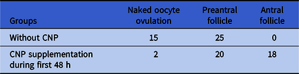
The objective of this experiment was to investigate the effect of early CNP supplementation on follicle development. Forty secondary follicles were allocated randomly into experimental and control groups. In the experimental group, CNP was supplemented into the medium in the first 48 h. In the control group, the only difference from the experimental group was absence of CNP in the culture medium. The culture was concluded when the dominant antral follicles reached 350–400 µm in diameter. Follicle stages in the experimental group and control group were compared.
Experiment 2
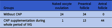
The objective of this experiment was to investigate if CNP could effectively prevent the oocyte from premature resumption of meiosis. Forty secondary follicles were allocated randomly into experimental and control groups. In the experimental group, CNP was supplemented into the medium for the whole period of in vitro culture. In the control group, the only difference from the experimental group was the absence of CNP in the culture medium. Follicle stages in the experimental group and control group were compared. To investigate the effect of CNP on maintaining meiotic arrest and the effect of ovulation induction on meiosis resumption, EGF and HCG were added to 50% of preovulatory follicles, but omitted in the other preovulatory follicles.
Experiment 3
The objective of this experiment was to investigate the fertilization potential of oocytes cultured in vitro. Eighteen preantral follicles were allocated randomly into experimental and control groups. The culture medium in the experimental and control groups during IVG was the same as in Experiment 2. After ovulation induced by EGF and HCG, sperm were added to COCs for IVF. The rate of 2-cell stage embryos after IVF was evaluated. In addition, follicle stages in the experiment group and control group were compared.
Statistical analysis
Statistical analysis was performed using GraphPad Prism 6.0 software (La Jolla, CA, USA). Data were analyzed with a contingency table (chi-squared test for Tables 1 and 2 and Fisher’s exact test for Table 3). A P-value < 0.05 was considered to be statistically significant.
The development of ovarian follicles in Experiment 1

The development of ovarian follicles between experimental group and control group was significantly different (P < 0.0001).
Development of ovarian follicles in Experiments 2 and 3 combined

Development of ovarian follicles between experimental group and control group was significantly different (P < 0.0001).
The maturity of oocytes in Experiment 2

Maturity of oocytes between group with ovulation induction and group without ovulation induction was significantly different (P < 0.0001).
Results
Supplementation of CNP improved follicle development
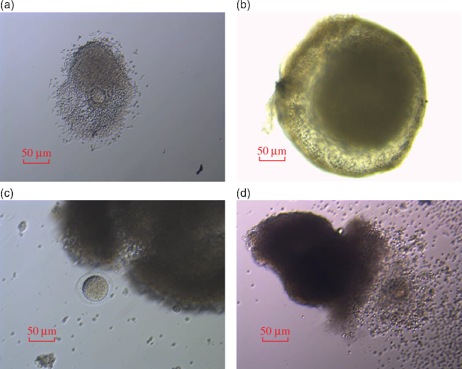
In all control groups in three experiments, the granulosa cells in all follicles migrated out from within follicles to different degrees and adhered to the plate. As a result, originally ball-shaped follicles flattened. In all experimental groups in three experiments, this phenomenon was not observed and all follicles grew as three dimensions (Fig. 1).
The effect of CNP on development of ovarian follicles. (a) Without CNP supplementation, the granulosa cells migrated from within follicle and the follicle flattened. (b) With CNP supplementation, the follicle grew three-dimensionally. (c) Without CNP supplementation, oocyte ovulated nakedly from preantral follicle. (d) With CNP supplementation, COC ovulated from antral follicle.

In all control groups in three experiments, 39.8% (39/98) follicles ovulated prematurely before the follicle reached the antral stage and the oocyte ovulation was not accompanied by granulosa cells. In all experimental groups in three experiments, the rate of premature ovulation was 3.1% (3/98) (Fig. 1).
In all control groups in three experiments, no follicles reached the antral stage. In the experimental groups, the rate of antral follicle was 45% (18/40) in Experiment 1, 75.9% (44/58) in Experiment 2 and Experiment 3 combined, and 63.3% (62/98) in all three experiments combined. Follicle development was significantly improved by supplementation with CNP and this effect was demonstrated even if CNP was only added at the early stage of IVG (P < 0.0001) (Tables 1 and 2).
Supplementation of CNP effectively maintained meiotic arrest of oocytes
In Experiment 2, 32 and 0 antral follicles were formed in the experimental group and control group, respectively. EGF and HCG were randomly added to 16 antral follicles to induce ovulation, but not to the other 16 antral follicles. These results demonstrated that 87.5% (14/16) of the oocytes from the follicles without ovulation induction remained at the GV stage. But 0% (0/16) of the oocytes from the follicles with ovulation induction maintained the meiotic arrest (Table 3).
Supplementation of CNP resulted in cleaved embryos after IVF
In Experiment 3, 12 and 0 antral follicles were formed in the experimental group and control group, respectively. In 12 oocytes retrieved from antral follicles, 11 were fertilized and cleaved into 2-cell stage embryos 20 h after IVF.
Discussion
Ovarian follicle cryopreservation and in vitro culture after warming is a potential strategy for fertility preservation. In 1977, Eppig and Schroeder successfully cultured murine ovarian follicles in vitro (Reference Eppig and Schroeder1977). In 1989, Eppig and Schroeder reported the first live offspring born after IVF and embryo transfer (ET) following in vitro culture of mouse ovarian follicles (Eppig and Schroeder, Reference Eppig and Schroeder1989). Then, the culture protocol was modified and live offspring were reported successively (Eppig and O’Brien, Reference Eppig and O’Brien1996; O’Brien et al., Reference O’Brien, Pendola and Eppig2003; Xu et al., Reference Xu, Kreeger, Shea and Woodruff2006; Higuchi et al., Reference Higuchi, Maeda, Horiuchi and Yamazaki2015; Wang et al., Reference Wang, Ge, Liu, Klinger, Dyce, De and Shen2017). However, the developmental competency of oocytes cultured in vitro was much lower than that in vivo (Eppig and O’Brien, Reference Eppig and O’Brien1996; O’Brien et al., Reference O’Brien, Pendola and Eppig2003; Appeltant et al., Reference Appeltant, Somfai, Maes, Van and Kikuchi2016; Zhang et al., Reference Zhang, Zhang, Fan, Li and Zhang2017a).
Because of environmental differences, ovarian follicle culture in vitro resulted in at least two unwanted aspects that might compromise the developmental competence of oocytes. Firstly, meiosis in oocytes is apt to resume prematurely. Secondly, the three-dimensional structure of the ovarian follicle is prone to change.
To prevent oocytes from premature resumption of meiosis, CNP was supplemented in the culture system of the ovarian follicle (Hiradate et al., Reference Hiradate, Hosshino, Tanemura and Sato2013; Franciosi et al., Reference Franciosi, Coticchio, Lodda, Tessaro, Modina, Fadini, Dal, Mariabeatrice, Mignini, Albertini and Luciano2014; Yang et al., Reference Yang, Wei, Li, Ge, Zhao and Ma2016; Wei et al., Reference Wei, Zhou, Yuan, Miao, Zhao and Ma2017; Zhang et al., Reference Zhang, Zhang, Fan, Li and Zhang2017a; Zhong et al., Reference Zhong, Fan, Li, Zhang and Zhang2018;). As the aims were to prevent premature meiosis of oocyte, CNP was mostly supplemented to oocytes retrieved from preovulatory antral follicles.
To retain the three-dimensional structure of the follicle and enhance the developmental competency of oocytes, many techniques such as using V-shaped microwell plates (Telfer and Zelinski, Reference Telfer and Zelinski2013), inverted hanging droplets (Wycheley et al., Reference Wycheley, Downey, Kane and Hynes2004; Choi et al., Reference Choi2013), alginate encapsulation (Mainigi et al., Reference Mainigi, Ord and Schultz2011; Kreegar et al., Reference Kreegar, Deck, Woodruff and Shea2016), collagen encapsulation (Mochida and Akatani-Hasegawa, Reference Mochida and Akatani-Hasegawa2013), fibrin–alginate interpenetrating encapsulation (Shikanov et al., Reference Shikanov, Xu, Woodruff and Shea2009; Jin et al., Reference Jin, Lei, Shikanov, Shea and Woodruff2010) and encapsulation with alginate plus amino acids (Brito et al., Reference Brito, Lima, Xu, Shea, Woodruff and Figueiredo2014) have been used. Compared with oocytes grown in vivo, spindle formation and chromosome alignment of oocytes cultured using some of the protocols mentioned above was abnormal and developmental competence was compromised (Mainigi et al., Reference Mainigi, Ord and Schultz2011).
In the present study, in addition to effective prevention of premature resumption of meiosis, the three-dimensional structure of the follicle was maintained and the rate of antral follicles was significantly enhanced by supplementation of CNP. Using this protocol, it might be unnecessary to take additional steps such as alginate encapsulation to maintain the three-dimensional structure of the ovarian follicles.
How could supplementation of CNP play the role described in the present study? Romero et al. (Reference Romero, Sanchez, Lolicato, Van and Smitz2016) reported that CNP-treated COCs showed a higher (P < 0.05) density of transzonal projections (TZP) between granulosa cells and oocyte. This might be helpful for the maintenance of the three-dimensional structure of the follicle. Franciosi et al. (Reference Franciosi, Coticchio, Lodda, Tessaro, Modina, Fadini, Dal, Mariabeatrice, Mignini, Albertini and Luciano2014) reported that the percentage of COCs with functionally open gap junctions in the experiment group with CNP supplementation for 6–8 h was significantly higher than that in control COCs, as measured by Lucifer yellow injection assay. Only with functional gap junctions, could cGMP flow into the oocyte. Then increased cAMP in oocytes induced by cGMP could prevent the premature resumption of meiosis. CNP could also stimulate ovarian follicle growth and increase ovarian follicle viability. In mice, Nppc and Npr2 are expressed in early preantral follicles and levels increase during early to late preantral follicle development. Treatment of cultured preantral follicles with CNP stimulated follicle growth and the treatment of cultured ovarian explants from infantile mice with CNP promoted the development of primary and early secondary follicles to the late secondary stage. In vivo studies indicated that, in infantile mice, a daily injection of CNP for 4 d promoted ovarian growth and follicles ovulated after ovulation induction. In prepubertal mice, CNP treatment alone promoted early antral follicle growth to the preovulatory stage, resulting in efficient ovulation by gonadotropin. Furthermore, mature oocytes retrieved after CNP treatment could be fertilized in vitro and develop into blastocysts. After ET, viable offspring were delivered (Sato et al., Reference Sato, Cheng, Kawamura, Takae and Hsueh2012; Xi et al., Reference Xi, Wang, Sarfaraz and Yao2019). This beneficial effect of CNP was more prominent in small follicles compared with large follicles in in vitro culture (Zhang et al., Reference Zhang, Wang, Liu, Yang, Wang, Zhang, Guo, Wang and Xia2017b). In addition, CNP could decrease levels of reactive oxygen species (Sato et al., Reference Sato, Cheng, Kawamura, Takae and Hsueh2012; Tiwari et al., Reference Tiwari, Prasad, Tripathi, Pandey, Ali, Singh, Shrivastav and Chaube2015; Xi et al., Reference Xi, Wang, Sarfaraz and Yao2019). The effects of CNP on preantral follicles in the present study might at least be partly explained by the mechanisms mentioned above.
Several chemicals, in addition to CNP, such as forskolin, 6-dimethylaminopurine (6-DMAP) have also been reported as substances that have the potential to improve nuclear–cytoplasmic synchronization of oocytes. It was reported that some of these chemicals may have a prolonged inhibitory effect due to a longer half-life or have detrimental effects by inducing cellular apoptosis (Bouhelan et al., Reference Bouhelan, Bockaert, Mermet-Bauvier, Guillon and Homburger1987; Samake and Smith, Reference Samake and Smith1997; Simli et al., Reference Simli, Pellerano, Pignllo, Tavosanis, Ottaggio, de Saint-Georges and Bonatti1997; Alexander et al., Reference Alexander, Coppola, DiBerardino, Rho, StJohn, Betts and King2006; Follin-Arbelet et al., Reference Follin-Arbelet, Misund, Hallan, Ugland, Sundan and Kiil2015). In contrast, CNP is a natural substance in vivo, and could orchestrate meiotic progression in cooperation with involved growth factors and hormones. More importantly, the inhibitory effect of CNP–NPR2 signalling is reversible and is inactivated soon after the luteinizing hormone surge. This make CNP a promising agent to improve the developmental competency of ovarian follicles cultured in vitro.
In conclusion, the supplementation of CNP in the culture system of murine preantral follicle during IVG period could sustain the three-dimensional structure of follicles, increase antral formation rate and maintain oocyte meiotic arrest. As the oocyte developmental competency was greatly improved, this protocol might benefit further research in the field of ovarian follicle culture.
Ethical approval and consent to participate
The experiment was performed in accordance with the guidelines of animal experiment in Shanxi Provincial Key Laboratory of Cell Regeneration and Birth Defects. The study protocol was approved by the ethics committee of Shanxi Provincial Key Laboratory of Cell Regeneration and Birth Defects.
Authors’ contributions
Wang HX and Guo XP jointly designed the experiment. Wang HX wrote the manuscript. Li A and Cao HX performed the experiments. Li HX and Li RJ worked as assistants in the experiments. All authors read and approved the final manuscript.
Conflict of interest
The authors declare that they have no competing interests.
Funding
The research did not receive any specific grant from funding agencies in the public, commercial or not-for-profit sectors.
Acknowledgements
Not applicable.