Introduction
Nasal septal perforation (NSP) is defined as a disruption of the composite integrity of the nasal septum—including the mucosa, bone or cartilage—resulting in direct communication between the nasal cavities.Reference Garaycochea, Santamaría-Gadea and Alobid1, Reference Santamaría-Gadea, Mariño-Sánchez, Arana-Fernández, Mullol and Alobid2 Its incidence is about 0.9 per cent.Reference Sonneveld and Sinha3, Reference Watson and Barkdull4 Aetiology is multifactorial, including inflammation, trauma, autoimmune disease and inhaled irritants.Reference Lanier, Kai, Marple and Wall5 It most commonly occurs after septoplasty, chemical cauterisation or nasal packing.Reference Watson and Barkdull4, Reference Goh and Hussain6, Reference Metzinger7
Both conservative and surgical options have been described, and surgical treatment generally relies on mucosal flaps with an inter-positional graft.Reference Sonneveld and Sinha3, Reference Metzinger7 However, no consensus exists on the ideal graft material or optimal technique.Reference Garaycochea, Santamaría-Gadea and Alobid1, Reference Kridel and Delaney8–Reference Delaney and Kridel10 Cartilage grafts have been used in nasal surgery since the 1940s in forms such as block, crushed, diced and shaved cartilage.Reference Trivisonno, Cohen, Magalon, Magalon, Sterodimas and Pascali11, Reference Tasman12 Diced cartilage grafts offer advantages over block grafts, including easier availability and improved shaping.Reference Liao, Chen, Xu, Cheng, Xie and He13 Experimental studies have also shown better viability of diced cartilage compared with other graft forms.Reference Liao, Chen, Xu, Cheng, Xie and He13, Reference Kayabasoglu, Ozbek, Yanar, Sahin, Keles and Yilmaz14 However, disadvantages such as reduced stability and a tendency to cluster at the implantation site have been reported.Reference Gode, Ozturk, Berber and Kısmalı15 To mitigate these issues, it has been suggested that diced cartilage be wrapped with materials such as oxidised cellulose, fascia, hyaluronic acid, fibrin glue or platelet-rich fibrin (PRF) before implantation.Reference Tasman12, Reference Gode, Ozturk, Berber and Kısmalı15–Reference Calvert and Kwon18 PRF, originally developed for oral and maxillofacial surgery, is rich in leukocytes and plateletsReference Dohan, Choukroun, Diss, Dohan, Dohan and Mouhyi19–Reference Dohan Ehrenfest, de Peppo, Doglioli and Sammartino21 and may enhance cartilage viability through growth factors.Reference Grzelak, Hnydka, Higuchi, Michalak, Tarczynska and Gaweda22 Leucocytes may also exert direct and immune-regulatory effects, providing anti-infective and anti-inflammatory benefits.Reference Dohan Ehrenfest, de Peppo, Doglioli and Sammartino21
Our study was based on two main hypotheses. The first hypothesis was that the use of diced cartilage as an inter-positional graft material could facilitate the surgical repair of NSP and potentially increase the success rate. Our second hypothesis was that using PRF as a membrane could help maintain the stability of diced cartilage fragments while also providing protection against potential infections in the nasal region, which is highly susceptible to infectious and inflammatory processes. In this study, we aimed to define the role of diced cartilage and diced cartilage combined with PRF in NSP surgery by evaluating their macroscopic and histopathological effects.
Materials and methods
In this study, 36 healthy adult New Zealand albino rabbits of similar age and weight (2500–3910 g) were used. The animals were obtained from the Experimental Medicine Research Centre of Selçuk University and maintained under standard housing conditions. Ethical approval was obtained from the Animal Experiments Committee (Approval No: 2023-43), and all procedures followed the Guide for the Care and Use of Laboratory Animals.
Experimental design and animal procedures
A total of 36 healthy New Zealand albino rabbits were included in the study and were divided into three equal groups. The groups were as follows: (1) Control group (C): A 10 × 10 mm septal perforation was created, and the defect was repaired using only bilateral mucoperichondrial flaps; (2) Diced Cartilage group (DC): A 10 × 10 mm septal perforation was created, and the defect was repaired using bilateral mucoperichondrial flaps together with diced cartilage fragments (1 × 1 mm) serving as the inter-positional graft material; (3) Diced Cartilage plus PRF group (DC plus PRF): A 10 × 10 mm septal perforation was created, and the defect was repaired using bilateral mucoperichondrial flaps together with diced cartilage fragments (1 × 1 mm) wrapped in a PRF membrane, which served as the inter-positional graft material.
Anaesthesia administration
Anaesthesia was achieved by intra-muscular (IM) administration of ketamine hydrochloride (Ketasol® 10 per cent, Richterpharma) at 35 mg/kg and xylazine hydrochloride (Xylazinbio® 2 per cent, Bioveta) at 5 mg/kg. The animals breathed spontaneously. Anaesthetic depth and analgesia were confirmed by assessing the limb withdrawal reflex, and once adequate anaesthesia was achieved, the experimental procedures were initiated.
Harvesting and preparation of cartilage grafts
The auricular vessels were identified by transillumination, and the donor site was marked. After local anaesthesia, a U-shaped incision was made, and a perichondrium-preserving skin flap was elevated. The cartilage was exposed, a 12 × 12 mm block harvested with bilateral perichondrium preserved and the donor site closed with polypropylene sutures.
Preparation of platelet-rich fibrin
Eight millilitres of blood were collected from the auricular veins opposite the cartilage donor side. Samples were placed into 10-mL glass tubes without anticoagulant and centrifuged at 3000 rpm for 10 minutes. PRF was prepared according to Dohan et al.,Reference Dohan, Choukroun, Diss, Dohan, Dohan and Mouhyi19 compressed into a membrane and made ready for use.
Creation and repair of nasal septal perforation
Nasal septal perforation was created according to the method described by Ulusoy et al.Reference Ulusoy, Uğraş, Uslu, Elsürer and Körez23 A mid-columellar skin incision was made, the nasal bones were exposed and medial osteotomies were performed to separate them. The nasal septum was then visualised, and a full-thickness 10 × 10 mm defect was created. Subsequently, the created perforations were repaired using mucosal flaps and inter-positional grafts in accordance with the assigned groups.
Following the surgical procedures, the rabbits were housed individually at 22 plus or minus 2°C, 40–60 per cent humidity, under a 12-hour light/dark cycle, with ad libitum access to standard feed and water. The animals were monitored daily for complications, body weight, temperature, respiratory rate and nasal bleeding. After a four-week follow-up period, the animals were euthanised using a high-dose anaesthetic, and necropsy was performed.
Macroscopic evaluation included assessment of septal perforation, infection and synechiae, graded according to the scoring system:
A. Perforation closure status:
0. perforation present.
1. no perforation.
B. Infected appearance:
0. no signs of infection.
1. signs of infection present.
C. Synechiae:
0. no synechiae.
1. synechiae present.
Histopathological Evaluation
Tissue samples from the rabbit nasal septum were fixed in 10 per cent neutral buffered formalin, routinely processed and paraffin-embedded. Sections 4-µm-thick were cut using a microtome and stained with haematoxylin–eosin (H&E) and Masson’s trichrome. Samples were evaluated for re-epithelialisation, vascularisation, necrosis, giant cells, abscess formation, fibrosis, new cartilage formation, foreign body reaction and inflammatory cell infiltration according to Ulusoy et al.Reference Ulusoy, Uğraş, Uslu, Elsürer and Körez23 Cartilage viability was assessed by chondroid matrix metachromasia and chondrocyte nuclei within lacunae; absence of both indicated necrotic cartilage.
The following scoring system was used for histopathological evaluation:
A. Cartilage viability:
0. absent.
1. present.
B. Fibrosis:
0. no fibrosis.
1. mild fibrosis not bridging the wound edges.
2. fibrosis bridging the wound edges and filling the perforation gap.
C. Other histopathological findings (re-epithelialisation, vascularisation, new cartilage formation, necrosis, neutrophils, abscess, lymphocytes/plasma cells, giant cells, foreign body reaction):
0. none (no findings).
1. mild (presence of findings, e.g., a small number of inflammatory cells).
2. marked (prominent findings, e.g., numerous obvious inflammatory cells).
Statistical Analysis
The number of animals to be included in the study was calculated using the Resource Equation Method to determine the minimum sample size needed to detect differences in perforation closure rates between groups. For a one-way analysis of variance (ANOVA) design assessing significant differences among the three groups, at least 20/3 plus 1 = 8 animals were required per group.
All statistical analyses were performed using R version 4.1.2 (The R Foundation for Statistical Computing, Vienna, Austria). Data normality was assessed with the Shapiro–Wilk test and variance homogeneity with Levene’s test. Parametric data, including baseline and post-sacrifice weights, were presented as mean plus or minus standard deviation and compared using the Welch F test followed by the Games–Howell post-hoc test and ANOVA followed by the Tukey HSD post-hoc test, respectively. Non-parametric variables, such as histopathological and macroscopic findings, were expressed as mean plus or minus standard error of the mean and analysed with the Kruskal–Wallis test followed by Dunn’s post-hoc test with false discovery rate adjustment. The Mann–Whitney U-test was used to compare cartilage viability between the DC and DC plus PRF groups. Subgroup comparisons of fibrosis scores were performed using the Fisher–Freeman–Halton test followed by the Two-Proportion Z test. Statistical significance was set at p less than 0.05.
Results
The baseline characteristics, macroscopic findings and histopathological results of the experimental groups are shown in Table 1. No significant differences were found among the groups in baseline or post-sacrifice weights (p = 0.987 and p = 0.775, respectively).
Basic characteristics of the study groups and their histopathological and macroscopic findings

Table 1 Long description
The table compares baseline characteristics, histopathological, and macroscopic findings across three study groups: Control, Diced Cartilage, and Diced Cartilage + PRF. Re-epithelialisation and perforation closure rates were significantly higher in the diced cartilage groups compared to the control group, with p-values less than 0.0012. Marked fibrosis was observed in 66.7% of the Diced Cartilage group and 81.8% of the Diced Cartilage + PRF group, while none was observed in the control group. Baseline and post-sacrifice weights showed no significant differences across groups. Vascularisation scores and necrosis levels were similar across groups, with no significant differences. The table highlights the effectiveness of diced cartilage treatments in enhancing certain histopathological and macroscopic outcomes.
Note: Data were presented was mean ± SD or mean ± SEM. Different small superscript letters in each row denote that statistically significant difference between groups according to the pairwise comparisons.
1 One-way ANOVA.
2 Kruskal–Wallis test followed by Dunn post-hoc test with FDR adjustment.
3 Mann–Whitney U test.
4 Fisher–Freeman–Halton test followed by Two Proportion Z-test.
ANOVA = analysis of variance; FDR = false discovery rate; PRF = platelet-rich fibrin; SD = standard deviation; SEM = standard error of the mean.
Macroscopic findings
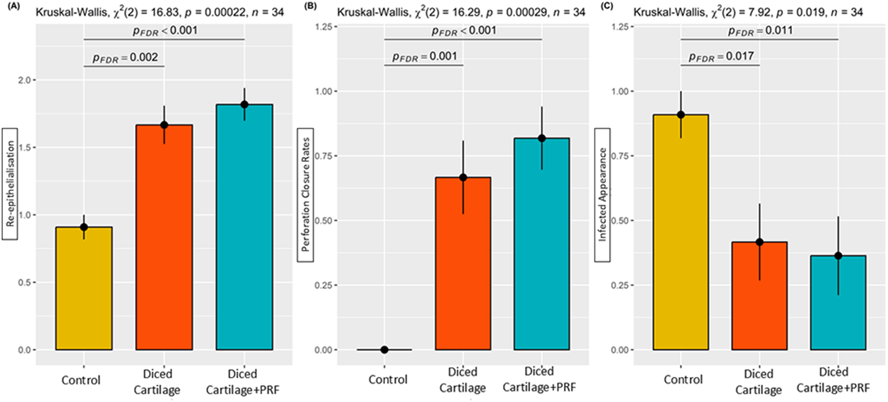
Perforation closure rates: The treatment success rate was significantly higher in both the DC group (66.7 per cent) and the DC plus PRF group (81.8 per cent) compared with the C group (0 per cent) (p < 0.001) (Table 1; Figs. 1 and 2B).
Macroscopic appearance of the completely closed nasal septum in the diced cartilage group (*).

Figure 1 Long description
This surgical photograph shows a close-up macroscopic view of an exposed nasal septum during an operative procedure in the diced cartilage treatment group. The surgical field is open, with surrounding pale white fur or soft tissue visible at the margins of the image. The central area contains moist red tissue consistent with the nasal septal mucosa and underlying septal structures. A metal surgical instrument enters from the left side of the image and points toward the reconstructed septal region. The septum appears fully closed and continuous, without a visible perforation or opening. In the center of the image, an asterisk marks the specific area of septal closure and reconstruction. The tissue surface around the marked area appears smooth, glistening, and intact, suggesting successful healing or repair after graft placement. The diced cartilage graft region blends with the surrounding tissue and does not show obvious gaps, defects, or exposed edges. Fine blood vessels and small amounts of surgical bleeding are visible throughout the operative field, which is typical of fresh intraoperative exposure. The image emphasizes the gross anatomical appearance and integrity of the repaired nasal septum following treatment with diced cartilage.
Comparison of the groups in terms of re-epithelialisation (A), perforation closure rate (B) and infected appearance (C).

Figure 2 Long description
The image contains three bar graphs. The first graph (A) compares re-epithelialisation among Control, Diced Cartilage and Diced Cartilage plus PRF groups, showing higher values for the latter two. The second graph (B) illustrates perforation closure rates, with Diced Cartilage and Diced Cartilage plus PRF groups having significantly higher rates than the Control. The third graph (C) depicts infected appearance, with the Control group showing higher values compared to the other two groups. Statistical values and significance levels are indicated above each graph.
Infected appearance score: The control group (0.909 ± 0.091) demonstrated a significantly higher score compared with both the DC group (0.417 ± 0.149) and the DC plus PRF group (0.364 ± 0.152) (p = 0.019) (Table 1; Fig. 2C).
Synechiae score: Although the DC plus PRF group (0.455 ± 0.157) exhibited higher scores compared with both the DC group (0.083 ± 0.083) and the C group (0.091 ± 0.091), the difference was not statistically significant (p = 0.051) (Table 1).
Histopathological findings
A significant difference was found among the groups in re-epithelialisation (Kruskal–Wallis χ2 = 16.83, p = 0.00022). Post-hoc analysis showed higher re-epithelialisation scores in the DC group (1.667 ± 0.142) and the DC plus PRF group (1.818 ± 0.122) compared with the control group (0.909 ± 0.091). No significant difference was observed between the two graft groups (Table 1, Figs. 2A, 3 and 4).
The diced cartilage group. Viable residual original cartilage (OC) and living inter-positional graft fragments (LC) are seen at the defect edge, together with fibrosis (F) and re-epithelialisation (black arrow) (haematoxylin–eosin stain, original magnification ×25).

Figure 3 Long description
A microscopic image showing a section of cartilage tissue. The image includes labels such as LC and OC, indicating different areas within the tissue. A black arrow points to a specific region, possibly highlighting a feature or area of interest. The tissue structure is visible with varying textures and densities.
The diced cartilage group. Centrally placed living cartilage fragments used as inter-positional graft material (LC), along with fibrosis (F) and re-epithelialisation (green arrow), are observed (Masson Trichrome stain, original magnification ×100).

Figure 4 Long description
The image shows a histological section with centrally placed living cartilage fragments labeled as LC. Fibrosis is indicated by the letter F and re-epithelialisation is marked with a green arrow. The section is stained using Masson Trichrome, highlighting different tissue components.
Although fibrosis scores in the DC and DC plus PRF groups were higher than in the control group, the difference was not statistically significant (p = 0.419). However, when the groups were compared in terms of fibrosis levels using the Fisher–Freeman–Halton test, the number of subjects exhibiting fibrosis that bridged the wound edges and filled the perforation gap was significantly higher in both the DC and DC plus PRF groups compared with the C group (p < 0.001) (Table 1; Figures 3, 4 and 5).
The diced cartilage plus platelet-rich fibrin group. Central bridging fibrosis (F) with marginal living cartilage (LC) (Masson trichrome stain, original magnification ×25).

Figure 5 Long description
The histological image shows a section with central bridging fibrosis labeled as F. Marginal living cartilage is labeled as LC on both sides of the fibrosis. The tissue is stained using Masson trichrome stain, highlighting different structures within the sample. The image provides a detailed view of the arrangement and interaction between the fibrosis and cartilage.
Although new cartilage formation was higher in the DC (0.583 ± 0.149) and DC plus PRF groups (0.364 ± 0.152) than in the C group (0.000 ± 0.000), the difference was not statistically significant (p = 0.303) (Table 1). Likewise, no significant difference in cartilage viability was found between the DC group (1.000 ± 0.000) and the DC plus PRF group (0.909 ± 0.091) (p = 0.338) (Table 1; Figures 3, 4 and 5).
No statistically significant difference was found among the groups in vascularisation scores (p = 0.129) (Table 1). Similarly, necrosis scores and neutrophil, lymphocyte and plasma cell infiltration scores did not differ significantly among the groups (Table 1). Abscess formation, foreign body reaction and giant cells were absent in all groups (Table 1).
Discussion
One of the main findings of our study is that the use of diced cartilage as an inter-positional graft material was associated with a significantly higher treatment success rate in nasal septal perforation repair. A further finding is that the anti-inflammatory and anti-infective properties of PRF appear to enhance healing and treatment success when used in combination with diced cartilage grafts; however, this effect was less pronounced than that reported in studies combining PRF with alloplastic materials.
A wide range of surgical techniques has been described for the management of nasal septal perforation.Reference Garaycochea, Santamaría-Gadea and Alobid1 Treatment success is influenced by multiple factors—such as perforation site and size, surgical technique, flap configuration and the use of inter-positional grafts—and reported success rates range widely from 30 to 100 per cent.Reference Kridel and Delaney8, Reference Lindemann, Goldberg-Bockhorn, Scheithauer, Hoffmann, Sommer and Stupp24, Reference Joo and Jang25 Overall, no surgical approach has yet been established as the gold standard for NSP management. A review of the literature shows that although follow-up periods are generally short and success rates appear high in retrospective studies, the inability to consistently reproduce these outcomes makes NSP repair a challenging procedure; consequently, research and the search for new techniques continue.Reference Lindemann, Goldberg-Bockhorn, Scheithauer, Hoffmann, Sommer and Stupp24, Reference Lindemann, Scheithauer, Hoffmann, Rettinger, Kobes and Sommer26
Because NSP is a three-layered defect, the notion that successful closure and physiological healing should be based on the principle of three-layer reconstruction has gained increasing acceptance.Reference Kim and Rhee9, Reference Kridel27, Reference Schultz-Coulon28 Considering that the primary goal of treatment is not only complete anatomical closure but also the restoration of nasal physiology, local intra-nasal flaps are widely regarded as a preferred option for repair, as they incorporate respiratory mucosa.Reference Metzinger7, Reference Kridel and Delaney8, Reference Kridel27 Numerous studies have demonstrated that using inter-positional graft materials with bilateral intra-nasal mucosal flaps significantly increases success rates.6,Reference Kim and Rhee9, Reference Kridel27, Reference Dedhia, Davis and Stephan29 In large perforations, it is thought that inter-positional graft materials may enable healing under reduced tension in flap-based repairs, preventing flap ischaemia, suture-line dehiscence and reperforation.Reference Kim and Rhee9, Reference Kridel27 Moreover, inter-positional grafts have been reported to contribute to perforation closure by guiding mucosal migration, increasing surface area and covering exposed mucoperichondrium.Reference Garaycochea, Santamaría-Gadea and Alobid1, Reference Goh and Hussain6, Reference Kim and Rhee9 Various materials have been used, including temporal muscle fascia, fascia lata, septal and conchal cartilage, costal cartilage, auricular perichondrium, ethmoid plate, mastoid periosteum, acellular dermal grafts, porcine mucosa and alloplastic materials such as polydioxanone plates, polypropylene mesh, titanium mesh, bioactive glass and hyaluronic acid.Reference Sonneveld and Sinha3, Reference Goh and Hussain6, Reference Kim and Rhee9, Reference Fermin, Bui, McCoul, Alt, Avila-Quintero and Chang30–Reference Daneshi, Mohammadi, Javadi and Hassannia33 Although autogenous tissues are preferred, alloplastic materials may be used when adequate tissue cannot be obtained.Reference Levin, Ziai, Shapiro and Roskies34 These materials can serve as inter-positional grafts with or without mucosal flaps and offer advantages such as no donor-site morbidity, reduced operative time, customisable size and shape and—especially for rigid materials—structural support.Reference Levin, Ziai, Shapiro and Roskies34 However, chronic foreign-body reactions may cause respiratory problems and loss of stability may lead to migration-related complications.Reference Hadford, Ciolek and Genther35 In a rabbit NSP model evaluating tissue compatibility, Mola et al. reported acute inflammation with alloplastic materials, whereas cartilage demonstrated the best compatibility.Reference Mola, Keskin, Ozturk and Muezzinoglu36 Moreover, the additional cost of alloplastic materials limits their use.Reference Levin, Ziai, Shapiro and Roskies34, Reference Jing, Li, Wu, Zheng, Zhang and Li37
In NSP repair, autologous inter-positional grafts are regarded as ideal due to their excellent biocompatibility and absence of additional cost.Reference Kim and Rhee9 However, they also present disadvantages, including prolonged operative time, donor-site morbidity, limited availability, potential resorption and variability in quality and quantity depending on clinical conditions. Except for costal cartilage, other cartilage grafts cause minimal donor-site morbidity, yet the limited amount that can be harvested restricts their use as intact grafts for large perforations. When harvested grafts are unsuitable for the repair site, they may be divided into appropriately sized segments. Cartilage grafts used in various configurations present distinct advantages and disadvantages. Block cartilage grafts provide structural support, contribute to contour and volume and show increased resistance to manipulation as single-piece grafts. However, block cartilage grafts also have disadvantages, including difficulty obtaining grafts of adequate size and shape, limited donor-site availability for harvesting cartilage of the desired dimensions and reduced flexibility and pliability once implanted.Reference Liao, Chen, Xu, Cheng, Xie and He13 Consequently, the use of cartilage in different forms has gained attention. Compared with block grafts, diced cartilage offers advantages such as easier shaping, simpler implantation, smoother contouring in rhinologic procedures and a more natural aesthetic appearance. Studies indicate that diced cartilage demonstrates superior viability and reduced resorption, attributed to increased surface area enabling diffusion-based nourishment at the implantation site.Reference Liao, Chen, Xu, Cheng, Xie and He13, Reference Kayabasoglu, Ozbek, Yanar, Sahin, Keles and Yilmaz14, Reference Kim, Suh and Jang38 The ability of an ideal graft to maintain viability, stability and resistance to resorption significantly affects long-term outcomes. Therefore, diced cartilage is frequently combined with various materials in rhinologic surgery.
Platelet concentrates are obtained by centrifuging blood plasma.Reference Pereira, Lago, RdAC, DdS and BSdE39 These easily accessible autologous products are widely used in surgical and dermatological procedures.Reference Pereira, Lago, RdAC, DdS and BSdE39–Reference Cl, Jeyaraman, Jeyaraman, Ramasubramanian, Khanna and Yadav41 PRF is a fibrin matrix rich in leucocytes and platelets.Reference Dohan, Choukroun, Diss, Dohan, Dohan and Mouhyi19–Reference Dohan Ehrenfest, de Peppo, Doglioli and Sammartino21 Its three-dimensional architecture, containing compact tetra- and trimolecular junctions, provides both strength and elasticity.Reference Dohan, Choukroun, Diss, Dohan, Dohan and Mouhyi19 By trapping numerous platelets, PRF contains platelet-derived cytokines, and its structure enables gradual release of growth factors.Reference Dohan, Choukroun, Diss, Dohan, Dohan and Mouhyi19, Reference Cl, Jeyaraman, Jeyaraman, Ramasubramanian, Khanna and Yadav41, Reference Dohan, Choukroun, Diss, Dohan, Dohan and Mouhyi42 PRF includes platelet-derived growth factor (PDGF-a/b/c), vascular endothelial growth factor (VEGF), hepatocyte growth factor (HGF), epidermal growth factor (EGF), connective tissue growth factor (CTGF), fibroblast growth factor (FGF), insulin-like growth factor (IGF) and transforming growth factor-β1 (TGF-β1).Reference Bai, VPT, Tang, Hung, Wang and Liang40, Reference Liu, Liu and Luo43 These factors regulate proliferation, growth, angiogenesis and wound healing.Reference Bai, VPT, Tang, Hung, Wang and Liang40, Reference Liu, Liu and Luo43 Studies show that PRF enhances diced cartilage graft viability in vivo.Reference Grzelak, Hnydka, Higuchi, Michalak, Tarczynska and Gaweda22 For cartilage, TGF-β and IGF-1 are key to viability and repair.Reference Grzelak, Hnydka, Higuchi, Michalak, Tarczynska and Gaweda22 Leucocytes within PRF become activated during preparation, and cytokines released during inflammation may modulate destructive processes in healing.Reference Dohan, Choukroun, Diss, Dohan, Dohan and Mouhyi44 Leucocytes also exert anti-infective effects via direct and immunoregulatory mechanisms.Reference Dohan Ehrenfest, de Peppo, Doglioli and Sammartino21 PRF may additionally exhibit bacteriostatic and bactericidal activity and act synergistically with antibiotics in wound infections.Reference Cl, Jeyaraman, Jeyaraman, Ramasubramanian, Khanna and Yadav41 Its matrix composition—containing glycosaminoglycans such as heparin and hyaluronic acid—supports cell migration and healing.Reference Dohan, Choukroun, Diss, Dohan, Dohan and Mouhyi42
In the literature, different cartilage graft forms—used alone or with autologous, heterologous or alloplastic materials—have been investigated for NSP repair. To our knowledge, this study is the first to histopathologically evaluate diced cartilage and its combination with PRF in NSP repair. Atespare et al. compared block cartilage, block cartilage wrapped in amniotic membrane and diced cartilage wrapped in amniotic membrane in rats, reporting the best viability and lowest resorption in the diced cartilage wrapped in amniotic membrane.Reference Atespare, Kara, Ilter, Boyaci, Çelik and Midi45 Since diced cartilage alone was not assessed previously, we examined diced cartilage and diced cartilage plus PRF separately and observed no difference in cartilage viability.
Because PRF contains growth factors, anti-inflammatory and anti-infective properties and a resilient fibrin structure that supports healing, it is considered an effective biomaterial when combined with diced cartilage in NSP repair. In the literature, several studies have investigated the histopathological changes in cartilage or surrounding tissues when PRF—an agent with notable resistance to infection—is combined with cartilage tissue. In our study, diced cartilage alone or with PRF reduced infection risk compared with no graft, likely due to cartilage’s inherent resistance and PRF’s anti-infective effects. Ulusoy et al. reported that combining polypropylene mesh with PRF improved closure and reduced inflammation, necrosis and abscess formation, whereas mesh alone caused marked inflammation.Reference Ulusoy, Uğraş, Uslu, Elsürer and Körez23 Because we used autologous grafts, foreign-body inflammation did not occur, so PRF’s anti-inflammatory effect was less evident. Based on these findings, PRF may be a more appropriate option for reducing the inflammatory response in cases where alloplastic inter-positional grafts are used rather than autologous grafts. Additionally, in our study, fibrosis evaluation showed higher bridging fibrosis (score 2) in both DC groups.
• Repair using mucosal flaps alone was found to be insufficient for nasal septal perforation closure
• Diced cartilage grafts may be successfully used as inter-positional graft materials in nasal septal perforation repair
• The combination of platelet-rich fibrin (PRF) with diced cartilage may positively influence healing through its anti-inflammatory effects and favourable fibrin architecture
• The beneficial effect of PRF combined with autologous grafts appears less pronounced than that reported with alloplastic materials
• When an adequately sized block cartilage graft is unavailable, diced cartilage, with or without PRF, may be successfully used for defect closure
Conclusion
The findings of this study indicate that diced cartilage grafts may be successfully used as inter-positional graft materials in the repair of nasal septal perforation. Furthermore, we believe that the combination of PRF with diced cartilage grafts may positively influence the healing process due to the anti-inflammatory properties and favourable fibrin architecture of PRF. However, the benefits of PRF when used with autologous grafts were found to be less pronounced than those reported in studies involving alloplastic materials. In conclusion, the findings of our study indicate that diced cartilage grafts may represent a biocompatible and suitable option for NSP repair and that, when a block graft of sufficient size cannot be obtained, diced cartilage—with or without PRF—has the potential to be used successfully for defect closure.
Author contributions
All authors contributed to the study conception and design. Literature review was conducted by Halis Çölkesen, Vedat Uslu and Bülent Ulusoy. Experimental applications were carried out by Halis Çölkesen, Çağdaş Elsürer, Vedat Uslu and Bülent Ulusoy. Histopathologic evaluation was performed by Serdar Uğraş. Statistical analysis was performed by Muslu Kazım Körez. Manuscript writing was performed by Halis Çölkesen. All authors reviewed and approved the final version of the manuscript and agreed to be accountable for all aspects of the work.
Competing interests
There is no conflict of interest between all authors.
Funding source
Financial support was taken from the Selçuk University Scientific Research Projects Support Office (Project Number: 23122028).