
Polymeric materials have been widely used in the last decade to investigate several biologic subjects such as protein adsorption[ Reference Luong-Van, Rodriguez, Low, Elmouelhi, Lowenhaupt, Natarajan, Lim, Prajapati, Vyakarnam and Cooper 1 ] and conformational changes,[ Reference Accardo, Di Fabrizio, Limongi, Marinaro and Riekel 2 , Reference Accardo, Burghammer, Di Cola, Reynolds, Di Fabrizio and Riekel 3 ] peptide amyloid fibrillation,[ Reference Accardo, Shalabaeva, Di Cola, Burghammer, Krahne, Riekel and Dante 4 ] cancer biomarkers,[ Reference Accardo, Trevisiol, Cerf, Thibault, Laurell, Buscato, Lenfant, Arnal, Fontaine and Vieu 5 ] cell membrane vesicles discrimination[ Reference Accardo, Tirinato, Altamura, Sibillano, Giannini, Riekel and Di Fabrizio 6 ] as well as cell adhesion, growth, and proliferation.[ Reference Ayala-Caminero, Pinzón-Herrera, Rivera Martinez and Almodovar 7 – Reference Pedron, Polishetty, Pritchard, Mahadik, Sarkaria and Harley 9 ] In the case of cancer cells, there is a continuous quest for tools able to extrapolate discrimination factors, which could be hidden in the mechanical characteristics[ Reference Swaminathan, Mythreye, Tim O'Brien, Berchuck, Blobe and Superfine 10 ] of cell external membranes and their adhesion properties. Some studies highlighted an enhancement in adhesion and proliferation of cells correlated to an increase in surface roughness.[ Reference Gentile, Tirinato, Battista, Causa, Liberale, di Fabrizio and Decuzzi 11 ] Here we present some morphological and immunofluorescence investigations on human colon carcinoma cells (HCT116)[ Reference Zhu, Zhao, Sheng, Hou, Hao, Yang, Zhu, Zhang, Han, Wei and Zhang 12 , Reference Huang, Greve, Nausieda, Nguyen and Domach 13 ] cultured on polymeric micro- and nanostructured biomimetic substrates.
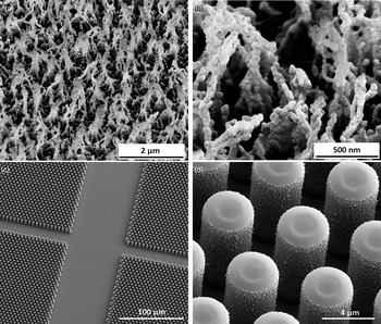
The two fabricated surfaces, inspired by the natural features of Lotus leaves,[ Reference Accardo, Di Fabrizio, Limongi, Marinaro and Riekel 2 , Reference Accardo, Trevisiol, Cerf, Thibault, Laurell, Buscato, Lenfant, Arnal, Fontaine and Vieu 5 ] were made of polymethylmethacrylate (PMMA),[ Reference Accardo, Shalabaeva, Di Cola, Burghammer, Krahne, Riekel and Dante 4 ] a fully biocompatible material, and SU-8, a photosensitive resist widely used in optical lithography for the development of lab on chip devices.[ Reference Marinaro, Accardo, De Angelis, Dane, Weinhausen, Burghammer and Riekel 14 ] Among the available techniques, a very effective approach to nanotexture polymeric surfaces is the use of plasma etching.[ Reference Phan, Yoon and Moon 15 ] The first kind of surface presented here included the texturing of flat PMMA sheets [Fig. 1(a)] by means of a one-step plasma oxygen approach using a Sentech ICP-RIE (inductively coupled plasma reactive ion etch) system, which allows to process several substrates at the same time. We used a 10 min plasma process at ambient temperature (21 °C) with a chamber pressure of 6.5 Pa, an ICP power of 100 W, a radiofrequency (RF) power of 50 W and an O2 flux of 30 sccm. The morphology resulted in highly oriented fibrils as shown in Fig. 1(b). The second type of surface included both an optical lithography process and a plasma roughening one to create a dense micropillars topology [Fig. 1(c)]. Silicon samples were first spin-coated with SU-8 negative tone resist at 4000 RPM, then baked at 65 °C for 15 min ramping up until 95 °C for 40 min and consequently exposed under UV light for 15 s using an optical mask with a hexagonal pillars lattice. Finally, a post-exposure baking at 65 °C for 5 min ramping up until 95 °C for 10 min and a chemical development in SU-8 developer for 5 min were performed. After this, a plasma process based on CF4/O2 (5/15 sccm) at a pressure of 9.06 Pa and an ICP/RF power of respectively 100 and 50 W for 10 min was used to enhance the roughness of the pillars as shown in Fig. 1(d). The pillars composing the microstructured surface had a topology of 4 µm edge, 2 µm gap, and 11 µm height.
(a, b) PMMA nanostructured morphology; (c, d) SU-8 microroughened pillars.

The SU-8 negative tone optical resist was provided by Microchem and used with no further modification, while 500 µm thick PMMA sheets were purchased from Goodfellow. HCT116 cells were obtained from the ATCC (American Type Culture Collection). The cells were cultured in Dulbecco's modified Eagle's medium (DMEM), supplemented with 10% fetal bovine serum (Invitrogen, Carlsbad, CA), penicillin (100 U/mL) and streptomycin (100 mg/mL) (Invitrogen). 3 × 10⁵ cells were inoculated onto PMMA nanostructured (nano-PMMA) surfaces and SU-8 micropillars, previously sterilized for 1 h under UV light without further surface treatment (i.e. coating with adhesion agents such as polylysine), for 48 h at 37 °C in a humidified 5% CO2/air atmosphere. After incubation, cells were washed twice in phosphate-buffered saline solution (PBS, Invitrogen) and fixed with 2% glutaraldehyde and 0.2 M of cacodylate buffer for 1 h at room temperature (RT). Cells were then washed with 0.1 M cacodylate buffer for 10 min and permeabilized with 0.5% Triton X-100 (Sigma Aldrich, St. Louis, MO) for 15 min at RT. Then, they were washed in PBS, incubated with 3% BSA for 20 min at RT, washed again in PBS and stained with vinculin primary antibody (Abcam, Cambridge, UK), a protein highly expressed in cellular focal adhesion plaques, for 30 min at 4 °C. To visualize vinculin, a secondary antibody (Alexa Fluor 488 chicken anti-mouse from Invitrogen) was added and incubated for 40 min at RT in the dark. Then, cells were stained with Alexa Fluor 635 phalloidin (Invitrogen), used to visualize F-actin, for 30 min at RT and finally were washed three times with PBS. Confocal three-dimensional (3D) images were acquired using an inverted Nikon A1 confocal microscope with a 60X objective. All fluorescence images were captured using a 488 nm excitation laser (for vinculin), a 635 nm excitation laser (for phalloidin) and processed with NIS-Elements Microscope Imaging Software. Concerning instead scanning electron microscopy (SEM) morphological investigation, the samples were dehydrated using different concentrations of ethanol diluted in deionized water (from 50% to 100%) and coated with a 15 nm layer of sputtered gold. The SEM micrographs were taken with a FEI Nanolab 600 setup.
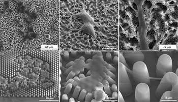
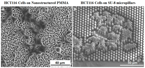
The SEM imaging revealed a good proliferation of the cells on the two fabricated surfaces and the formation of solid links with the nano- and micropatterned structures composing the surfaces (Fig. 2). Concerning PMMA nanostructured surfaces, the cells interconnections were deeply anchored to the nanometric hairy protrusions making the whole cellular body very adherent to the substrate [Figs. 2(a)–2(c)]. On the other hand, the SU-8 micropillars showed a partial bending because of the force exerted by the filopodia (plasma-membrane protrusions that act as antennae for cells to probe their environment[ Reference Jacquemet, Hamidi and Ivaska 16 , Reference Mattila and Lappalainen 17 ]) while interconnecting cells among each other [Figs. 2(d)–2(f)]. This mechanism highlights how such microstructured surfaces could be of interest for studies focusing on the mechanical forces exerted by cancer cells during their growth and proliferation processes.
SEM micrographs of HCT116 cells cultured for 48 h on PMMA nanostructured surfaces (a–c) and on SU-8 micropillars substrates (d–f).

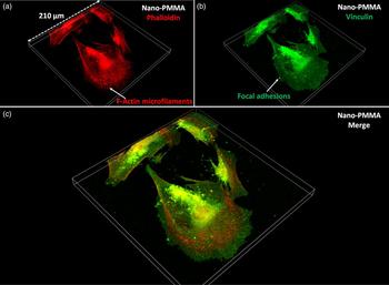
Furthermore, as mentioned earlier, a confocal microscopy investigation was performed to highlight the focal adhesions and the overall cytoskeletal conformation of HCT116 cells. In particular, we focused our attention on vinculin, a membrane-cytoskeletal protein in focal adhesions that is involved in the transmission of mechanical forces and it has a key role in promoting cell migration on a substrate.[ Reference Ziegler, Liddington and Critchley 18 ] Indeed, the phenotypic change of reduced cell adhesion and growth are associated with the loss of this protein within the cell.[ Reference Carisey and Ballestrem 19 ] Furthermore, vinculin has also a direct interaction with the actin cytoskeleton and it is mediated by two regions located in the protein tail.[ Reference Nikkhah, Edalat, Manoucheri and Khademhosseini 20 ] Starting from these considerations, we probed the expression of cytoskeletal immunofluorescent markers of vinculin and phalloidin in HCT116 cells cultured on both flat and nanostructured PMMA highlighting how the nanostructured PMMA surface sensitively influenced, through its nanofeatures, the degree of adhesion of the entire cell. By producing 3D confocal images (obtained from single slices acquired at several z-heights), a cell thickness of 2.6 and 15.3 µm was detected, respectively, for the case of the nanotextured PMMA (nano-PMMA, Fig. 3) and for the flat one (Fig. S1 of the Supporting Information). This, indeed, confirms how the presence of the PMMA nanoroughness induced by plasma reactive ion etching plays a fundamental role in the adhesion between the cells and the substrate favoring their anchoring [Figs. 2(a)–2(c)]. The higher degree of cell adhesion is particularly evident looking at the expression and conformation of phalloidin in HCT116 cells on nano-PMMA [Figs. 3(a) and 3(c)] where it is possible to recognize the presence of spread cytoskeletal F-actin microfilaments, which are not detectable instead in HCT116 cells cultured on flat PMMA [Fig. S1(a), Supporting Information]. Further, the expression of vinculin [Figs. 3(b) and 3(c)] highlights the presence of focal adhesions, that are usually found under the cell or at the edge of the plasma membrane where the cell is attached to the surface, and are visible as green dots at the end of the red F-actin microfilaments.[ Reference Baldo, Lopes, Faquim-Mauro, Jacysyn, Niland, Eble, Clissa and Moura-Da-Silva 21 ] HCT116 cells cultured on flat PMMA [Fig. S1(b), Supporting Information] on the other hand show a globular and less spread/adherent morphology in agreement with their phalloidin expression. Although the nanoroughness of the substrate plays a major role in favoring the tight anchoring of cell interconnections [Figs. 2(a)–2(c)], the oxygen plasma treatment is known to induce a further chemical makeup of the surface that, put together with the tailoring of other factors, can modulate cell adhesion.[ Reference Bacakova, Filova, Parizek, Ruml and Svorcik 22 ] As observed in previous studies, polymeric surfaces,[ Reference Kim, Seo and Min 23 , Reference Borges, Benetoli, Licínio, Zoldan, Santos-Silva, Assreuy, Pasa, Debacher and Soldi 24 ] and specifically PMMA,[ Reference Ozcan and Hasirci 25 , Reference Ozcan, Zorlutuna, Hasirci and Hasirci 26 ] exposed to plasma modification exhibit surface chemistry changes which are reflected in an augmentation of oxygen content and an enhancement of surface energy associated with an increase in wettability that influences cell attachment. Therefore, the enhancement of HCT116 cells adhesion on nano-PMMA surfaces, compared with pristine flat PMMA ones, can be ascribed to the combination of the PMMA surface chemistry modification and the nanofibrillar roughness morphology.
3D Confocal microscopy of HCT116 cells cultured for 48 h on nanostructured PMMA surfaces (a, phalloidin; b, vinculin; c, merge).

A good adhesion of mammalian cells is critical to determine cell viability and to study their proliferation on surfaces. Moreover, it is known that cell migration, differentiation, and adhesion are influenced by the geometrical features of surfaces.[ Reference Gentile, Tirinato, Battista, Causa, Liberale, di Fabrizio and Decuzzi 11 , Reference Cesca, Limongi, Accardo, Rocchi, Orlando, Shalabaeva, Di Fabrizio and Benfenati 27 , Reference Biggs, Richards and Dalby 28 ] Here we proposed a straightforward fabrication and imaging protocol to highlight colon cancer cell adhesion properties through the study of both focal adhesions and cellular cytoskeleton of cells grown on biomimetic nanostructured polymeric substrates without any pre-treatment of the surfaces with adhesion agents. As a model system, HCT116 cell line[ Reference Zhu, Zhao, Sheng, Hou, Hao, Yang, Zhu, Zhang, Han, Wei and Zhang 12 , Reference Huang, Greve, Nausieda, Nguyen and Domach 13 , Reference Kai, Nagano, Sugihara, Arima, Sampetrean, Ishimoto, Nakanishi, Ueno, Iwase and Saya 29 ] was used and the changes in cell adhesion were observed by using confocal fluorescence and SEM. The results showed the enhanced adhesion of the cancer cells on nanostructured PMMA compared to pristine one as well as a better expression of focal adhesions and F-actin microfilaments. The combination of the reported nanostructured surfaces with SEM morphological analysis and 3D immunofluorescence imaging provide indeed an interesting platform for advancing the understanding of mechanisms behind cancer cell growth and in further investigations may include the monitoring of cell migration dynamics as well as the characterization of other proteins involved in cell adhesion and spreading processes such as focal adhesion kinase.[ Reference Golubovskaya, Kweh and Cance 30 ] On the other hand, the pulling behavior of the cells on the SU-8 micropillars suggests that this kind of device could be useful for quantitative studies of the forces exerted by cancer cells during their proliferation.
Supplementary material
The supplementary material for this article can be found at https://doi.org/10.1557/mrc.2017.128.