Acute rheumatic fever, and its long term sequel, rheumatic heart disease, is a major problem in children, adolescents and young adults.1 Despite the tremendous progress made in cardiology, the menace of morbidity and mortality in India due to acute rheumatic fever and its consequences remain very high.2 Because of the preoccupation of many cardiologists with cardiac diseases seen in adults, such as ischaemic heart disease, the problems of acute rheumatic fever and rheumatic heart disease have tended to be sidelined. Studies on prevalence, treatment, and prevention receive only scant attention. In India, only exotic palliative methods, such as balloon mitral valvotomy or valvar replacement, have achieved the centre of the stage.3, 4
Precise diagnosis of acute rheumatic fever has presented problems since Hippocrates, who provided the first written description of arthritis in man 400 years before the birth of Christ.5 Lack of specific criterions had led to diagnostic chaos until the publication of the listings of Duckett Jones in 1944.6 Despite the four revisions and modifications of the Jones criterions,7, 8 acute rheumatic fever remains either underdiagnosed, leading to nearly half of the patients with established rheumatic heart disease not receiving prophylaxis, or overdiagnosed, leading to unnecessary treatment with steroids and other drugs depending on traditional characteristic auscultatory findings. It is rare for modern techniques like echocardiography to be used when establishing the Jones criterions,9 despite the fact that carditis is diagnosed more frequently and reliably using echocardiography than using traditional auscultatory findings.10 Early diagnosis of acute rheumatic fever, though difficult, is very important if we are to prevent the serious consequences in young patients. The aim of our study, therefore, was to establish the role of echocardiography in the precise diagnosis of acute rheumatic fever, and to determine the various echocardiographic features of carditis or subclinical valvitis seen in patients with acute rheumatic fever diagnosed by the Jones criterions.
Material and methods
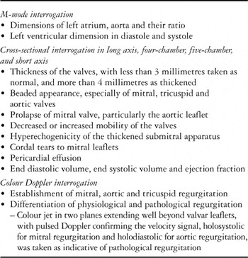
We studied prospectively 492 consecutive patients diagnosed clinically with acute rheumatic fever, referred to us by paediatricians from two reputed paediatric centres in Bangalore, namely VaniVilas Children's Hospital, and Indira Gandhi Institute of Child Health. All the patients had been examined thoroughly by the paediatricians, who had used appropriate pathological tests. Echocardiography was performed in detail by the well trained and experienced echocardiographer, who was unaware of the diagnosis. A single cardiologist then reexamined the patients, and made the final diagnosis, using the revised Jones criterions of 1992. Out of 492 patients referred, only 452 fulfilled these criterions. They formed the study group. The echocardiogram was done in detail, and various echocardiographic parameters were meticulously recorded (Table 1). The cardiologists then analyzed the results with the assistance of a statistician, using D-base and EP inf. The cost of investigations and repeated tests in poor patients were borne by the Medical Education and Research Trust, Bangalore, Karnataka.
Table 1. Echocardiographic investigations.

Results
Out of 452 patients with acute rheumatic fever diagnosed strictly by the Jones criterion of 1992, 200 came from a rural background, and 252 from the urban area. Of the patients, 230 were males and 222 were females. Their age ranged from 1 year 11 months to 20 years, with a mean age of 11.7 plus or minus 5.4 years. We were also referred 10 exceptional patients who suffered their first attack of acute rheumatic fever as adults. They ranged in age from 28 to 51 years, and along with a 65-year-old female who suffered a recrudescence of rheumatic activity, they were not included when calculating the mean age. Of 239 patients (52.8%) with polyarthritis, 164 were diagnosed clinically as having carditis by traditional auscultation, but only 141 of these showed echocardiographic evidence of carditis (85.97%), the remaining 23 cases (14%) having no evidence of carditis. Among 40 patients with rheumatic chorea, 28 (70%) had echocardiographic evidence of carditis or valvitis. Polyarthralgia was seen in 213 patients (47.12%), of whom 38 (17.8%) had carditis clinically, albeit that 88 patients (41.3%) showed echocardiographic evidence of subclinical carditis or valvitis.
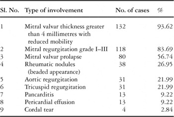
The incidence of the varied echocardiographic findings in 141 patients with carditis is shown in Table 2.
Table 2.

The flitting type of migratory polyarthritis was present in 239 patients (52.87%), whereas migratory polyarthralgia was seen in 213 cases (47.12%). Out of these 213 cases of arthralgia, 38 patients (17.84%) had clinical evidence of carditis. Echocardiographic evidence of subclinical carditis or valvitis, such as thickening and beaded appearance of the valvar leaflets, was present in 88 cases (41.3%).
Out of 164 patients diagnosed clinically with carditis, only 141 had supporting echocardiographic findings (85.97%), with the remaining 23 patients (14%) having functional murmurs, tachycardia, anaemia, fever of some other aetiology, or congenitally malformed hearts, the latter found in patients with an atrial septal defect and a subaortic fibrous shelf, respectively. We found four patients with severe mitral regurgitation due to cordal tears and flail valvar leaflets. They were referred for surgery.
Rheumatic chorea was found in 40 (8.85%) patients, with 8 having bilateral chorea. In one patient, the chorea lasted for one year, and in another it had recurred after 2 years. Two other screened siblings of one child with chorea showed no evidence of acute rheumatic fever at the time of screening, but developed rheumatic chorea after 3 months. The titres of Antistreptolysin O were negative in 17 cases (42.5%), but ranged from 400 to 800 international units in the remaining 23 cases. The mean duration of onset of chorea after the initial fever was 3 months. We were surprised to find that 28 of the patients with rheumatic chorea (70%) had echocardiographic evidence of valvar involvement, although this had not been detected clinically.
Subcutaneous nodules (Fig. 1) were seen in only 7 cases (2.4%). All these patients had high titres of antistreptolysin O, ranging between 800 and 1600 international units, but c-reactive protein was positive in only 4 cases. All seven patients had both clinical and echocardiographic evidence of carditis.

Figure 1. (a,b) show the appearance of subcutaneous rheumatic nodules.
Erythema marginatum was seen in only 2 (0.4%) of our patients (Fig. 2). The youngest child was 1 year 11 months old. As erythema marginatum occurs transiently and disappears, it is easily missed at the time of examination. Both of our patients had clinical evidence of carditis, confirmed by the echocardiogram.

Figure 2. (a,b) show erythema marginatum.
Among the minor criterions, a raised erythrocytic sedimentation rate was the commonest, found in 314 cases (69.46%). The highest rate was 140 millimetres in the first hour. The antistreptolysin O titer was elevated in 311 cases (68.8%). Fever was present in 224 cases (49.55%). C-reactive protein was positive in only 181 cases (40%). Culture for β-haemolytic streptococcus was positive in 79 cases (17.47%), and these children showed marked rising titers of antistreptolysin O, with neither the erythrocytic sedimentation rate nor symptoms subsiding subsequent to treatment with aspirin. We did not discover a history of scarlet fever, a preceding β-haemolytic streptococcal infection, or prolongation of the P-R intervals on the electrocardiogram in any of our patients.
Discussion
Diagnosis of carditis, considered a major criterion for recognition of acute rheumatic fever, traditionally depends on the clinical auscultatory findings.4 Of our 164 patients diagnosed clinically with carditis, nonetheless, only 141 (85.9%) had echocardiographic evidence of carditis or valvitis, as judged on the basis of a beaded appearance of the valvar leaflets, which is equivalent to rheumatic nodules as seen on the valves at autopsy (Fig. 3). The remaining 23 patients either had functional murmurs, fever, tachycardia, or anaemia, with 2 patients proving to have congenitally malformed hearts. A 12-year-old boy with a discrete subaortic shelf, wrongly diagnosed as having acute rheumatic fever with carditis, had been treated with steroids by his paediatrician. He subsequently developed tubercular meningitis, as the primary complex flared up subsequent to the steroids. This episode demonstrates graphically the potential error in diagnosis that can be made if clinical carditis, if unconfirmed by echocardiography, is taken as a major criterion for acute rheumatic fever when combined with two minor criterions such as fever and a raised erythrocytic sedimentation rate. Indeed, one-sixth of our cohort, wrongly diagnosed with acute rheumatic fever, were treated unnecessarily and placed on secondary prophylaxis. As we have shown, pancarditis can be diagnosed precisely using echocardiography (Fig. 4), thus avoiding false positive diagnoses. On the other hand, in 88 of our patients with polyarthalgia, almost half of those with this minor criterion, there was no clinical evidence of carditis, but echocardiographic interrogation revealed subclinical carditis or valvitis. If these patients had not been investigated echocardiographically, they would have gone undiagnosed, would not have received secondary prophylaxis beyond 5 years, and could end up with rheumatic heart disease, allegedly without any past history of acute rheumatic fever! Our study shows that two-fifths of our patients, had we relied on traditional auscultatory findings, would not have been diagnosed with carditis had they not also been investigated with echocardiography (Fig. 5). We also identified four patients with severe mitral regurgitation who were referred for surgical repair. Cotrim et al.11 concluded that echocardiography, if performed early, is very important in all children suspected to have acute rheumatic fever, particularly because mitral regurgitation can be demonstrated by colour flow mapping in the absence of any cardiac murmur. Wilson and Neutze12 supported this conclusion, endorsing the fact that pulsed and colour Doppler echocardiography provide a method with which to detect minor degree of pathological regurgitation in the absence of characteristic clinical signs. The addition of echocardiographic interrogation before diagnosing carditis, therefore, not only prevents clinical overdiagnosis, but also identifies those subclinical cases of carditis and valvitis that would otherwise pass undiagnosed and without prophylaxis. The nodules seen on the mitral and aortic valvar leaflets are likely the equivalents of the verrucous lesions seen at autopsy in patients dying of acute rheumatic fever with carditis.13 We contend, therefore, that echocardiography can play an important role in the early and precise diagnosis of carditis, and should be included as part of the Jones criterions.

Figure 3. Mitral valvar verrucous nodules as seen (a) at autopsy, and compared (b) to an echocardiogram showing nodules on the mitral leaflets.

Figure 4. Pancarditis with mitral regurgitation.

Figure 5. The echocardiograms show (a) hyperechogenic submitral valvar apparatus as seen in the parasternal long axis view, and thickening of the leaflets, beyond 4 millimetres (b), again as seen in parasternal long axis view.
We recognize, nonetheless, the limitations of our study. We were able to correlate the clinical and echocardiographic findings only at the time of diagnosis of acute rheumatic fever, and hence could not shed any light on its sequels. Furthermore, we have not conduced any prospective long term follow-up to demonstrate the potential significance of the identified subclinical valvitis. We recognize the need to expand out study, using tissue harmonics to strengthen the criterions with which to diagnose subclinical carditis. Finally, we did not establish either the sensitivity or specificity of cardiac auscultation when taking into consideration the echocardiogram as the “gold standard” test. Despite these limitations, we concluded that uncritical adherence to the revised Jones criterions for the diagnosis of acute rheumatic fever may lead to gross underdiagnosis among the children with polyarthralgia, and overdiagnosis in those in whom carditis is reported on the basis of clinical examination. Hence, we suggest that echocardiographic evidence of carditis or valvitis should be given its due importance. If we are to avoid morbidity, mortality, and unnecessary expenditure on sophisticated treatment, an early and precise diagnosis of carditis is very important. The echocardiogram is a very simple, noninvasive, reproducible and a useful tool for making such an early and precise diagnosis of carditis in the setting of acute rheumatic fever. We submit that, when the Jones criterions undergo their next revision, then echocardiography should be included as necessary for the diagnosis of carditis.
Acknowledgements
We express our grateful thanks to the late Dr. K. S. Shadaksharappa, Ex Chairman, and Dr. M. Maiya, the present Chairman of the Medical Education and Research Trust, Bangalore for encouragement and financial assistance. Our thanks also go to Dr. D. G. Benakappa, Director, Indira Gandhi Institute of Child Health, and Dr. T. A. Veena, Professor of Pediatrics, Vani Vilas Children's Hospital Bangalore. Source of Grant: Late Dr. K. S. Shadaksharappa, the then Chairman of Medical Education Research Trust, Bangalore, Karnataka, gave financial assistance to the poor patients.