INTRODUCTION
The international effort to develop malaria vaccines has focused on a number of targets in the parasite's life-cycle, including the sporozoite stage which holds much promise as a source of immunogens to block transmission at the hepatic phase of infection (Waters, Reference Waters2006; Matuschewski, Reference Matuschewski2006). Our understanding of Plasmodium sporozoite biology is much less advanced than that of the blood stages, and the recent advances in vaccine development emphasize the need to explore this stage of the parasite's life-cycle in greater depth. Electron microscopical studies of Plasmodium berghei, P. gallinaceum, P. yoelii and P. falciparum in the 1960s and 1970s established the general pattern of ultrastructural organization in mature malaria sporozoites (Garnham et al. Reference Garnham, Bird and Baker1963; Aikawa, Reference Aikawa1971; Cochrane et al. Reference Cochrane, Aikawa, Jeng and Nussenzweig1976) and showed that they possess many features similar to those of malaria merozoites, as expected from the ability of both forms to invade the cells of their hosts. Recent molecular studies have emphasized the distinctive stage-specific nature of gene expression in sporozoites (Florens et al. Reference Florens, Washburn, Raine, Anthony, Grainger, Haynes, Moch, Muster, Sacci, Tabb, Witney, Wolters, Wu, Gardner, Holder, Sinden, Yates and Carucci2002; Le Roch et al. Reference Le Roch, Zhou, Blair, Grainger, Moch, Haynes, De La Vega, Holder, Batalov, Carucci and Winzeler2003; Hall et al. Reference Hall, Karras, Raine, Carlton, Kooij, Berriman, Florens, Janssen, Pain, Christophides, James, Rutherford, Harris, Harris, Churcher, Quail, Ormond, Doggett, Trueman, Mendoza, Bidwell, Rajandream, Carucci, Yates, Kafatos, Janse, Barrell, Turner, Waters and Sinden2005), reflecting the much more complex life-history of this parasite stage.
The production of sporozoites (sporogony) within oocysts in the mosquito mid-gut wall has also been described ultrastructurally by several authors (Terzakis et al. Reference Terzakis, Sprinz and Ward1967; Sinden and Strong, Reference Sinden and Strong1978; Meis et al. Reference Meis, Wismans, Jap, Lensen and Ponnudurai1992). During this process, the developing cellular mass (sporoblast) has a centrally-placed nucleus that undergoes a sequence of repeated cryptomitoses (endomitoses) to produce many thousands of genomic centres beneath the periphery of a single nuclear envelope (Schrével et al. Reference Schrével, Asfaux-Foucher and Bafort1977). These separate into individual nuclei which move into the sporozoite buds beginning to enlarge at the sporoblast surface. At the same time, the other sporozoite organelles begin to appear within the buds. When fully assembled, the mature, elongate sporozoites pinch off from the parent sporoblast and break through the sporocyst wall to enter the mosquito's haemocoel en route to its salivary glands.
The synthesis and assembly of organelles to form the mature sporozoite are at present poorly understood, although recent reports of the effects of genetic modification on sporozoite development are giving new insights into the molecular requirements for sporozoite function (Ménard et al. Reference Ménard, Sultan, Cortes, Altsuler, van Dijk, Janse, Waters, Nussenzweig and Nussenzweig1997; Persson et al. Reference Persson, Oliveira, Sultan, Bhanot, Nussenzweig and Nardin2002) and development (Thathy et al. Reference Thathy, Fujioka, Gantt, Nussenzweig, Nussenzweig and Ménard2002; Khater et al. Reference Khater, Sinden and Dessens2004). Trafficking pathways for the formation and placement of micronemes and rhoptries have been reported for malaria merozoites (Bannister et al. Reference Bannister, Hopkins, Fowler, Krishna and Mitchell2000, Reference Bannister, Hopkins, Dluzewski, Margos, Williams, Blackman, Kocken, Thomas and Mitchell2003) and for Toxoplasma gondii tachyzoites (Karsten et al. Reference Karsten, Qi, Beckers, Reddy, Dubremetz, Webster and Joiner1998; Hager et al. Reference Hager, Striepen, Tilney and Roos1999; Hoppe et al. Reference Hoppe, Ngo, Yang and Joiner2000). In the present paper we have analysed the structures associated with vesicle trafficking in malaria sporozoites, using Plasmodium berghei berghei as a model system. We find that such trafficking is essentially similar to that of merozoites, but has an additional, novel mechanism for vesicle translocation. This work is based on data from a study performed in the 1970s by one of the authors (G. A.-F.), supplemented by recent 3-dimensional reconstructions from serial sections of the same strain of Plasmodium berghei.
MATERIALS AND METHODS
Oocysts of Plasmodium berghei berghei, strain Anka, were grown as previously described (Schrével et al. Reference Schrével, Asfaux-Foucher and Bafort1977) or were provided by the Institut Pasteur, Paris. Mosquitoes (Anopheles stephensi) were allowed to feed on murine blood, and then at intervals between 6 and 12 days later were killed and their guts dissected out into physiological saline (6% (w/v) NaCl). After different assays with double fixation procedures for transmission electron microscopy, method number 5 described by Terzakis (Reference Terzakis1968) was selected. Briefly, fixation was carried out at 4°C, in 2·5% (v/v) glutaraldehyde in 0·1 m sodium citrate buffer, pH 7·4 for 45 min, followed by 1% (w/v) osmium tetroxide in the same buffer (pH 7·4), for 45 min. After a brief wash (2 min) in 0·1 m sodium acetate (pH 7·4) samples were block stained in 5% (w/v) aqueous uranyl acetate (pH 3·4) for 9–20 min. They were then briefly washed in the 0·1 m sodium acetate buffer (pH 7·4), dehydrated in an acetone series and embedded in Araldite epoxy resin. Sections were further stained with uranyl acetate and lead citrate and imaged in Hitachi HU 11C and HU 7600 electron microscopes, or for serial sections, in a Jeol 1010 electron microscope.
The 3-D images were obtained from 90 nm serial sections performed with an Ultracut S (Leica) and mounted on copper slot grids, coated with Parlodion and a carbon film. The selected images were processed by Adobe Photoshop software and the IMOD program for 3-D reconstruction of EM serial sections from the Boulder Laboratory for 3-D Electron Microscopy of Cells (http://bio3D.colorado.edu). Due to the distortion effects of the electron beam on the ultrathin sections, an initial superposition of the images was performed with Adobe Photoshop before the application of IMOD. Video recording was achieved with Hyper Cam 2 and subsequent encoding with the DrDivX program.
RESULTS
Sporozoite budding
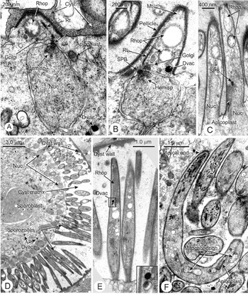
The major events of this process have been described (Vanderberg and Rhodin, Reference Vanderberg and Rhodin1967; Vanderberg et al. Reference Vanderberg, Rhodin and Yoeli1967; Sinden and Garnham, Reference Sinden and Garnham1973; Sinden and Strong, Reference Sinden and Strong1978) and will be outlined only briefly here (Fig. 1A–F, Fig. 7C–G). Essentially, sporozoite buds first appear as a series of conical elevations at the sporoblast surface, each opposite one of the numerous spindle pole bodies located in a nuclear pore and associated with a hemispindle (Fig. 1A), at the surface of the sporoblast nucleus (Fig. 1A, B). As they elongate, each bud is invaded by a nucleus, mitochondrion and apicoplast (Fig. 1A, C). At the apex of the bud, 3 polar rings are formed immediately underneath the flat apical membrane, and attached apically to the external surface of the largest, basal (the third) of the rings (Fig. 2D), the inner membrane complex (IMC) extends underneath the plasma membrane, so creating the 3-layered pellicle (Fig. 1B, Fig. 2F, G) characteristic of apicomplexan zoites. An assemblage of longitudinal subpellicular microtubules is also formed, attached apically to the third polar ring and extending basally down into the sporoblast cytoplasm beyond the bud (not shown). The bud elongates and becomes more cylindrical though eventually narrower at both ends (Fig. 1D, E), the basal end constricting into a short pedicle connecting the bud to the sporoblast. At this stage the bud is straight or very slightly curved, so that the numerous sporozoite buds project radially out from the parent sporoblast (Fig. 1D). When the sporozoites finally detach from the sporoblast, they become much more curved (Fig. 1F).
(A–F) Sporozoite buds at different stages of development. (A) First signs of bud formation, with an early conical elevation at the sporoblast surface close to a spindle pole body attached to a hemispindle of the last endomitosis. An early rhoptry, a single Golgi cisterna and part of a rootlet fibre are also visible. (B) The bud has lengthened and 2 spheroidal rhoptries have appeared. A rootlet fibre and 2 subpellicular microtubules are present, and at the bud surface the triple membranes of the pellicle define the outline of the bud, as throughout the sequence. In a later stage (C) 2 longer (8 μm) buds are shown, each with a nucleus invading its base; on the right, the most apical rhoptry is beginning to adopt a club-like shape. (D) Lower magnification shows the surface of a later sporoblast, with numerous buds at its surface. In (E), a group of sporozoite buds similar to those in (D) are shown at higher magnification, illustrating the change in shape and the narrowing of the attachment zone at their bases; the inset shows 2 dense vacuoles at higher magnification. In (F), the sporozoites have detached from the sporoblast, one of them showing its typical curved form, with micronemes and rhoptries visible apically. Abbreviations: Dvac, dense vacuole (acidocalcisome-like); Golgi, Golgi cisterna(e); Hemisp, hemispindle; Mt, microtubule; Nuc, nucleus; Rhop, rhoptry; Rt, rootlet fibre; SPB, spindle pole body.

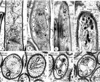
(A–H) Endoplasmic reticulum and free ribosomes in developing sporozoites. (A and B) Confluence of the nuclear envelope and rough endoplasmic reticulum in the apical part of the bud (arrowhead). Also visible are the coated vesicle budding zone of the outer nuclear envelope (A), a Golgi cisterna (A and B) and (A) a group of post-Golgi vesicles. Numerous free ribosomes are present in the cytosol (A). (C) and (D) Apical extension of the endoplasmic reticulum which reaches almost to the extreme apical end, indicated by the presence of the polar rings (D). (E–H) Transverse sections of sporozoite buds showing the nuclear envelope studded with ribosomes (E), 2 profiles of rough endoplasmic reticulum (F), which is quite distinct from the smooth membranes of the invagination of the inner membrane complex (G, H). Subpellicular microtubules beneath the inner membrane complex (IMC) and plasma membrane are also visible. Abbreviations: CV, coated vesicle budding from the nuclear envelope; ER, endoplasmic reticulum; Golgi, Golgi cisterna(e); IMC, inner membrane complex; IMCi, inner membrane complex invagination; Mic, micronemes; Mt, microtubule; Nuc, nucleus; Plas, plasma membrane of parasite; Tgves, trans-Golgi vesicle.

Organelles related to protein synthesis and trafficking
These include free ribosomes, the endoplasmic reticulum (ER), nuclear envelope, Golgi complex and post-Golgi vesicles of various kinds (Fig. 2A–F).
Ribosomes, endoplasmic reticulum and nuclear envelope
The cytosol around and apical to the nucleus contains free ribosome clusters whose numbers increase considerably as the bud elongates (Fig. 2B, E) and dominate much of the cytoplasm in free sporozoites. The ER develops as an apically-directed flat cisternal extension of the nuclear envelope (Fig. 2A, B, arrowheads), first as a single or double flat cisterna (Fig. 2F), later becoming a highly branched compartment pervading the apical cytoplasm almost to the polar rings towards bud maturity (Fig. 2C, D) then, as development procedes, dwindling to a much reduced organelle at full maturation. The presence of attached ribosomes and a dilated lumen distinguishes the ER from another membranous compartment in the apical region, described by Sinden and Strong (Reference Sinden and Strong1978) ; a smooth cisterna with no ribosomes that forms a curved plate in mature sporozoites. Transverse sections of the sporozoite bud show this to be an invagination of the IMC (Fig. 2G, H).
Golgi complex
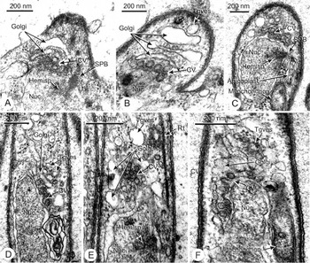
From the earliest appearance of the sporozoite bud, a group of 1–3 smooth cisternae similar to the Golgi complex of the merozoite stage (Bannister et al. Reference Bannister, Hopkins, Fowler, Krishna and Mitchell2000, Reference Bannister, Hopkins, Dluzewski, Margos, Williams, Blackman, Kocken, Thomas and Mitchell2003) is present (Fig. 1A, B, Fig. 2A, B, Fig. 3A–F). This lies close to a zone of 40–50 nm diameter coated vesicles budding from the nuclear envelope (Fig. 3A–F) indicating that the Golgi complex receives these vesicles by fusion (Fig. 2A, Fig. 3A, B). The coated vesicles bud off from the nucleus' apical surface close to the spindle pole body (Fig. 3C), an arrangement that persists into the mature elongated bud stage, although in later times the spindle pole body and budding zone usually migrate to the side of the nucleus (Fig. 3D). At later stages of bud formation, the Golgi cisternae become quite extensive and highly folded, and numerous trans-Golgi vesicles of various sizes (50–150 nm) and irregular smooth cisternae appear around and apical to the Golgi complex (Fig. 3D–F). Some of these larger vesicles are likely precursors of rhoptry-directed and pro-micronemal vesicles (see below).
(A–F) Development of the sporozoite Golgi complex and related structures. (A and B) Early budding stages show the Golgi complex above the nucleus before nuclear migration into the sporozoite bud. (A–C) Clusters of coated vesicles are visible at the nuclear envelope, budding from it in (C), and closely related to the spindle pole body and related hemispindle in (A) and (C). In (B), the oblique section through a bud shows 3 Golgi cisternae and a post-Golgi vesicle. (D–F) More advanced sporozoite buds showing an elaboration of the Golgi complex and the shifting of the coated vesicle budding zone more to the side of the nucleus. Abbreviations: CV, coated vacuole budding from the nuclear envelope; Golgi, Golgi cisterna(e); Hemisp, hemispindle; Nuc, nucleus; Rt, rootlet fibre; SPB, spindle pole body; Tgves, trans-Golgi vesicle.

The development of rhoptries and micronemes
Rhoptries
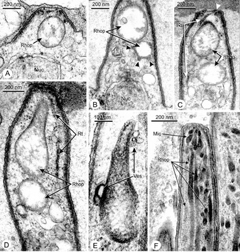
These organelles appear at the same time as the first structural signs of sporozoite bud differentiation (Fig. 1A, Fig. 4A). Initially, they are spherical and generally electron lucent, with denser flocculent material lying close to the rhoptry membrane or scattered within the rhoptry lumen (Fig. 1A–C, Fig. 4A). As the bud elongates, new spheroidal rhoptries are formed close to the Golgi complex, while more apically the older, larger rhoptries become pear-shaped, with a basal bulb and a narrower neck (Fig. 4D, E). With further bud elongation the rhoptry contents become more compact and granular in appearance (Fig. 2C, Fig. 4F, Fig. 5A, B), although within a single bud, the more apical rhoptries are more advanced than others in shape and overall density (Fig. 4C, D) until a late stage in bud development.
(A–E) Rhoptry development in elongating sporozoite buds. (A) Early conical bud containing a young rhoptry beneath the immature polar ring complex. (B) In a more advanced bud, 2 or perhaps 3 rhoptries have appeared, one of them indicating fusion with small vesicles (arrowheads). (C–D) As the bud elongates further, the most apical rhoptry becomes pear–shaped; a rootlet fibre lies close to the rhoptries and is attached apically to the first polar ring (arrowhead). (E) The rhoptry has elongated, increasing in density, and vesicle clusters accumulate close to the rhoptry base and narrower apical region. (F) The apical region of a late-stage bud showing 4 long narrow dense rhoptries and a group of micronemes converging on the apical surface. Abbreviations: Mic, microneme; Nuc, nucleus; Rhop, rhoptry; Rt, rootlet fibre; Ves, vesicle.

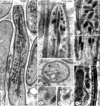
(A–H) Microneme development in late-stage sporozoite buds. (A) A survey micrograph of the apical region showing numerous mature bottle-shaped micronemes surrounding one or more rhoptry profiles. (B) Higher magnification of the apical extremity of a less mature bud where a series of dense-cored promicronemes and a few bottle-shaped mature micronemes surround multiple rhoptries; the 3 polar rings are also visible in section. (C) A group of mature micronemes at a higher magnification. (D–F) Transitional stages between the dense-cored promicronemes and the mature bottle-shaped micronemal form. (G, H) Longitudinal sections through sporozoite bud apices showing evidence of interaction between micronemes and subpellicular microtubules, with micronemes aligned close to and parallel to microtubules, seen in grazing section through the pellicle (G), and in longitudinal section, linked to microtubules by thin filaments (H, arrowheads). Abbreviations: IMC, inner membrane complex; Mic, microneme; Mt, microtubule; Plas, plasma membrane of parasite; Prmic, promicroneme; Rhop, rhoptry.

Rhoptry enlargement is associated with the appearance of spheroidal and ellipsoidal vesicles 70–80 nm in diameter which cluster around the perimeters of growing spheroidal and club-shaped rhoptries (Fig. 4A–E), sometimes closely apposed (Fig. 4E) or apparently in the act of fusion with these organelles (Fig. 4B). In later stages of bud elongation, such vesicles become very numerous and form a column stretching from the Golgi cisternae to the bases and necks of growing rhoptries (see later). Finally, rhoptries, elongate considerably (up to 4 μm), become quite narrow (0·1 μm) and internally dense (Fig. 4F). In mature buds rhoptries number up to 4 per sporozoite (Fig. 4F), and have the form of sinuous blunt-ended sacs that taper apically and end blindly separately from each other close to the centre of the apical plasma membrane (Fig. 4F, Fig. 5A, B).
Micronemes
These organelles are formed late in sporozoite development, when they accumulate in considerable numbers in the sporozoite's apical region (Fig. 4F, Fig. 5A–H). When mature, each microneme is shaped like a long-necked bottle about 150 nm long by 80 nm at its widest diameter, with an elliptical or cylindrical bulbous part and a narrow apical duct (Fig. 5A, C, G, H). At the bud's extreme apical end a small cluster of micronemes is positioned around the apical tips of the rhoptries, their narrow ends converging on the centre of the apical membrane (Fig. 4F). Elsewhere in the apical cytoplasm, micronemes generally appear to be orientated at random except where they lie close to the subpellicular microtubules (Fig. 5G, H). In this case, micronemes are usually orientated with their long axes parallel to the pellicle. Filamentous connections occur between these micronemes and subpelliclular microtubules (Fig. 5H), recalling a similar situation in developing merozoites (Bannister et al. Reference Bannister, Hopkins, Dluzewski, Margos, Williams, Blackman, Kocken, Thomas and Mitchell2003).
In addition to the bottle-like micronemes, maturing buds contain substantial numbers of 120 nm-diameter spheroidal vesicles with irregular outlines and moderately dense cores set in a less dense, granular interior (Fig. 5B–F). These appear in the region between the Golgi complex and the more apical zone of mature micronemes and are absent from fully mature buds and free sporozoites. Some of them show transitional stages (Fig. 5D–F) towards the elongate, uniformly dense bottle form, indicating that they are an early stage of microneme development, maturing to the bottle-like form as they move apically from the trans-surface of the Golgi complex. For this reason we here name them pro-micronemes.
Dense vacuoles
In our study we found variable numbers of spheroidal vesicles about 100 nm in diameter with very dense contents, scattered in the cytoplasm at all stages of bud formation and in mature sporozoites (Fig. 1A, B, D, E, Fig. 6C). They are also present in the sporoblast at all stages. In some instances they are completely filled with dense material, but often they have a dense core eccentrically placed in an otherwise pale interior, an appearance typical of acidocalcisomes described in other apicomplexan genera (Ruiz et al. Reference Ruiz, Luo, Moreno and Docampo2004; Docampo et al. Reference Docampo, De Souza, Miranda, Rohloff and Moreno2005). However, as vesicles with very dense interiors may include more than one type of organelle, we give them here the provisional title of dense vacuoles, pending chemical analysis.
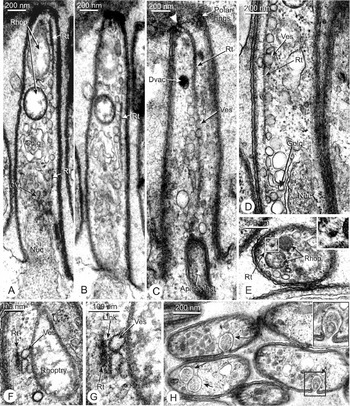
(A–G) Evidence of post-Golgi trafficking along the rootlet fibre to the rhoptries (for rootlet fibre attachments see also Fig. 1 (A, B) and Fig. 4 (C, D)). (A and B) The long track of the rootlet fibre and numerous associated small vesicles can be traced in these two consecutive sections, from the Golgi complex to the developing rhoptries. (C) A similar section from another bud shows numerous vesicles around the rootlet fibre, which is attached apically to the first polar ring (arrowhead); a dense vacuole (of likely acidocalcisome identity) and an apicoplast are also visible. (D) Higher magnification of the trafficking pathway along the rootlet fibre; note the variety of vesicle sizes and shapes, and their moderate opacity. (E) Transverse section of a sporozoite bud showing the rootlet fibre positioned close to 2 rhoptries; the inset depicts the fibre at higher magnification, and demonstrates the thin filaments directed radially outwards at its periphery. (F) Oblique section through the rootlet fibre and a vesicle cluster associated with a developing rhoptry. (G) Higher magnification shows filamentous connections between the rootlet fibre and vesicles (Link). (H) Large micropore vesicles (arrows) in an oblique section through a group of late-stage sporozoite buds, containing heterogenous material. Some of these vesicles open to the exterior. Abbreviations: CV, coated vesicle; Dvac, dense vacuole (acidocalcisome-like); Golgi, Golgi cisterna(e); Nuc, nucleus; Rhop, rhoptry; Rt, rootlet fibre; Ves, vesicle associated with the rootlet fibre.

Cytoskeletal structures related to trafficking
The rootlet fibre (tigelle)
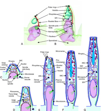
This structure, described briefly by Sinden and Strong (Reference Sinden and Strong1978; see also Sinden and Matuchewski, Reference Sinden, Matuschewski and Sherman2005) is a longitudinal solid fibre about 30 nm thick attached at its base to the spindle pole body on the side of the nucleus (Fig. 1A, B, Fig. 3E, Fig. 4D, Fig. 6A–G) and apically to the first polar ring. It forms a straight rod along one side of the bud, except apically where it arches over to the opposite side to make its polar ring attachment (Fig. 1A, B, Fig. 6A–F, Fig. 7A–F). This arrangement is clearly visible in 3-D reconstructions (Fig. 7A, B). In this study, we did not find evidence of the cross-striations described by Sinden and Strong (Reference Sinden and Strong1978). Numerous short filaments extend out radially from the fibre's surface, giving it a rather hairy appearance (Fig. 6E–G). The fibre continues to elongate as the sporozoite grows but in mature buds it is no longer detectable. In the intermediate stages of sporozoite bud elongation, numerous rounded or irregularly-shaped vesicles cluster along its margins between the Golgi cisterna as far as the developing rhoptries (Fig. 6A–G, Fig. 7E). The vesicles are about 100 nm in diameter and are moderately dense. They are absent from the most mature sporozoite buds.
(A–G) Vesicle trafficking system in sporozoite bud development: reconstruction and summary diagrams. (A and B) Two reconstructions of early and mid-stage sporozoite bud formation prepared from serial sections, showing the arrangement of the rootlet fibre attached at its base to the spindle pole body at the nuclear envelope, and above to the polar rings; for clarity the other membranous organelles are not depicted here; in (B) the trafficking of vesicles is indicated. (C–F) Interpretations of the ultrastructural evidence from this study are illustrated, indicating the relationship between the endoplasmic reticulum (ER), nucleus, Golgi complex, secretory vesicle (rhoptry and microneme) biogenesis, rootlet fibre and microtubules from the earliest bud stage (C) to a late stage (G) before final sporozoite detachment (not shown). At the bud surface, the inner membrane complex (IMC) and plasma membrane form a 3-membraned pellicle, underlain by microtubules. The pathway from the ER to the Golgi complex is conserved throughout the sequence, during which the nucleus migrates from the sporoblast into the bud (E–F). The biogenesis of a series of rhoptries in each bud is shown to entail the formation of post-Golgi vesicles and their movement to their points of fusion with the rhoptry base and neck (D, E) along the rootlet fibre (connected basally to the spindle pole body (SPB) and apically to the first polar ring). After this, (F), micronemes begin to be formed by budding from the Golgi complex of spheroidal pro-micronemes which elongate into mature bottle-shaped micronemes; these are finally moved and positioned at the apical extremity by interaction with subpellicular microtubules. In the mature bud (G), other structures appear including an invagination of the inner membrane complex and a series of large complex vesicles, representing either exocytic or endocytic vacuoles. At the base of the bud a constriction ring appears where the mature sporozoite eventually detaches from the sporoblast (not shown here). During this final stage the rootlet fibre disappears. Note that the development of mitochondria and apicoplasts is not depicted here.

Subpellicular microtubules
These are the dominant cytoskeletal elements of the sporozoite stage from the first appearance of sporozoite buds to maturity (Fig. 1B, Fig. 2F, G). As described previously (Vanderberg et al. Reference Vanderberg, Rhodin and Yoeli1967), they form a longitudinal cage-like assembly attached by numerous short side filaments (Fig. 5H) to the IMC along the length of the bud and apically to the inner surface of the third polar ring, transverse sections showing the 15 (or sometimes 16) +1 configuration typical of this species (Fig. 2E–F, Fig. 5D). Apart from micronemes, as described above, there are no clear indications of other vesicular structures linked to the microtubules. At their basal ends, they are not connected to the spindle pole body but, in earlier stages before the bud begins to pinch off from the sporoblast, some of them extend beyond the bud for up to 100 nm into the sporoblast.
Micropores
In late-stage buds, conspicuous spheroidal vesicles up to 250 nm in diameter appear in the region between nucleus and apex (Fig. 6H), containing many rounded masses of granular material similar to those present in the oocyst matrix between the buds. Some images show vacuoles of this type opening to the exterior (Fig. 6H and inset), although in our material we were unable to definitively locate a cytostomal ring around their apertures. These organelles correspond to structures described as micropores by previous authors (see Discussion section).
DISCUSSION
The ER-nuclear envelope-Golgi trafficking route is similar to that of the Plasmodium merozoite stage
It has previously been shown that in P. falciparum blood-stage merozoite development the route from the ER to the Golgi cisterna lies through the nuclear envelope, with which the ER is confluent (Bannister et al. Reference Bannister, Hopkins, Fowler, Krishna and Mitchell2000, Reference Bannister, Hopkins, Dluzewski, Margos, Williams, Blackman, Kocken, Thomas and Mitchell2003), a situation which also exists in Toxoplasma gondii tachyzoites (Hager et al. Reference Hager, Striepen, Tilney and Roos1999). This situation is clearly also present in developing sporozoites, and is likely to be an arrangement typical of apicomplexans in general. The Golgi cisternal complex is broadly similar to the P. falciparum merozoite equivalent, although it becomes much more extensive in later stages of sporozoite development, presumably reflecting the much larger overall size and much greater numbers of secretory organelles. However, the number of cisternae is still minimal, reflecting the distinctive pattern seen in Plasmodium, which contrasts with the much larger Golgi stacks of most eukaryotes.
Multiple rhoptries are formed sequentially during bud development
As in merozoites (Etzion et al. Reference Etzion, Murray and Perkins1991; Bannister et al. Reference Bannister, Hopkins, Fowler, Krishna and Mitchell2000), sporozoite rhoptries begin to form as spheroidal structures early in bud development, closely associated with the Golgi cisterna adjacent to the spindle pole body, and after a period of increasing diameter, they change shape as they develop an apical neck or duct.
The present results also support the concept that Plasmodium rhoptries grow by fusion of small vesicles derived from the Golgi complex (Etzion et al. Reference Etzion, Murray and Perkins1991). Our evidence includes (1) images where vesicles are apparently fixed in the act of fusing with an early rhoptry, as also shown for merozoite rhoptry formation (Bannister et al. Reference Bannister, Hopkins, Fowler, Krishna and Mitchell2000), or are closely apposed to the rhoptry membrane, and (2) the increasingly large number of vesicles present between the Golgi body and rhoptries as they enlarge and become more numerous.
The rhoptries become quite dense internally as they mature, indicating that water is removed from them to concentrate their contents. This poses the question of the fate of the large amount of membrane that would be added to the rhoptries by the fusion of numerous vesicles carrying less dense (i.e. more hydrated) cargoes. Excess membrane could be retrieved from rhoptries into other vesicles then shuttled back to the Golgi body or elsewhere, or alternatively, membrane could be internalized within rhoptries and metabolized to form a rhoptry component, for example to provide lipid, which has been demonstrated as a significant feature of T. gondi rhoptries (Foussard et al. Reference Foussard, Leriche and Dubremetz1991), although our observations did not show any indication of internal uptake or elaboration of membranes, for example, like the lamellar structures described in sporozoite rhoptries after aldehyde fixation in the presence of tannic acid (Stewart et al. Reference Stewart, Schulman and Vanderberg1985).
The data also show that, unlike merozoites where the rhoptry number (2) does not increase after merozoite buds begin to form, new rhoptries are initiated in sequence in the elongating sporozoite bud. The greater number (4) and much larger sizes of sporozoite rhoptries can be correlated with the much longer duration and complex invasive life of the sporozoite, and suggests that all the secretory equipment the sporozoite needs is created in the oocyst rather than being continually added in its later life.
Micronemes undergo a period of post-Golgi maturation
Micronemes begin to be formed quite late in bud maturation and accumulate within the apical region distant from the Golgi complex. The results reported above indicate that they are budded from the Golgi cisterna in an immature form with a rounded, dense cored structure which gradually transforms into a more elongate bottle-like form with a duct at one end. The change in appearance suggests that there is, in the microneme membrane, a molecular mechanism for water removal to create the final dense, elongate mature organelle, as must also be the present in rhoptries.
The rootlet fibre (tigelle) is implicated as a novel organelle for vesicle trafficking to the rhoptries
Our results show that as the bud elongates, large numbers of small vesicles congregate around the rootlet fibre between the Golgi complex and the rapidly growing rhoptries, indicating that the fibre forms a major post-Golgi trafficking axis to these organelles, although immuno-EM is clearly needed to confirm this destination. It has been suggested previously that the rootlet fibre could be a mechanism for pulling the nucleus (to which it is attached) from the sporoblast into the growing bud (Sinden and Matuschewski, Reference Sinden, Matuschewski and Sherman2005) but, although this cannot be ruled out, it seems unlikely since the rootlet continues to elongate rather than shorten as the nucleus enters the bud. The solid cross-section of the fibre shows clearly that it is not microtubular, and its molecular composition awaits further analysis. The presence of this putative pathway may be essential for the delivery of vesicles from the Golgi complex over the extended length of the maturing bud to create, by sequential fusion, the 4 large rhoptries of the mature sporozoite.
There is evidence for micronemal trafficking along subpellicular microtubules
The presence of filamentous links between micronemes and adjacent subpellicular microtubules resembles the situation in merozoite development (Bannister et al. Reference Bannister, Hopkins, Dluzewski, Margos, Williams, Blackman, Kocken, Thomas and Mitchell2003), and suggests that sporozoite micronemes also reach their final apical destination by propulsion along microtubular tracks. This may be a general feature of apicomplexan zoites where accurate placement of micronemes at their point of secretion is important, and gives extra significance to the microtubule arrays typical of these invasive forms.
Micropore vesicles are formed actively in late-stage sporozoite buds
A number of investigators have described structures corresponding to the large vesicles containing vesicular material found in this study, termed by them micropores and interpreted as endocytic vesicles (Garnham et al. Reference Garnham, Bird, Baker and Bray1961, Reference Garnham, Bird and Baker1963; Sinden and Garnham, Reference Sinden and Garnham1973; Sinden and Strong, Reference Sinden and Strong1978). Endocytic vacuoles with similar vesicular contents have also been described in T. gondii (Nichols et al. Reference Nichols, Chiappino and Pavesio1994). The considerable increase in their numbers late in sporozoite development suggests an important function in parasite maturation, although clearly, further work is needed to establish their detailed structure and role in sporozoite development.
We thank Colette Besse and Evelyne Caigneaux, SGEMAB, University of Poitiers, for the efficient help with electron microscopy. L. H. B. was a recipient of a fellowship from the Muséum National d'Histoire Naturelle (MNHN), Paris. The support of Dr Brigitte Arbeille-Brassart, Head of Laboratoire de Biologie Cellulaire et Microscopie Electronique, University of Tours, in the preparation of serial electron microscope sections and 3-D reconstructions is gratefully acknowledged. We are also grateful to members of the CEPIA for providing Anopheles mosquitoes, to Sabine Thiberge who also provided some of the infected mosquitoes, and to Medhi Labaied (MNHN) for mosquito dissections. We also thank Mr Doahn Baccam (MNHN) who provided exceptional help with the preparation of electron micrographs for publication.
Note added in proof
The reader's attention is drawn to a paper on electron tomography of mature Plasmodium berghei sporozoites which has been published since the present paper was accepted.