Introduction
Kawasaki disease is an acute vasculitis that predominantly affects children under 5 years of age. Coronary artery aneurysms are the most concerning complication, occurring in approximately 20% of children who receive delayed or inadequate treatment. Reference Newburger, Takahashi and Burns1,Reference McCrindle, Rowley and Newburger2 Long-term follow-up after resolution of Kawasaki disease depends on the degree of coronary artery dilation, typically assessed by coronary artery z-scores. For patients with a history of coronary artery aneurysms (z-score ≥ 2.5), multimodal surveillance with echocardiography and either computed tomography angiogram or cardiac MRI is recommended. Reference Jone, Tremoulet and Choueiter3 Stress echocardiography offers a noninvasive assessment of myocardial dysfunction and has been shown to predict major adverse cardiovascular events in adolescents with persistent coronary artery aneurysms. Reference Noto, Kamiyama and Karasawa4 Although stress echocardiography and stress cardiovascular magnetic resonance are increasingly used across a range of paediatric and CHD populations, no dedicated paediatric guidelines for stress imaging currently exist, resulting in substantial practice variation. Reference Hatipoglu, Voges and Pushparajah5
To date, few studies have evaluated whether patients with a history of coronary artery aneurysms develop systolic or diastolic impairment during stress testing compared to healthy controls. We aimed to assess left ventricular systolic and diastolic function at rest and during recovery after exercise stress echocardiography in patients with a history of Kawasaki disease-related coronary artery aneurysm. We hypothesised that Kawasaki disease patients with a history of coronary artery aneurysm would demonstrate impaired systolic and diastolic function following exercise stress echocardiography compared to controls.
Materials and methods
We evaluated exercise stress echocardiography in 34 patients at a single institution with a history of a coronary artery aneurysm secondary to Kawasaki disease, defined as a maximum coronary artery z-score ≥ 2.5 in the left main, left anterior descending, left circumflex, or right coronary arteries on initial or follow-up imaging. A maximal z-score ≥ 2.5 coincides with the American Heart Association Kawasaki disease coronary risk levels 2–5. Reference Jone, Tremoulet and Choueiter3 The results were compared to an age-matched control cohort. The inclusion and exclusion criteria of the age-matched control cohort have been outlined in a prior study. Reference Azar, Capovilla and Gutierrez6 In short, the control cohort consisted of healthy, asymptomatic volunteers who did not have an indication for exercise testing. These volunteers underwent a baseline transthoracic echocardiogram prior to recruitment to rule out cardiac pathology.
Two-dimensional echocardiography from the apical 4-chamber, 3-chamber, and 2-chamber views was obtained at rest and within 5 minutes of recovery from standardised ergometer exercise stress echocardiography. Left ventricle strain, left atrial strain, and mitral valve inflow velocities were measured. The mitral valve E/e’ was measured by taking the mitral E velocity and dividing by the average of lateral and septal e’ velocities. This study was approved by the institutional review board.
Paired t-tests compared pre- and post-exercise values within each group. Linear mixed-effects modelling was used to evaluate significant pre- to post-exercise differences between the Kawasaki disease and control cohorts. Inducible ischaemia was defined by traditional visual assessment for new or worsening regional wall motion abnormalities using the standard segmental model, consistent with current American Society of Echocardiography guidelines. Reference Pellikka, Arruda-Olson and Chaudhry7 Inter- and intraobserver agreement was assessed using intraclass correlation coefficient from 10 randomly selected post-exercise stress echocardiography studies. Analyses were performed using Stata version 19 (StataCorp, College Station, TX).
Results
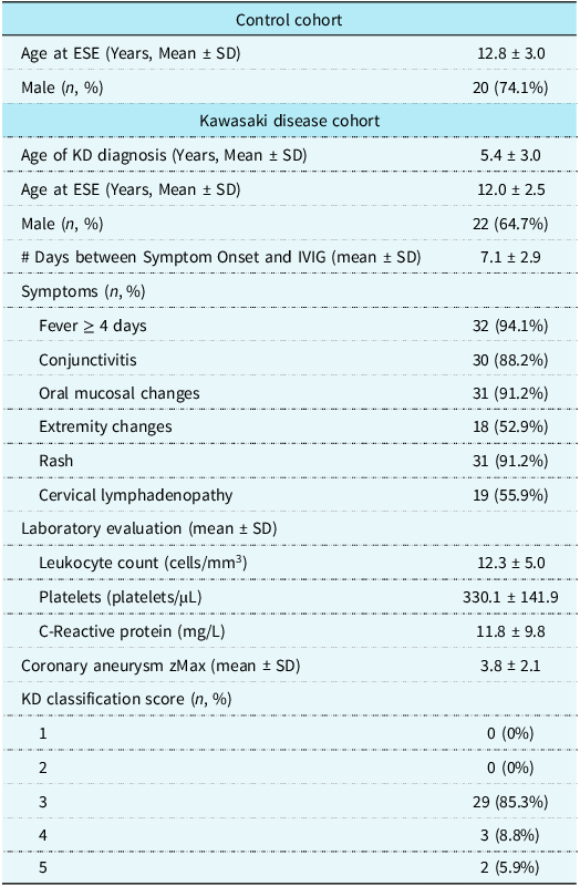
There were 34 patients in the Kawasaki disease cohort, of which, 22 (64.7%) were male (Table 1). The average age at diagnosis of Kawasaki disease was 5.4 ± 3.0 years. The average maximal coronary z-score was 3.8 ± 2.1. Most of the Kawasaki disease cohort had coronary artery risk level 3 (29/34, 85.3%). For the other 5 patients, 3 (8.8%) were at risk level 4 and 2 patients (5.9%) were at risk level 5. An average of 7.1 ± 2.9 days elapsed between symptom onset and start of intravenous immune globulin. Exercise stress echocardiography occurred an average of 6.7 ± 3.1 years following Kawasaki disease diagnosis. There were no patients in the Kawasaki disease cohort who had regional wall motion abnormalities suggestive of inducible ischaemia. There were 27 patients in the control cohort, of which, 20 (74.1%) were male. The average age at exercise stress echocardiography was 12.0 ± 2.5 years and 12.8 ± 3.0 years for the Kawasaki disease and control cohorts, respectively.
Demographic and laboratory values of the control and Kawasaki disease cohorts

Table 1. Long description
The table compares demographic and laboratory values of control and Kawasaki disease cohorts. It has two main sections: Control cohort and Kawasaki disease cohort. The Control cohort section includes two rows: Age at ESE (Years, Mean ± SD) with a value of 12.8 ± 3.0, and Male (n, %) with a value of 20 (74.1%). The Kawasaki disease cohort section includes multiple rows: Age of KD diagnosis (Years, Mean ± SD) with a value of 5.4 ± 3.0, Age at ESE (Years, Mean ± SD) with a value of 12.0 ± 2.5, Male (n, %) with a value of 22 (64.7%), Days between Symptom Onset and IVIG (mean ± SD) with a value of 7.1 ± 2.9, Symptoms (n, %) with values for Fever ≥ 4 days (32 (94.1%)), Conjunctivitis (30 (88.2%)), Oral mucosal changes (31 (91.2%)), Extremity changes (18 (52.9%)), Rash (31 (91.2%)), Cervical lymphadenopathy (19 (55.9%)), Laboratory evaluation (mean ± SD) with values for Leukocyte count (cells/mm³) (12.3 ± 5.0), Platelets (platelets/µL) (330.1 ± 141.9), C-Reactive protein (mg/L) (11.8 ± 9.8), Coronary aneurysm zMax (mean ± SD) (3.8 ± 2.1), and KD classification score (n, %) with values for 1 (0 (0%)), 2 (0 (0%)), 3 (29 (85.3%)), 4 (3 (8.8%)), and 5 (2 (5.9%)).
ESE = exercise stress echocardiography; IVIG = intravenous immunoglobulin; KD = Kawasaki disease; n = number; SD = standard deviation.
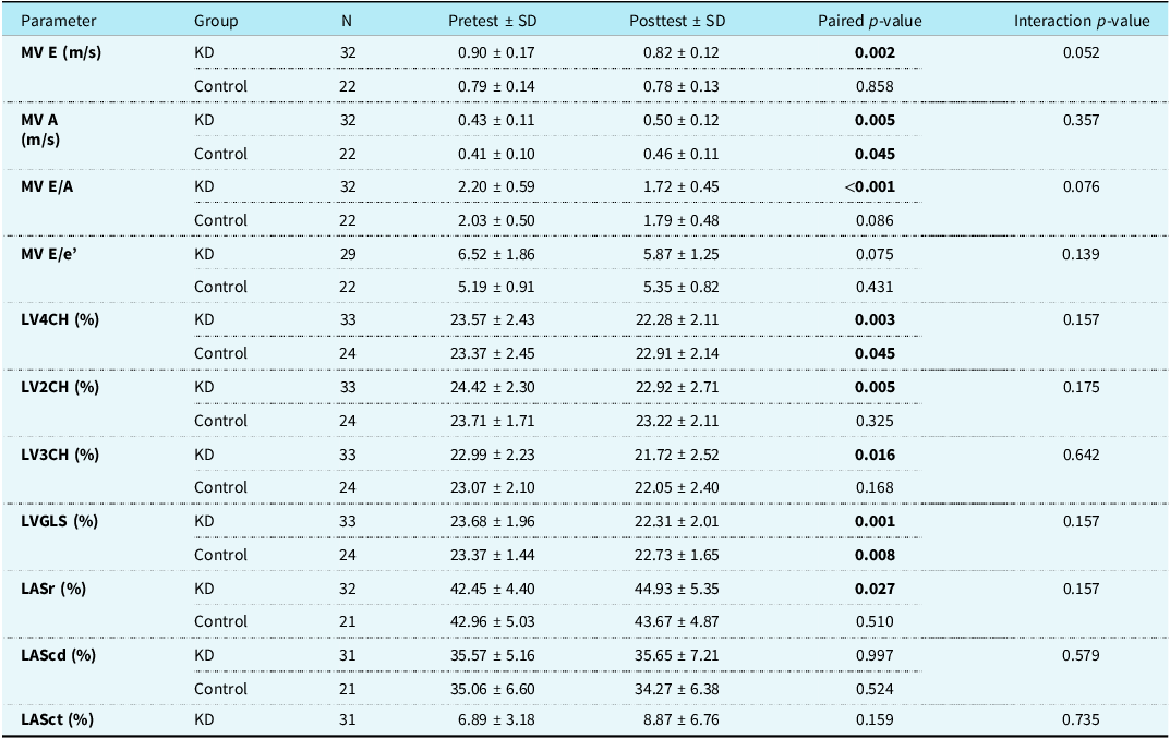
The average heart rate at the time of the pre-stress echocardiogram was 67 ± 10.8 bpm and 71 ± 16.1 bpm for the aneurysm and control cohorts, respectively. The average heart rate at the time of the post-stress echocardiogram was 88 ± 11.3 bpm and 91 ± 11.3 bpm for the aneurysm and control cohorts, respectively. There were significant pre- to post-exercise differences in both groups for average left ventricle global and 4-chamber longitudinal strain and mitral A velocity (Table 2). In the Kawasaki disease cohort, additional significant changes were observed for mitral valve E velocity, mitral valve E/A, left ventricular 2 and 3 chamber strain, and left atrial reservoir strain. In mixed-effects models, there were no significant differences in the magnitude of pre- to post-exercise change between the Kawasaki disease and control cohorts (Table 2). There was good to excellent intraobserver and acceptable to good interobserver agreement by intraclass correlation coefficient for all strain measurements (Supplemental Table S1). Individual data descriptions for the 5 Kawasaki disease patients who were risk level 4 or 5 are available in Supplemental Table S2.
Paired pre- and post-exercise stress echocardiography t-test for the aneurysm and control cohorts. Interaction p-values were obtained from linear mixed-effects models with subject-specific random intercepts and test whether pre- to post-exercise changes differ between groups

Table 2. Long description
A table comparing pretest and posttest values for various cardiac parameters in KD and control groups. The table has 12 rows and 6 columns. The columns are labeled Parameter, Group, N, Pretest ± SD, Posttest ± SD, Paired p-value, and Interaction p-value. The rows are labeled with different cardiac parameters and their respective values for the KD and control groups. Each row provides the pretest and posttest values along with their standard deviations, paired p-values, and interaction p-values. Notable parameters include MV E (m/s), MV A (m/s), MV E/A, MV E/e', LV4CH (%), LV2CH (%), LV3CH (%), LVGLS (%), LASr (%), LAScd (%), and LASct (%). The table shows significant changes in various parameters pre- to post-exercise for both groups, with specific significant changes observed in the Kawasaki disease cohort.
LAScd = left atrial conduit strain; LASct = left atrial contractile strain, LASr = left atrial reservoir strain; LV2CH = left ventricular 2 chamber strain; LV3CH = left ventricular 3 chamber strain; LV4CH = left ventricular 4 chamber strain; LVGLS = left ventricular global longitudinal strain; MV A = mitral valve A velocity; MV E = mitral valve E velocity, MV E/e’ = ratio of the mitral valve E velocity and the average of the lateral and septal e’ velocity.
Discussion
In this study, we evaluated left atrial and ventricular functional responses to exercise in patients with a remote history of Kawasaki disease and prior coronary artery aneurysm compared with age-matched healthy controls. While the Kawasaki disease cohort had a greater number of systolic and diastolic parameters that changed significantly from rest to post-exercise recovery, mixed-effects modelling revealed no significant difference in the magnitude of exercise-induced change between the two cohorts.
Exercise stress echocardiography is a safe and noninvasive evaluation of cardiac function. It has been shown to detect myocardial ischaemia with better sensitivity than electrocardiographic stress alone in paediatric patients with a history of Kawasaki disease. Reference Pahl, Sehgal and Chrystof8 A prior study demonstrated exercise-induced diastolic abnormalities on regional tissue Doppler-based indices in Kawasaki disease patients with coronary aneurysms. Reference Arnold, Goebel and Ulmer9 This study was published in an earlier era where the coronary artery aneurysm classification scheme was based on absolute coronary diameters rather than z-scores. Reference Arnold, Goebel and Ulmer9,Reference Newburger, Takahashi and Gerber10 In that cohort, the majority of patients met criteria for giant coronary aneurysms based on the prior classification scheme (diameter >10 mm). In the present study, we evaluated diastolic function using approaches not employed by Arnold et al., including mitral valve E/e’ and left atrial strain. More recently, using contemporary z-score criteria, Tedla et al. demonstrated abnormal exercise stress echocardiograms in 3/29 patients with a history of Kawasaki disease, all 3 were classified as American Heart Association coronary artery risk level 4 or 5. Reference Tedla, Burns and Tremoulet11 Notably, none of the 16 patients who were classified as risk level 2 to 3 had abnormal stress findings.
In contrast to these prior studies, our cohort was characterised by milder coronary involvement defined using contemporary z-score- based criteria, with an average maximal z-score of 3.8. The American Heart Association’s Kawasaki disease guideline recommends periodic assessment of inducible myocardial ischaemia via stress testing in patients with Kawasaki disease risk classification level ≥ 3 (coronary artery z-score ≥ 2.5). Reference Jone, Tremoulet and Choueiter3 The guideline acknowledges that while testing for inducible ischaemia is suggested starting at risk classification 3, the patients at most risk for stenosis and obstruction are those with a coronary artery z-score ≥ 10. Reference McCrindle, Manlhiot and Newburger12 There were no patients in the present study who had inducible ischaemia during the exercise stress echocardiography. Furthermore, the absence of a differential exercise response between groups in our study may reflect a lower burden of microvascular dysfunction and adverse remodelling in patients with less severe coronary involvement. Reference Suzuki, Miyagawa-Tomita and Komatsu13
Deformation imaging via left ventricular longitudinal strain and left atrial strain has become popular over the past decade due to its ability to provide a more sensitive assessment of subclinical myocardial dysfunction than conventional markers of myocardial function such as ejection fraction. Reference Thomas, Edvardsen and Abraham14 In patients with acute Kawasaki disease, left ventricular longitudinal strain was decreased compared to controls despite no statistically significant differences in conventional systolic indices. Reference McCandless, Minich and Wilkinson15 Abnormal stress imaging may have prognostic implications in higher-risk Kawasaki disease populations. In a study by Noto et al., Kawasaki disease patients with inducible regional wall motion abnormalities on dobutamine stress echocardiography had an approximately threefold higher risk of major adverse cardiovascular events compared to those without wall motion abnormalities over a 15-year follow-up period. Reference Noto, Kamiyama and Karasawa4 The absence of significant differences between the Kawasaki disease and control cohorts, despite the use of deformation imaging following exercise stress, may provide additional evidence of preserved global systolic and diastolic function in Kawasaki disease patients with mild to moderate coronary artery involvement. These findings were corroborated by the lack of difference in MV E/e’ and further support the notion that routine exercise stress imaging may be of limited yield for surveillance in lower-risk Kawasaki disease patients. Reference Jone, Tremoulet and Choueiter3,Reference Tedla, Burns and Tremoulet11
Left atrial reservoir strain increased significantly from rest to post-exercise recovery in the Kawasaki disease cohort but not in the control group. Left atrial functional remodelling is recognised as an important compensatory mechanism in the early stages of left ventricular diastolic dysfunction to preserve cardiac output. Reference Thomas, Marwick and Popescu16 Left atrial strain is divided into three phases. Reservoir strain measures left atrial filling during ventricular systole. Conduit strain measures passive left atrial emptying during ventricular diastole. Contractile strain measures active left atrial emptying during late ventricular diastole when the atrium contracts. In the early stages of left ventricular diastolic dysfunction, both left atrial reservoir and conduit strains decrease due to increased atrial afterload and decreased passive ventricular filling, respectively. Reference Thomas, Marwick and Popescu16,Reference Sabatino, Di Salvo and Prota17 The left atrial contractile strain increases in early stages of diastolic dysfunction to compensate for poor passive left ventricular filling. Reference Singh, Addetia and Maffessanti18 Rest to post-exercise left atrial conduit and contractile strain did not differ significantly between the two cohorts. Given the well-described inverse relationship between left atrial reservoir strain and diastolic dysfunction severity, Reference Singh, Addetia and Maffessanti18 the observed increase in left atrial reservoir strain in the Kawasaki disease cohort is unexpected and likely a chance finding, as mixed-effects modelling demonstrated a nonsignificant interaction.
There are a few limitations of our retrospective study. The relatively small sample size limits statistical power and raises the possibility of type II error, particularly for detecting subtle differences in the magnitude of exercise-induced strain changes. Our sample size, however, is consistent with previously published paediatric Kawasaki disease stress echocardiography literature, where single-centre cohorts have ranged from roughly 20 to 60 patients. Reference Pahl, Sehgal and Chrystof8,Reference Arnold, Goebel and Ulmer9,Reference Tedla, Burns and Tremoulet11 Our cohort had mild to moderate coronary artery involvement, which may limit generalisability in patients with more severe coronary artery aneurysms. Due to our cross-sectional design, we cannot address longitudinal changes in myocardial mechanics over time. Importantly, stress echocardiography in our study was performed during early to mid-recovery rather than peak exercise. Immediate post-exercise imaging is the most widely used protocol in published Kawasaki disease stress echocardiography studies, Reference Pahl, Sehgal and Chrystof8,Reference Tedla, Burns and Tremoulet11 and the American Society of Echocardiography recommends post-exercise imaging be completed within 1–2 minutes of peak exercise Reference Pellikka, Arruda-Olson and Chaudhry7 as transient strain impairments and diastolic filling abnormalities are most apparent at peak stress. Our delay in obtaining stress imaging, as evidenced by a post-stress heart rate around 90 beats per minute, may have precluded our ability to find meaningful differences. Notably, left ventricular longitudinal strain has been shown to increase with exercise, Reference Cifra, Mertens and Mirkhani19 yet our cohorts demonstrated lower systolic strain indices post-exercise. We suspect that this finding may not reflect true systolic dysfunction in either cohort, but rather, the strain values at mid-recovery may be attenuated by increased parasympathetic tone and reduced preload. Future studies employing supine bicycle protocols with peak stress imaging may provide a more sensitive assessment of myocardial function.
In paediatric patients with remote history of Kawasaki disease and prior coronary artery aneurysm, exercise stress echocardiography demonstrated no significant differences in the magnitude of exercise-induced change in patients with mild to moderate coronary artery involvement. Further studies are needed to determine whether patients with more severe coronary artery disease develop long-term abnormalities in myocardial function during exercise.
Supplementary material
The supplementary material for this article can be found at https://doi.org/10.1017/S1047951126123786.
Acknowledgements
None.
Financial support
This research received no specific grant from any funding agency, commercial, or not-for-profit sector.
Competing interests
None.
Ethical standard
The authors assert that all procedures contributing to this work comply with the Helsinki Declaration of 1975, as revised in 2008, and have been approved by the Institutional Review Board.