Case report
A 32-year-old nulliparous pregnant woman presented for foetal echocardiography due to suspicion of a large right atrial aneurysm visualised at 18 weeks gestational age on prenatal ultrasound. Maternal history was notable for Graves’ disease in full remission. Family history was negative for CHD, although maternal family history was unknown since mother was adopted. Initial foetal echocardiogram at 22 weeks gestational age confirmed a dilated right atrium and right atrial aneurysm with to-fro flow as well as a small global pericardial effusion (Fig 1a). The aortic annulus and ascending aorta were hypoplastic (z score of −3.24 and −3.04, respectively) and aortic coarctation was suspected. The left ventricle appeared slender but apex forming. Due to the uncertain natural history of the right atrial aneurysm and potential progression of left heart hypoplasia, delivery at a tertiary care centre with a multidisciplinary foetal team management was arranged.
(a) Foetal echocardiogram showing the right atrial aneurysm (RAA). (b) Postnatal transthoracic echocardiogram showing coarctation of the aorta (white arrow).

Foetal surveillance at 26 weeks gestational age demonstrated foetal tachycardia at 188 beats/minute, presumably sinus tachycardia, with 1:1 atrioventricular relationship, preserved biventricular function, and no signs of foetal distress. Subsequent foetal echocardiograms showed increasing right atrial aneurysm dilation from an initial 9.5 × 9.0 mm to 13.5 × 15.7 mm. Foetal supraventricular tachycardia >220 beats/minute developed at 36 weeks gestational age in conjunction with escalation of the ductus venosus pulsatility index for veins and “a” wave reversal. As a result, she was delivered at 36 weeks gestational age by low segment transverse caesarean section for non-reassuring foetal status.
Her postnatal course was complicated by haemodynamically unstable chaotic atrial tachycardia to 250 beats/minute. Postnatal echocardiography revealed right atrial aneurysm, mildly hypoplastic aortic isthmus without coarctation, severely diminished biventricular systolic function and possible non-compaction of the left ventricle. Despite aggressive postnatal management with multiple antiarrhythmics, including amiodarone and digoxin, atrial tachycardia persisted, and ventricular function worsened, requiring veno-arterial extra corporeal membrane oxygenation. By 6 days of life, the infant was decannulated from veno-arterial extra corporeal membrane oxygenation due to improvement in biventricular function and control of atrial tachycardia with amiodarone, digoxin, and esmolol. However, at 2 weeks of age, she developed moderate left ventricular dysfunction secondary to the development of a discrete aortic coarctation (Fig 1b). She underwent surgical repair of the aortic coarctation with end-to-end anastomosis. Biventricular function subsequently normalised, and rate control of atrial tachycardia was again achieved with multiple antiarrhythmics.
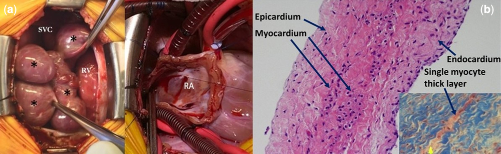
Recurrence of complex atrial tachycardia refractory to medical therapy occurred at 6 weeks of age, with the patient presenting in near hemodynamic collapse. She was taken to the operating room where multiple aneurysms of the right atrium were resected, the presumed focus of atrial ectopy. Intraoperatively, findings included multi-lobular aneurysmal outpouchings of the right atrium free wall and a similar but smaller lesion of the left atrial appendage (Fig 2a). The resected lesions were fibrotic with focal severe thinning of the wall consistent with aneurysmal tissue. Histopathologic studies revealed an aneurysm of the atrial wall on Haematoxylin and Eosin staining (Fig 2b). Areas of disruption in the atrial wall were also noted (Fig 2b). On Masson’s trichrome staining, a significant amount of connective tissue was noted encasing very thin myocardial layer, which in some areas was only one myocyte thick. Immediately following resection of these lesions, atrial tachycardia resolved. She has since developed recurrent atrial tachycardia that has been controlled on propranolol and flecainide. Her single nucleotide polymorphism microarray was normal and the family declined additional genetic testing.
(a) Intraoperative image showing right atrium before and after resection of RAA (*). (b) Haemotoxylin and Eosin (H&E) stain of resected RAA. RAA wall has a very thin myocardial layer, in some areas only one myocyte thick (shown by yellow arrow in Masson’s trichome stained inset).

Discussion
Isolated right atrial aneurysms are unusual malformations in children, rarely diagnosed in foetal life.Reference Nagata, Stambach, Wilson, Menon and Jaeggi1 Patients often remain asymptomatic until the third decade when atrial flutter/fibrillation and thromboembolic events may occur.Reference Ishii, Inamura and Kayatani2, Reference Faqeeh, Alsaadi, Aselan, Almuzaini and Algamdi3 Thinning of the atrial wall increases the risk of atrial dilation, potentially leading to disruption of normal electrical conduction from the sinus node and hence increased vulnerability to atrial tachyarrhythmias.
Few foetal cases of right atrial aneurysms are reported in the current literature. The congenital type is believed to be caused by dysplasia of the muscular wall of the right atrium.Reference Aryal, Hakim and Giri4 Subendocardial, pericardial, and patchy interstitial fibrosis and lipomatous degeneration of the right atrial aneurysms without inflammation have also been noted.Reference Aryal, Hakim and Giri4 Concurrent left atrial appendage aneurysm in patients with right atrial aneurysms is rare but was present in our case.Reference Aryal, Hakim and Giri4 Fibrosis affects 23% of cases and degenerated myocardium occurs in about 12% on histopathological examination.Reference Aryal, Hakim and Giri4
Atrial tachycardia requiring antiarrhythmics developed in the majority of reported cases.Reference Aryal, Hakim and Giri4 Patients commonly presented with respiratory distress due to postnatal airway compression, not present in our case. Rarely does atrial tachyarrhythmia result in left ventricular dysfunction. In our case, the foetus was noted to have an elevated resting heart rate at the end of the second trimester, relatively earlier in pregnancy than typical foetal supraventricular tachycardia. This may have contributed to the ventricular dysfunction noted in the newborn. We present the first reported case requiring veno-arterial extra corporeal membrane oxygenation secondary to left ventricular dysfunction induced by medically refractory atrial tachyarrhythmia. Surgical resection of the right atrial aneurysms led to resolution of atrial tachycardia. When foetal supraventricular tachycardia develops in these cases, prenatal management options may be limited compared to other forms of primary supraventricular tachycardia, as illustrated by the difficult postnatal course in this case.
A prior case of a foetus with right atrial aneurysm and pericardial effusion was reported by Oztunc et al and associated with hydrops fetalis.Reference Oztunc, Ugan Atik and Koka5 In our case, a pericardial effusion was noted in the absence of hydrops. There is no known association of pericardial effusions with right atrial aneurysms. The aetiology of the pericardial effusion which persisted throughout foetal life for this case remains unexplained, and interestingly resolved spontaneously postnatally.
The most common type of congenital cardiac defect noted in cases of right atrial aneurysm is an ostium secundum atrial septal defect. To our knowledge, the development of an aortic coarctation in postnatal life has not previously been reported. There is no clear association between the right atrial aneurysm and the development of an aortic coarctation. This patient had small left heart structures in foetal life, which could be predictive of aortic coarctation in postnatal life.
We present a case of right atrial aneurysm of foetal onset with associated atrial tachycardia that improved following postnatal right atrial aneurysm resection. Unlike prior reported cases, our patient additionally had a pericardial effusion without idiopathic hydrops fetalis and developed an aortic coarctation requiring surgical repair due to the development of left ventricular dysfunction.
Author ORCIDs
Edem Binka 0000-0002-9940-2506
Acknowledgements
We would like to acknowledge Katherine Gibson RDCS (AE, FE, PE) and Catherine Eskey RDCS for their important contributions in fetal echocardiographic image acquisition for this case.
Financial support
Dr. Binka was supported by the T32 HL125239 from NIH/NHLBI while conducting this study.
Conflicts of interest
None.
Ethical standards
This case report does not involve human and/or animal experimentation.