INTRODUCTION
The family Otoplanidae (Plathelminthes: Rhabditophora: Proseriata) is represented by a group of typically marine flatworms inhabiting the sandy-breaker zone of sea coasts known as the ‘Otoplanen-Zone’ of Remane (Reference Remane1933). This group of neoophoran Plathelminthes is the dominant taxon in the surf-zone, where it moves rapidly among the sand grains. Behaviour and reproduction are quite obscure; recently, new findings (Lanfranchi & Melai, Reference Lanfranchi and Melai2008) suggest that these animals cross the sandy layers driven by two primordial instincts. The quiet deeper layers are those where the Otoplanidae take refuge to lay eggs and probably to rest and escape predators. The superficial sand is presumably the zone where they mate and search for food.
Otoplanidae species have been collected in different globe zones:
(1) Seawater:
Arctic Ocean (Steinböck, Reference Steinböck1932).
Atlantic Ocean (Graff, Reference Graff von1913; Steinböck, Reference Steinböck1931, Reference Steinböck1932; Marcus, Reference Marcus1949, Reference Marcus1950, Reference Marcus1952; Karling, Reference Karling1973; Sopott-Ehlers & Ehlers, Reference Sopott-Ehlers and Ehlers1980; Sopott-Ehlers, Reference Sopott-Ehlers1985; Ax & Sopott-Ehlers, Reference Ax and Sopott-Ehlers1987; Ax & Armonies, Reference Ax and Armonies1990).
Baltic, Black and Mediterranean Seas (Calandruccio, Reference Calandruccio1897; Du Plessis, Reference Du Plessis1889; Giard, Reference Giard1904; Hallez, Reference Hallez1910; Meixner, Reference Meixner, Grimpe and Wagler1938; Ax, Reference Ax1951, Reference Ax1956, Reference Ax1959; Luther, Reference Luther1960; An der Lan, Reference An der Lan1964; Riemann, Reference Riemann1965; Lanfranchi, Reference Lanfranchi1969, Reference Lanfranchi1978; Sopott-Ehlers, Reference Sopott-Ehlers1972, Reference Sopott-Ehlers1976; Ax et al., Reference Ax, Sopott-Ehlers and Weidemann1978; Martens & Schockaert, Reference Martens and Schockaert1981; Delogu & Curini-Galletti, Reference Delogu and Curini-Galletti2007; Delogu et al., Reference Delogu, Casu and Curini-Galletti2008; Lanfranchi & Melai, Reference Lanfranchi and Melai2007, Reference Lanfranchi and Melai2008).
Pacific Ocean (Karling, Reference Karling1964; Ax & Ax, Reference Ax and Ax1967, Reference Ax and Ax1974; Tajika, Reference Tajika1983a, Reference Tajikab, Reference Tajikac, Reference Tajika1984; Miller & Faubel, Reference Miller and Faubel2003).
(2) Brackish and fresh water:
Elba River (Riemann, Reference Riemann1965).
Oka River (Gieysztor, Reference Gieysztor1938).
Oriental Pyrenees (Ax, Reference Ax1951).
Paraná and Colastiné Rivers (Noreña et al., Reference Noreña, Damborenea and Brusa2005).
The aim of the present study is to gain extensive knowledge of the mesopsammic fauna concerning the family Otoplanidae.
The new species Otoplana proxima is attributed to the subfamily Otoplaninae on the basis of its partially ciliate body, its ciliate creeping sole and above all its cylindrical pharynx situated horizontally along the ventral body zone. Moreover, it possesses a male genital pore for discharging surplus spermatozoa, typical of the genus.
MATERIALS AND METHODS
The specimens were collected in May 2005 at Marina di Bibbona (Leghorn, Italy), 43°14′13″N 10°31′36″E, where the ‘Otoplanen-Zone’ is characterized by fine sand, by scooping up the superficial layer of sediment. The meteorological conditions were optimal with a temperature of 28–29°C, a calm sea and a light breeze.
Each organism was first anaesthetized with a solution of 1/3 of MgCl2 21% and 2/3 tap water. Subsequently, at least 30 specimens were studied in vivo by slight squeezing under the coverslip, in order to draw the habitus with the aid of the camera lucida. Finally, by compressing the coverslip more forcefully, the spines of the sclerotic apparatus were examined.
For histological procedures, five specimens were fixed in Stieve solution. The sections were stained with Heidenhain's haematoxylin, using eosin as counterstain.
A graphical elaboration was used to support the microscopic study.
TAXONOMY
TYPE MATERIAL
At least 15 specimens were studied in vivo, including drawings and photographs. Three specimens were fixed and sectioned.
Holotype: one sagittally-sectioned specimen is deposited in the Electron Microscopy Laboratory Collection of the Dipartimento di Biologia, Unità di Etologia (Università di Pisa).
DIAGNOSIS
Sexually mature organism measures about 5 mm in length. The body is fusiform, dorsally convex, ventrally flat, colourless and transparent (Figures 1, 2, 3 & 6).
Habitus of Otoplana proxima sp. nov.: amp, accessory male pore; ap, adhesive papillae; b, brain; esv, external seminal vesicles; gc, glandular complex; ge, germaries; i, intestine; s, sclerotic apparatus; sta, statocyst; tb, tactile bristles or ‘Tastborsten’; te, testes; th, tactile hairs; vg, vesicula granulorum; vi, vitellaries; ph, pharynx; vs, vesicula seminalis.

Photographs of Otoplana proxima sp. nov. in vivo: 2, 3 and 6 living animals; 4 anterior end; 5 posterior end; 7 post-pharyngeal zone; 8 pre-pharyngeal zone with glandular complex and testes.

The anterior end is marked by two couples of robust tactile bristles or ‘Tastborsten’ (tb) retractable into the respective wide pockets. Tactile hairs (th) are present on the lateral and especially frontal sides (Figures 1, 2 & 4).
The rabdoids are present as true rhabdites, grouped into longitudinal lines along the body, with the exception of the anterior end, where they are randomly scattered.
Characteristic glandular complexes (gc), constituted of 5–8 structural units are present on the ventral and dorsal surfaces, generally distributed along a few longitudinal rows (Figures 1 & 8).
The ovoidal brain is at some distance from the small statocyst (sta) (Figures 1, 2, 3, 4 & 6).
The testes (te), starting not far from the brain, consist of two series of small follicles along the longitudinal axis. They are numerous and reach the two germaries (ge) at about 2/3 of body length (Figures 1, 2, 3 & 6).
Two rows of small vitellaries (vi) are present laterally to the testes. They begin abreast of the testis follicles and reach the pharynx opening maintaining a regular distribution (Figures 1, 2 & 3).
Two germaries (ge) are present in front of the pharynx, posteriorly to the last testis follicles, at 2/3 of body length. They are globoid, larger than the testes and vitellaries, and contain numerous egg-cells (Figure 1).
The pharynx (ph) shows the so-called bell-shaped organization or ‘Glöckchen’, typical of the genus. The sacciform intestine (i) is a caecum at both ends (Figure 1).
In the postpharyngeal zone, the sacciform vesicula seminalis (vs) is connected distally with a relatively large vesicula granulorum (vg). The external seminal vesicles (esv) and the accessory male pore (amp), typical of the genus, are clearly visible (Figures 1 & 7).
The caudal end is characterized by a tiny plate and provided with a few small adhesive papillae (ap). These bi-glandular structures are also present in the lateral epidermis (Figures 1, 2 & 5).
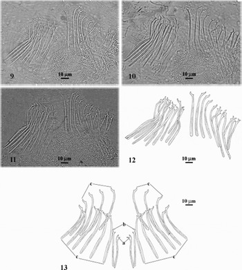
The male copulatory organ is characterized by a sclerotic apparatus (s) with 23–24 spines of variable shape and length (Figures 9, 10, 11, 12 & 13):
• one couple (a) of specular spines, practically straight, 42 µm long, placed in the centre of the complex, with a rounded proximal end and a forked tip bent medially;
• one couple (b), similar and external to the previous, 44 µm long;
• 19–20 spines (c) distally curved outwards, equally subdivided along both sides, 63–70 µm long, with a more or less forked distal end.
Photographs (9–11), tracing (12) and spatial distribution (13) of the spines of male sclerotic apparatus of Otoplana proxima sp. nov.: a, b and c, groupings of similar spines.

CONCLUDING REMARKS
As reported in the literature (Lanfranchi & Melai, Reference Lanfranchi and Melai2007), there are at present four known species in the genus Otoplana: O. intermedia Du Plessis, Reference Du Plessis1889 (Ax, Reference Ax1956) collected in the Ligurian and Tyrrhenian Seas, O. bosporana Ax, Reference Ax1959 sampled in the Bosphorus (Black Sea), O. truncaspina Lanfranchi, Reference Lanfranchi1969 discovered at Monte Rosso al Mare (Ligurian Sea) and O. oxyspina Lanfranchi & Melai, Reference Lanfranchi and Melai2007 collected at Caletta Beach (Ligurian Sea). With the addition of O. proxima, the effective species of the taxon Otoplana amount to five.
The habitus of our species evidences a body length (5 mm) shorter than in O. intermedia (8 mm) and longer than in O. bosporana (2.5–3 mm) and O. oxyspina (3.3–4 mm). Otoplana truncaspina presents a body length of 4.5–5 mm, similar to that observed in O. proxima.
The organization of the cephalic zone and the distribution of the rhabdithes show similarity with the previously described species.
The pharynx, located in the end of the second body half, is characteristic of the subfamily.
The yolk follicle path, in a single longitudinal row from the anterior end to the pharynx opening on each side of the body, is similar to that observed in O. intermedia, although the follicle dimensions are smaller.
The position of the testes, as well as their extension, is shared with all the species of the genus. The dimensions and distribution not in single line of the testes in our species correspond to those of O. intermedia and O. truncaspina.
In O. proxima the locations of the vesicula seminalis, vesicula granulorum and penis papilla appear to coincide with that of the species already described.
The spines of the male copulatory organ of the new species display a different organization from that of all the other species. Furthermore, all the bristles appear more pliable distally and canaliculated proximally.
The total number of bristles (23–24) observed in the new species is lower than that of O. bosporana (30–33) and slightly higher than that of O. oxyspina (21). Otoplana intermedia and O. truncaspina present respectively 24 and 23 spines, similarly to O. proxima.
The position of the (a) couple of spines in the central zone is compatible with that of the small bristles of O. oxyspina and O. truncaspina. This couple possesses a clearly forked tip and it is longer (42 µm) than that of O. oxyspina (36 µm) and O. truncaspina (28–36 µm).
The new species does not present the ‘Medianstachel’ described in O. oxyspina, O. bosporana and O. truncaspina. On the contrary, it is similar to O. intermedia, which is free of the funnel-shaped sting.
The presence of the (b) couple on both sides of the (a) spines is a peculiar character of O. proxima. This couple is absent in all the other species. Its length (44 µm) is compatible only with that of some bristles of O. bosporana ranging from 33–59 µm. The spines of the other species are longer.
The (c) bristles, with a forked tip, a curved axis and a canaliculated proximal end, bear a closer resemblance to (d) type of O. oxyspina and O. intermedia. Their length (63–70 µm) is similar to that of O. oxyspina (58–65 µm) and O. truncaspina (60–66 µm). Otoplana intermedia shows spines longer (80–90 µm), while those of O. bosporana are shorter (33–59 µm).
On the basis of the data presented, we conclude that our species differs clearly from the species already described. As far as habitus, distribution of testes and vitellaries, and pharynx position are concerned, the more similar species is O. intermedia. The body length of O. proxima is comparable to that of O. truncaspina, and its sclerotic apparatus is devoid of ‘Medianstachel’ like O. intermedia, which is also lacking in the central spine couple. The pliable spines of the new species are particular for their canaliculated proximal end. These dissimilarities allow the constitution of a new valid species of otoplanid.