Introduction
Plastic bronchitis is a rare, serious condition with a mortality rate of up to 50 per centReference Stiller, Riedel, Paul and van Landeghem1 due to obstruction of the tracheo-bronchial tree by solid casts of lymphatic origin. The onset of symptoms can be acute and rapidly progressive, leading to acute respiratory failure. Otolaryngologists may become involved in the management of plastic bronchitis as the symptoms and radiological findings can mimic foreign body inhalation. It is essential to have a high index of clinical suspicion for plastic bronchitis, in order to include it in the differential diagnosis in patients presenting with unilateral or bilateral complete chest radiograph opacification, with absence of air bronchograms.
We present two cases of plastic bronchitis which illustrate the benefits of bronchoscopy in the diagnosis and definitive management of this condition.
Case report
Case one
We present the case of a previously well, two-year-old boy born at 29 weeks' gestation. He had been ventilated for 24 hours following his premature birth, but had no other respiratory history. His vaccinations were up to date, and there were no developmental concerns or significant family history.
The child presented with a three-day history of non-productive cough, lethargy, pyrexia and poor oral intake. He was admitted to hospital with exacerbation of cough and respiratory distress. His left hemithorax was dull on percussion, with reduced air entry on auscultation.
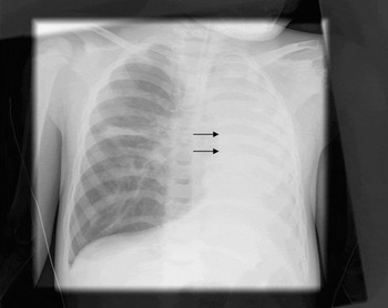
A chest radiograph showed complete opacification of the left hemithorax, with volume loss and mediastinal shift, consistent with a lung collapse (Figure 1).
Postero-anterior chest radiograph showing complete opacification of the left hemithorax, with volume loss and mediastinal shift (arrows).

The child's oxygen saturations deteriorated, leading to intubation and intensive care admission. High pressure settings were required initially for ventilation.
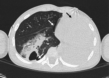
A high resolution computed tomography (CT) scan showed complete collapse of the left lung, with mediastinal shift and a left main bronchus tapering 1 cm distal to the carina. There was complete obliteration of the distal bronchus, and a right pneumothorax had developed (Figure 2) requiring chest drain insertion.
Axial, high resolution computed tomography scan of the thorax, showing left lobe collapse (black arrow), right lobe consolidation and two pneumothoraces (white arrows).

A flexible bronchoscopy was performed, showing complete blockage of the left main bronchus (Figure 3). We then proceeded to direct, rigid bronchoscopy. A friable, mucoid, plug-like substance was suctioned from the left main bronchus.
Three-dimensional computed tomography reconstruction showing complete occlusion of the left main bronchus (arrow). H = head; F = feet; R = right; L = left; A = anterior

The child underwent aggressive lavage and suctioning for three days.
A repeated chest radiograph showed partial resolution of the left lung collapse.
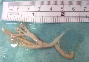
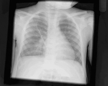
Repeated rigid bronchoscopy two days following intubation removed a thick, cast-like substance which was sent for analysis (Figure 4). Both lungs re-expanded well after removing the cast, a clinical improvement was consistent with radiological findings (Figure 5).
Bronchial cast removed during the first patient's second rigid bronchoscopy.

Chest X-ray after the first patient's second rigid bronchoscopy, showing complete resolution.

The child was weaned off the ventilator and extubated on the fourth day of admission. His chest drains were removed on day six.
All microbiological cultures were negative, including a broncho-alveolar lavage, as were virology and sickle cell screening tests. Histological analysis of the specimen showed acute inflammatory cells, predominantly neutrophils with some eosinophils, consistent with a type I cast.Reference Seear, Hui, Magee, Bohn and Cutz2
Case two
We also treated a 15-year-old boy who presented with myocarditis and required extra-corporeal membrane oxygenation due to poor cardiac output. Within three days of hospital admission, he developed complete ‘whiteout’ of the right lung field. Opacification of both lung fields is not uncommon whilst patients are receiving extra-corporeal membrane oxygenation.
As the chest radiograph did not show any improvement following regular chest physiotherapy and suctioning, flexible bronchoscopy was performed. Plugs of bronchial casts were found to be occluding the lumen of the right main bronchus. After several attempts at removing the plastic casts using flexible bronchoscopy, perfluorocarbon was instilled to help dissolve the thick plugs.
Discussion
Plastic bronchitis is uncommon in children, with only 40 cases reported prior to 1985Reference Bowen, Oudjhane, Odagiri, Liston, Cumming and Oh3 and 72 cases before 1989.Reference Pérez-Soler4 Although seen infrequently, plastic bronchitis can be serious, and requires early diagnosis to prevent a potential fatal outcome from respiratory failure.Reference Cajaiba, Borralho and Reyes-Múgica5
Plastic bronchitis is most commonly associated with asthmaReference Morgan and Bogomoletz6 and allergic bronchopulmonary aspergillosis.Reference Werkhaven and Holinger7 Other associations include haematological disorders such as α thalassaemiaReference Veras, Lannes, Piva, Stein, Jones and Silva8 and sickle cell disease.Reference Raghuram, Pettignano, Gal, Harsch and Adamkiewicz9 Cystic fibrosisReference Waring, Brunt and Hilman10 and respiratory tract pathogens (such as Haemophilus influenzae, Corynebacterium diphtheriae and viruses) have also been linked.Reference Kao and Richmond11 Acquired cases have been reported following cardiac surgery for congenital heart disease (such as Fontan's operation),Reference Colloridi, Roggini, Formigari, Ventriglia and Giglioni12 whilst other cases have no clear aetiology.Reference Eberlein, Drummond and Haponik13 The cases presented here did not have a clear association with any established risk factors, and therefore fall into the idiopathic category.
The pathogenesis underlying the formation of a bronchial cast remains poorly understood. Postulated mechanisms include post-infection ciliary dysmotility, resulting in a decreased ability to expectorate,Reference Raghuram, Pettignano, Gal, Harsch and Adamkiewicz9 and microvascular occlusion in a sickle cell crisis.Reference Raghuram, Pettignano, Gal, Harsch and Adamkiewicz9 Neoplastic processes have resulted in bronchial cast formation,Reference Kuperman, Wexler, Shoseyov, Weintraub, Revel-Vilk and Kerem14 which may be linked to increased cell turnover or, in the case of lymphoma, disorder of the lymphatic system. Quasney et al. Reference Quasney, Orman, Thompson, Ring, Salim and Schoumacher15 have postulated that the increased incidence of plastic bronchitis following Fontan's procedure for congenital cyanotic heart disease could be due to the univentricular circulation resulting in third space fluid losses.
The histological classification of bronchial casts represents a significant advance in the understanding of plastic bronchitis pathogenesis,Reference Seear, Hui, Magee, Bohn and Cutz2 and may have a potential role in enabling more focused therapy. Type I casts are inflammatory in nature and contain cells such as eosinophils, together with Charcot–Leiden crystals. However, type II casts, associated with cardiac surgery, are acellular and have been associated with a significantly different mortality rate.Reference Brogan, Finn, Pyskaty, Redding, Ricker and Inglis16
The clinical presentation of patients with plastic bronchitis can mimic foreign body inhalation or status asthmaticus,Reference Noizet, Leclerc, Leteurtre, Brichet, Pouessel and Dorkenoo17 against a background of acute respiratory distress. Seventy per cent of patients have a wheeze, upper respiratory tract infection and chronic cough.Reference Cairns-Bazarian, Conway and Yankelowitz18 A high index of clinical suspicion is therefore required. Chest radiographs typically show unilateral opacification. Computed tomography has the additional benefit of locating the cast, and has the potential to direct bronchoscopy.Reference Goo, Jhang, Kim, Ko, Park and Park19
• Plastic bronchitis is uncommon but has a mortality rate of up to 50 per cent
• Risk factors for the condition include asthma, allergic bronchopulmonary aspergillosis, haematological disorders, cystic fibrosis and respiratory tract pathogens
• Acquired cases have been reported following cardiac surgery for congenital heart disease (e.g. Fontan's operation)
• Clinical symptoms and radiological signs are non-specific and may mimic foreign body inhalation or status asthmaticus, against a background of acute respiratory distress
• Computed tomography has the benefit of locating the cast, and has the potential to direct bronchoscopy
• Rigid bronchoscopy has a dual role of assisting diagnosis and enabling definitive treatment
Treatment of plastic bronchitis focuses on supportive measures to optimise alveolar ventilation: oxygen, physiotherapy (which may also help the patient to expectorate casts), β2 agonists and analgesia.Reference Aldrich, Dhuper, Patwa, Makolo, Suzuka and Najeebi20 Intravenous steroids have also been given to treat the underlying inflammatory process.Reference Raghuram, Pettignano, Gal, Harsch and Adamkiewicz9
In our second patient, we used perfluorocarbon as a lavage agent to help dissolve the bronchial casts. Perfluorocarbon tends to circulate in dependent areas and regions where gas exchange is most diminished. This characteristic may be useful in the removal of foreign bodies and bronchial casts. Persisting pulmonary opacification on chest radiographs is expected for several days after perfluorocarbon instillation, and hence caution should be used when interpreting chest radiograph films.
Flexible bronchoscopy has a role in the diagnosis of plastic bronchitis.Reference Verghese, Jackson, Vaughns and Preciado21 However, rigid bronchoscopy, as used in our first case, has a dual role in both assisting diagnosis and providing definitive treatment resulting in a marked clinical improvement. The benefits of cast removal were reported by Raghuram et al. Reference Raghuram, Pettignano, Gal, Harsch and Adamkiewicz9 in three cases of acute coronary syndrome associated with sickle cell disease, which were all successfully treated by rigid bronchoscopy. In patients receiving extra-corporeal membrane oxygenation, rigid bronchoscopy may pose problems in view of the need to maintain the integrity of the neck cannulae whilst attempting to position the rigid bronchoscope; hence, flexible bronchoscopy is preferred in such cases.
Other reported treatments for plastic bronchitis include aerosol-administered urokinase,Reference Quasney, Orman, Thompson, Ring, Salim and Schoumacher15 the use of acetylcysteine plus a bronchodilator,Reference Urschel, Paulson and Shaw22 and vasodilatation using sildenafil citrate following Fontan's operation for a hypoplastic heart.Reference Haseyama, Satomi, Yasukochi, Matsui, Harada and Uchita23 Lobectomy has been reported in one case of plastic bronchitis when casts extended into the right middle lobe.Reference Park, Elshami, King and Jung24 These treatments have not been widely employed, since the condition is so uncommon. Other possible future therapies include the use of tissue plasminogen activator, the potential of which has been demonstrated in vitro.Reference Quasney, Orman, Thompson, Ring, Salim and Schoumacher15 The application of intratracheal therapies during bronchoscopy, such as recombinant human deoxyribonuclease (rhDNase),Reference Manna, Shaw, Tibby and Durward25 has also been postulated. A clear understanding of the pathogenesis of plastic bronchitis may facilitate the development of further potential interventions.
Conclusion
Although plastic bronchitis is uncommon, awareness amongst otolaryngologists is important as the condition may present with non-specific acute respiratory distress which may mimic foreign body inhalation both clinically and radiologically. We suggest that rigid bronchoscopy has the dual benefit of assisting diagnosis whilst enabling therapeutic intervention for this potentially life-threatening condition. We also propose the use of perfluorocarbon as a lavage agent in the management of plastic bronchitis.