

Neuroimaging has provided insight into numerous neurological disorders in children, such as epilepsy and cerebral palsy. Many clinicians and investigators believe that neuroimaging holds great promise, especially in the areas of behavioral and cognitive disorders. However, concerns about the risks of various neuroimaging modalities and the potential for misinterpretation of imaging results are mounting. Imaging evaluations also raise questions about stigmatization, allocation of resources, and confidentiality. Children are particularly vulnerable in this milieu and require special attention with regards to safety guidelines and modality adaptations. This article examines pediatric neuroimaging practice through an ethics lens. Most authors in the field of neuroethics focus on the future concerns of neuroimaging. In contrast, our paper examines ethical matters surrounding current clinical applications in the pediatric population. We first provide a brief overview of the neuroimaging technologies most commonly used in a pediatric clinical context and then discuss a variety of ethical issues arising from the use of these technologies.
Neuroimaging Technology Overview
Structural Neuroimaging
Presently, structural, anatomic neuroimaging is the most commonly employed modality in the clinic.1
Accardo J, Kammann H, Hoon A. Neuroimaging in cerebral palsy. The Journal of Pediatrics 2004;145:S19–S27.
Derdeyn CP. Physiological neuroimaging: Emerging clinical applications. JAMA 2001;285:3065–8.
The strengths of CT are its low cost, examination speed, ready accessibility, and easy use.3
Kuzniecky RI, Knowlton RC. Neuroimaging of epilepsy. Seminars in Neurology 2002;22:279–87.
MRI is the most widely used imaging modality in clinical pediatrics.4
See note 1, Accardo, Kammann, Hoon 2004;145:S19–S27.
Shellock FG. Magnetic resonance safety update 2002: Implants and devices. Journal of Magnetic Resonance Imaging 2002;16:485–96.
See note 3, Kuzniecky, Knowlton 2002.
Santosh PJ. Neuroimaging in child and adolescent psychiatric disorders. Archives of Disease in Childhood 2000;82:412–9.
See note 5, Shellock 2002.
Functional Neuroimaging
Functional imaging is broadly defined as those techniques used to provide measures of brain activity.9
D'Esposito M. Functional neuroimaging of cognition. Seminars in Neurology 2000;20:487–98.
Frank Y, Pavlakis SG. Brain imaging in neurobehavioral disorders. Pediatric Neurology 2001;25:278–87.
PET is based on the detection of photons arising from the decay of injected radiotracer (i.e., radiolabeled molecules).11
See note 7, Santosh 2000.
Turner R, Jones T. Techniques for imaging neuroscience. British Medical Bulletin 2003;65:3–20.
Pacak K, Eisenhfer E, Goldstein DS. Functional imaging of endocrine tumors: Role of positron emission tomography. Endocrine Reviews 2004;25:568–80; Sudhir K, Rajshekher G, Subhashini P. Positron emission tomography in neurological diseases. Neurology India 2005;53:149–55.
Anand A, Shekhar A. Brain imaging technologies in mood and anxiety disorders. Annals of the New York Academy of Science 2003;985:370–88; see note 13, Pacak et al. 2004.
See note 3, Kuzniecky, Knowlton 2002.
See note 7, Santosh 2000;82:412–9.
fMRI is a technique based on MRI that detects small fluctuations in the magnetic signal resulting from changes in blood oxygen level associated with brain activation.17
See note 10, Frank, Pavlakis 2001.
Wilke M, Holland SK, Myseros JS, Schmithorst VJ, Ball WS. Functional magnetic resonance imaging in pediatrics. Neuropediatrics 2003;34:225–33.
See note 9, D'Esposito 2000.
Desmond JE, Chen SHA. Ethical issues in the clinical application of fMRI: Factors affecting the validity and interpretation of activations. Brain and Cognition 2002;50:482–97.
See note 7, Santosh 2000.
See note 18, Wilke et al. 2003.
See note 20, Desmond, Chen 2002.
Ethical Issues in the Pediatric Clinical Context
Risk Reduction and Description
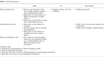
In the pediatric clinical context, there are two very important risk-related ethical imperatives—to reduce risk where possible and to describe risks as accurately and completely as possible to those with the authority to provide consent to neuroimaging (most commonly the parents). Some specific risks are associated only with a specific neuroimaging modality, whereas unexpected findings, lack of child-appropriate protocols and equipment, and potential overuse of imaging technologies are problems common to all of the technologies. These risks have implications for clinical practice. In this section, we explore some of the risks of each technology and then some of the risks common to all of the technologies through the lens of risk reduction and/or risk description. The risks discussed are those on which we believe the ethical imperatives mentioned above would require action (Table 1).
Risk Description

MRI
Direct physical risk MRI scanners use a strong static magnetic field, a pulsed gradient magnetic field, and a radiofrequency field to obtain images of the body in selected planes.24
Health Protection Branch, Environmental Health Directorate. Safety Code–26: Guidelines on Exposure to Electromagnetic Fields from Magnetic Resonance Clinical Systems, Ottawa, On: Minister of National Health and Welfare; 1987.
Center for Devices and Radiological Health. Magnetic Resonance Working Group. A Primer on Medical Device Interactions with Magnetic Resonance Imaging Systems. Washington, D.C.: Food and Drug Administration; 1997. Available at: http://www.fda.gov/cdrh/ode/primerf6.html.
Detailed patient screening guidelines have been developed for the MR environment (www.MRIsafety.com). These guidelines help to reduce the risk of such harms as are encountered when a patient with a metal implant undergoes an MRI. Unfortunately, these guidelines do not specifically address pediatric concerns. Therefore, screening guidelines specific to children should be developed and disseminated.
According to Shellock and Crues,26
Shellock FG, Crues JV. MR procedures: Biologic effects, safety and patient care. Radiology 2004;232:635–52.
See note 25, Center for Devices and Radiological Health 1997.
Landrigan C. Preventable deaths and injuries during magnetic resonance imaging. New England Journal of Medicine 2001;345:1000–1.
Further research into risks associated with MRI is also necessary. Only a few epidemiological studies into MR exposure have been carried out; consequently, there is an insufficient scientific basis for the assumption that MR exposure has no long-term effects.29
Levine D. MR imaging of fetal central nervous system abnormalities. Brain and Cognition 2002;50:432–48; Feychting M. Health effects of static magnetic fields—A review of the epidemiological evidence. Progress in Biophysics and Molecular Biology 2005;87:241–6.
Indirect physical risk The risks of MRI may not be directly related to the properties of the MRI magnet and environment. For example, under certain clinical circumstances, an MRI exam may require the administration of sedation or a contrast agent. These procedures introduce risk due to potential serious side effects.
The absence of patient motion is necessary for optimal MRI. Sedation has frequently been used to eliminate motion in children. Indeed, historically, sedation has been required in almost half of the pediatric patients who are imaged.30
Harned RK, Strain JD. MRI-compatible audio/visual system: Impact on pediatric sedation. Pediatric Radiology 2001;31:247–50.
American Academy of Pediatrics. Prolonged recovery and delayed side effects of sedation for diagnostic imaging studies in children. Pediatrics 2000;105:42–6.
Mayo Clinic. Anesthesia: Options and Considerations. Rochester, MN: Mayo Clinic; 2004. Available at: http://www.mayoclinic.com/health/anesthesia/SC00026.
See note 30, Harned, Strain 2001.
American Academy of Pediatrics. Guidelines for monitoring and management of pediatric patients during and after sedation for diagnostic and therapeutic procedures. Pediatrics 1992;89:110–5.
Additional guidelines are also needed. Guidelines have been developed by the American Academy of Pediatrics in its Monitoring and Management of Pediatric Patients During and After Sedation for Diagnostic and Therapeutic Procedures. However, disabled children may have unpredictable responses to sedation.35
Hinton VJ. Ethics of neuroimaging in pediatric development. Brain and Cognition 2002;50:455–68; see note 1, Accardo et al. 2004.
MRI delivery guidelines are also needed. For example, consider the use of gandolinium in a pediatric population. Gandolinium is a commonly used MRI contrast agent. It has a slower clearance in fetuses, neonates, and infants compared to adults.36
See note 29, Levine 2002.
Gooden CK, Dilos B. Anesthesia for magnetic resonance imaging. International Anesthesiology Clinics 2003;41:29–37.
Direct psychological risk The MRI procedure may produce considerable fear, distress, and potentially long-term psychological effects for a significant proportion of patients.38
McIsaac HK, Thordarson DS, Shafran R, Rachman S, Poole G. Claustrophobia and the magnetic resonance imaging procedure. Journal of Behavioral Medicine 1998;21:255–68.
Rosen AC, Gur RC. Ethical considerations for neuropsychologists as functional magnetic imagers. Brain and Cognition 2002;50:469–81.
See note 7, Santosh 2000.
See note 18, Wilke et al. 2003.
CT
A significant risk associated with CT scanning is the negative effect of X-ray radiation. This risk is generally considered quite small compared to the benefits of accurate diagnosis and treatment. However, one report has suggested that CT scans of children are often done without adjusting dose to weight, resulting in up to 50% of the dose being unnecessary.42
Ron E. Ionising radiation and cancer risk: Evidence from epidemiology. Pediatric Radiology 2002;32:232–7.
Picano E. Sustainability of medical imaging. British Medical Journal 2004;328:578–80.
Fox AJ. Use of the lowest necessary radiation dose. American Journal of Neuroradiology 2004;25:519.
Food and Drug Adminstration. Reducing Radiation Risk from Computed Tomography for Pediatric and Small Adult Patients. FDA Public Health Notification. Washington, D.C.: FDA; 2001. Available at: www.fda.gov/cdrh/safety.html.
See note 43, Picano 2004.
PET/SPECT
The radiation hazard posed by PET is said to be low and equivalent to the hazard posed by CT scan47
See note 13, Sudhir et al. 2005.
Jadvar H, Alavi A, Mavi A, Shulkin BL. PET in pediatric diseases. Radiologic Clinics of North America 2005;43:135–52.
Stoll AL, Renshaw PF, Yurgelun-Todd DA, Cohen BM. Neuroimaging in bipolar disorder: What have we learned? Biological Psychiatry 2000;48:505–17.
In addition, PET and SPECT imaging often require intravenous administration of radiotracers. PET/SPECT technologists do not always have experience establishing intravenous access in children.50
Roberts EG, Shulkin BL. Technical issues in performing PET studies in pediatric patients. Journal of Nuclear Medicine Technology 2004;32:5–9.
See note 50, Roberts, Shulkin 2004.
Risks common to various technologies
Both magnetic and radiological imaging share additional risks that may be of particular concern in the pediatric population. Special precautions need to be taken when reading the brain images of children to ensure detection of any anomalies as well as appropriate follow-up.52
Illes J, Racine E. Imaging or imagining? A neuroethics challenge informed by genetics. The American Journal of Bioethics 2005;5:5–18.
Mamourian A. Incidental findings on research functional MR images: Should we look? American Journal of Neuroradiology 2004;25:520–2.
Reports of unexpected MRI findings in brain imaging are not uncommon in the clinical domain and have been reported in pediatric populations.54
Kim BS, Illes J, Kaplan RT, Reiss A, Atlas SW. Neurologic findings in healthy children on pediatric fMRI: Incidence and significance. American Journal of Neuroradiology 2002;23:1674–7.
Illes J, Desmond JE, Huang LF, Raffin TA, Atlas SW. Ethical and practical considerations in managing incidental findings in functional magnetic resonance imaging. Brain and Cognition 2002;50:358–65.
There have been many complaints in the literature that child-appropriate neuroimaging protocols have not been developed. These types of protocols would ensure that pediatric subjects comprehend the procedure and experience a minimum of fear.56
See note 35, Hinton 2002.
Rumsey J, Filipek P, Kennedy D, Lange N, Reiss A, Evans A, et al. Tools for pediatric neuroimaging workshop: Report of the working groups. 1997. Vienna, VA: Inter-institute Group for Pediatric Neuroimaging: NIMH, NINDS, NICHD; 1997.
Ment LR, Bada HS, Barnes P, Grant PE, Hirtz D, Papile LA, et al. Practice parameter: Neuroimaging of the neonate. Neurology 2002;58:1726–38.
Another risk common to neuroimaging technologies is the potential overuse. Up to a third of all radiological examinations have been reported as totally or partially inappropriate.59
Hall EJ. Lessons we have learned from our children: Cancer risks from diagnostic radiology. Pediatric Radiology 2002;32:700–6; see note 45, Food and Drug Administration 2001.
Ball WS. Safety of pediatric neuroimaging. American Journal of Neuroradiology 1996;17:1037.
Interpretation
Several articles have warned of the potential for misinterpretation or overinterpretation of imaging examination results.61
See note 35, Hinton 2002; see note 52, Illes, Racine 2005.
The limits of interpretative powers
Conventional MRI and CT image interpretation requires knowledge of normal brain anatomy and development, recognition of findings in specific disorders, and an awareness of potential study artifacts. Although neuroanatomical imaging measures the volume and shape of brain structures, the underlying cause of any differences may not be determinable.62
Durston S, Pol HEH, Casey BJ, Giedd JN, Buitelaar JK, van Engeland H. Anatomical MRI of the developing human brain: What have we learned? Journal of the American Academy of Child and Adolescent Psychiatry 2001;40:1013–20.
See note 10, Frank, Pavlakis 2001.
Functional neuroimaging has encountered criticisms based on the interpretation of functional–behavioral associations and the lack of standardization in the field. For example, functional neuroimaging cannot determine that a particular cognitive process causes imaged brain activity because this association has not been proven to be causal.64
See note 35, Hinton 2002.
Additionally, the structure and content of neuroimaging data sets currently have no universally accepted standards.65
Governing Council of the Organization for Human Brain Mapping. Neuroimaging databases. Science 2001;292:1673–6.
See note 39, Rosen, Gur 2002.
The interpretation of neuroimaging examination results faces further challenges when children are imaged. Because the developing brain is constantly changing, a certain degree of normal variability must be taken into consideration when interpreting pediatric neuroimaging data.67
See note 35, Hinton 2002.
See note 7, Santosh 2000.
See note 7, Santosh 2000; see note 35, Hinton 2002; see note 18, Wilke et al. 2003.
See note 7, Santosh 2000; see note 20, Desmond, Chen 2002.
See note 7, Santosh 2000; see note 10, Frank, Pavlakis 2001; see note 35, Hinton 2002; see note 20, Desmond, Chen 2002; see note 18, Wilke et al. 2003.
Neuroimaging is still at a relatively early stage of development. Although it has allowed researchers and clinicians to view the human brain more thoroughly than ever before, the comprehensive analysis, interpretation, and standardization of imaging data are daunting tasks.
The ethical implications
The present technological and interpretive limitations directly influence the ethical aspects of neuroimaging in the clinic. Although some structural changes (e.g., discrete tumors or lesions) can be unambiguously identified via neuroimaging, other structural abnormalities can be much more subtle.72
Canli T, Amin Z. Neuroimaging of emotion and personality: Scientific evidence and ethical considerations. Brain and Cognition 2002;50:414–31.
See note 57, Rumsey et al. 1997.
Functional imaging is very important for presurgical planning—especially in epilepsy surgery—and has the potential to provide insight into neurological and psychiatric disorders.74
See note 18, Wilke et al. 2003.
Shevell M, Ashwal S, Donley D, Flint J, Gingold M, Hirtz D, et al. Practice parameter: Evaluation of the child with global developmental delay. Neurology 2003;60:367–80; see note 58, Ment et al. 2002.
Stigmatization
A noticeable feature of some neuroimaging studies is the use of value-laden language to describe various aspects of brain structure and function even though normal brain anatomy and function has yet to be determined. The language implies the existence of normal and abnormal brain templates and increases the potential for the stigmatization of pathologically labeled individuals. Special precautions will need to be taken in this regard in respect of cognitive profiling, intelligence tests, and behavioral disorders diagnosis. Despite this, Giedd76
Giedd JN. Neuroimaging of pediatric neuropsychiatric disorders: Is a picture really worth a thousand words? Archives of General Psychiatry 2001;58:443–4.
American Academy of Pediatrics. Clinical practice guideline: Diagnosis and evaluation of the child with Attention-Deficit/Hyperactivity Disorder. Pediatrics 2000;105:1158–70; American Academy of Neurology. Practice parameter: Screening and diagnosis of autism. Neurology 2000;55:468–79.
Research on children's mental disorders has lagged behind parallel efforts for adults.78
Hinshaw SP. The stigmatization of mental illness in children and parents: Developmental issues, family concerns and research needs. Journal of Child Psychology and Psychiatry 2005;46:714–34.
Mash EJ, Barkley RA, eds. Child Psychopathology. New York: Guilford Press; 2003; see note 78, Hinshaw 2005.
Illes J, Raffin TA. No child left without a brain scan? Toward a pediatric neuroethics. Cerebrum 2005;7:33–46.
DiPietro JA. Baby and the brain: Advances in child development. Annual Review of Public Health 2000;21:455–71.
See note 35, Hinton 2002.
See note 78, Hinshaw 2005.
Allocation of Resources
Serious ethical issues also arise when examining neuroimaging and allocation of resources. At the societal level, pediatric neuroimaging raises several questions surrounding its potential exclusivity, expense, and the medicalization of social phenomena. For example, with the advent of private MRI clinics, imaging modalities may permit queue jumping so that any potential benefits will favor those with the resources to afford access. Additionally, the expense of neuroimaging may restrict access to wealthy communities and regions. This potential inequality needs to be acknowledged, and investigations into the best use of public funds for neuroimaging techniques should be undertaken. Another potential concern relates to the massive funds allowed for the development of neuroimaging modalities to the detriment of basic community-based and family services.84
See note 78, Hinshaw 2005.
See note 35, Hinton 2002; see note 75, Shevell et al. 2003.
Neurobiological explanations may be viewed as more objective than environmental descriptions. However, environmental factors, although more complex and nuanced, may provide insight into the deeper social causes of particular neurological disorders. A focus on neuroimaging procedures could channel resources away from environmental concerns, such as the consequences of poverty on children. For instance, not only is prematurity more common in low-income populations, but the developmental outcomes for these infants born into poverty are significantly worse than for middle-class preterm infants.86
See note 81, DiPietro 2000.
Confidentiality
The importance of the privacy and confidentiality of children's health information has been recognized by several professional and international organizations.87
American Academy of Pediatrics. Privacy protection of health information: Patient rights and pediatrician responsibilities. Pediatrics 1999;104:973–7; Canadian Coalition for the Rights of Children. Rights of the Child: Health Care and the UN Convention on the Rights of the Child. 2002. Available at: www.rightsofchildren.ca.
See note 35, Hinton 2002.
Secondary information obtained in the process of patient screening also raises ethical concerns. Specifically with regards to MRI procedures, patients must be screened for such personal information as presence of IUDs, diaphragms, tattoos, and body piercings.89
See note 26, Shellock, Crues 2004.
Concern about confidentiality is one of the primary reasons young people delay seeking healthcare services for sensitive issues.90
Tharp M, Meier P. Confidentiality of Care for Teens Allows Parental Involvement. 2002. Available at: www.aap.org/advocacy/washing/confidentiality_care.htm.
Ford CA, English A. Limiting confidentiality of adolescent health services: What are the risks? JAMA 2002;288:752–3.
Acquavella AP, Braverman P. Adolescent gynecology in the office setting. Pediatric Clinics of North America 1999;46:489–503.
Conclusion
Neuroimaging is a developing field that holds great promise in aiding diagnosis, prognosis, and treatment of pediatric neurological disorders. However, as outlined in this paper, many unknowns and numerous complex risks surround neuroimaging technologies. Children, especially disabled children, are particularly vulnerable groups that require special attention. Clinicians within the field of neuroimaging who work with the pediatric population ought to be aware of the particular needs of children. Additionally, clear guidelines and standards need to be developed around the use of neuroimaging technologies to provide diagnostic and prognostic accuracy and confidence. The creation of standards for a technology still in development could be a difficult task. However, given the fact that these technologies are presently being used in the clinic and perhaps imprudently, clinicians need guidance.
In our paper, we have highlighted the areas in need of further clarification and research, including:
- Development of detailed guidelines with respect to pediatric neuroimaging, especially of disabled children.
- Adaptation of neuroimaging modalities to accommodate the particularities of the pediatric population.
- Clinician assurance that risks are accurately and completely disclosed to parents and mature minors.
- Development of guidelines for assessing risk versus benefit (e.g., when pediatric neuroimaging is obligatory or optional).
- Development of best practices for handling unanticipated findings.
- Restraint in interpretation of results to mitigate inappropriate diagnoses and prognoses.
- Advancement of research into the environmental (including psychosocial environment) influences on pediatric neurological disorders alongside the development of neuroimaging technologies.
- Development of clear confidentiality policies and privacy protection practices for the children undergoing neuroimaging examinations.
Through the resolution of these issues, neuroimaging could better achieve its promise for the pediatric population. Safe and responsible use of neuroimaging technologies and analysis of neuroimages promote the best interests and well-being of the pediatric patient.