Introduction
Ultrastructural studies of spermatogenesis, spermiogenesis and spermatozoa have been studied by various workers in different groups of the Platyhelminthes. Studies which have focused on digenetic trematodes include those of Justine et al. (Reference Justine, Jamieson and Southgate1993), Gracenea et al. (Reference Gracenea, Ferrer and Gonzalez-Moreno1997), Iomini & Justine (Reference Iomini and Justine1997), Ashour et al. (Reference Ashour, Stietieh and Khalil2001), Baptista-Farias et al. (Reference Baptista-Farias, Kohn and Cohen2001), Pamplona-Basilio et al. (Reference Pamplona-Basilio, Baptista-Farias and Cohn2001), Ndiaye et al. (Reference Ndiaye, Miquel, Ba, Feliu and Marchand2002, Reference Ndiaye, Miquel, Fons and Marchand2003, Reference Ndiaye, Miquel, Ba and Marchand2004) and Levron et al. (Reference Levron, Ternengo and Marchand2004). However, ultrastructural studies on spermatogenesis and sperm production in paramphistomes are rare and include those of Nollen & Pyne (Reference Nollen and Pyne1979), Gupta et al. (Reference Gupta, Parshad and Guraya1986), Li & Wang (Reference Li and Wang1997) and Taelab (Reference Taelab2000).
The present paper describes the ultrastructure of spermiogenesis in two paramphistomes, Sandonia sudanensis and Basidiodiscus ectorchis from the Nile fish Synodontis schall in Egypt, to further our knowledge of this process in this group of trematodes.
Materials and methods
Specimens of Synodontis schall were collected from fishermen at Bahr El Azam, Giza Governorate, south of Cairo during the spring of 2003. Fish were killed by a blow to the head, and a post-mortem examination was carried out. The alimentary canal was removed and the posterior part of the large intestine was examined under a binocular microscope for the two trematode species, Sandonia sudanensis and Basidiodiscus ectorchis. Live worms were cooled in 0.7% NaCl solution and then fixed in 3% glutaraldehyde in 0.1 m cacodylate buffer, pH 7.2 for 3 h at 4°C, then post-fixed in 1% cacodylate buffered OsO4 at room temperature for 2 h. Fixed worms were dehydrated in a graded ethanol series and propylene oxide and embedded in epoxy resin. A series of ultrathin sections were cut using a diamond knife of an LKB 4800 ultratome, mounted on uncoated copper grids and double-stained with alcoholic uranyl acetate and aqueous lead citrate. Grids were viewed in a JEOL 1200 EX2 transmission electron microscope.
Results
Basidiodiscus ectorchis exhibits an elongated bell-shaped body with a tapering anterior end. Both the anterior testis, near the anterior third of the body, and the posterior testis, near the middle of the body, are oval, diagonal, smoothly contoured and mostly extracaecal. The body of S. sudanensis is elongate and oval-shaped with an anterior rounded end. The testes are diagonal, globular, smoothly contoured and intracaecal.
Spermatogonia formation
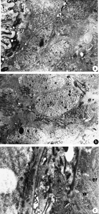
Cells below the basal lamina of the testis in both species represent the spermatogonia and spermatocytes. Spermatogonia form a peripheral layer, characterized by a prominent nucleus and a high nucleo-cytoplasmic ratio (fig. 1a). Spermatocytes are the largest cells formed and show an increase in the nucleo-cytoplasmic ratio (fig. 1b).
Transmission electron micrographs of the testis of: (a) Basidiodiscus ectorchis to show the peripherally-located, electron-dense spermatogonia (sg) resting on the undulating basal membrane (bm) of the whole testis (mitochondrion (m) ( × 500); (b) Sandonia sudanensis to show a group of large sized spermatocytes (sc) with nucleus (n), mitochondria (m), Golgi bodies (g), synaptonemal complex (sy), spermatozoa (sz) ( × 3000); (c) B. ectorchis to show an early spermatid (sp) an elongated nucleus (n) with condensed lamellae of chromatin, centriole (c) surrounding the nucleus from each side, axoneme (ax), endoplasmic reticulum (er) ( × 4000).

Spermatid formation
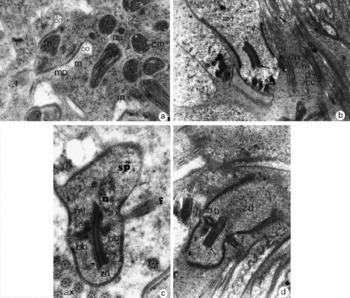
Differentiation of the spermatid begins with the elongation of the cell and its nucleus. As a result, the nucleus changes from being round in shape and distal in position to becoming elongate, cylindrical and oriented along the longitudinal axis of the cell (fig. 1c). The chromatin becomes condensed and granular. These granules form lamellar-like structures near the centre of the nucleus providing a longitudinally striated appearance in longitudinal sections and a honeycomb one in transverse sections (fig. 2a). The formation of a zone of differentiation at the distal end of the cell with a thickening of the cell membrane (fig. 2b,c) accompanies nuclear elongation and orientation.
Transmission electron micrographs of the testis of: (a) Basidiodiscus ectorchis to show the formation of the median process (mp), the migrated nucleus (n) and mitochondria (m) and the invagination of the plasma membrane to form the groove-like collars (co); note the presence of both longitudinal and transverse spermatid nuclei (nu) and a few mitochondria (m) in the common cytoplasmic mass (cm) ( × 12,000 K); (b) Sandonia sudanensis to show the formation of the zone of differentiation (zd) and the microtubule organizing centre (MTOC) (arrowed); note the longitudinal sections through the parallel helices of the central unit of the axoneme (ax); central core (cc), doublet tubules (d) and sheath (h) ( × 15,000 K); (c) B. ectorchis presenting a late spermatid (sp) through the zone of differentiation (zd) with the nucleus (n), the MTOC (arrowed) with its characteristic striation, two basal bodies (bb), one extending flagellum (f), cortical microtubules (mt), rootlets (r) and free axonemes (ax) ( × 20,000); (d) S. sudanensis to show the zone of differentiation (zd) and the MTOC (arrowed), a basal body (bb) perpendicular to the MTOC and a striated rootlet (r) parallel to the MTOC in addition to an extended flagellum (f) ( × 15,000).

Two centrioles appear within the protrusion; at first, they are formed on both sides of the nucleus (fig. 2c) and then at right angles to each other. They multiply producing two new centrioles at a short distance from the old ones. The latter act as an organizing centre for the assembly of the constituents of the new ones. From the four structures, two longitudinally-oriented centrioles join together and form an elongate structure called the microtubule organizing centre (MTOC), which develops close to the nucleus and possesses a characteristic pattern of striation. This structure is made up of seven layers of plates of dense material lying parallel to the long axis of the spermatid. The more dense elements are situated on each side of the less dense central elements (fig. 2b,c).
The two remaining centrioles are situated on either side of the central body and each forms a basal body. From the two basal bodies, two flagellar processes emerge laterally from the zone of differentiation. Each basal body consists of an axial unit surrounded closely by an extension of the plasma membrane (fig. 2d). At this stage, the basal bodies with their extensions are attached to the MTOC in a perpendicular fashion.
Tapering striated rootlets, each with an elongated triangular form and associated with the basal bodies extend along each side of the nucleus below the zone of differentiation (fig. 2d), thus anchoring the basal bodies and the developing axial units. The major transverse periodicity of the rootlet repeats at intervals with minor striations (fig. 3a).
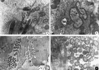
Transmission electron micrographs of the testis of: (a) Sandonia sudanensis to show the elongated median process (mp) with the migrated nucleus (n) and mitochondria (m) accompanied by striated rootlets (r) plus the invagination of the plasma membrane forming groove-like collars (co), and spermatid nuclei (nu) ( × 12,000); (b) S. sudanensis to show two transversely cut median processes (mp) after the fusion, each with two free flagella marked by two dense plaques (dp) representing the points of fusion; note the arrangement of microtubules (mt) in a continuous ring below the plasma membrane ( × 25,000); (c) Basidiodiscus ectorchis to show two groups of spermatozoa (sz), each consisting of 32 spermatozoa ( × 4000); (d) B. ectorchis to show various transverse sections through the median process (mp) with microtubules (mt) and dense plaques (dp) in addition to free axonemes (ax) ( × 25,000).

The differentiating zone is clearly set apart from the spermatid proper. At its basal (proximal) end, the plasma membrane is curled back upon itself forming two fibrous arching membranes (figs 2a, 3a). At the distal end of the spermatid a median cytoplasmic process, (fig. 2a) which encloses nuclear elements, mitochondria and rootlets is formed and provided with cortical microtubules. The latter are arranged in a continuous ring lying immediately below the plasma membrane forming a cytoskeleton (fig. 3b).
The basal bodies and flagellar processes are, at first, at right angles to the MTOC in its early stage of formation (fig. 2c), but as development proceeds, their orientation changes and they rotate gradually through an angle of 90° about the MTOC. Eventually, the basal bodies and flagellar processes come to lie parallel to the longitudinal axis of the developing spermatozoon, one on each side of the median cytoplasmic process (MCP). The nucleus migrates into the MCP accompanied with numerous mitochondria and striated rootlets (fig. 3a). The nucleus comes to rest in the distal end of the median cytoplasmic process, which will develop into the head region of the fully developed spermatozoon.
Formation of spermatozoa
Following migration of the nucleus and mitochondria, the two flagella fuse with the median cytoplasmic process to form the monopartite shaft of the mature sperm. This fusion, referred to as proximo-distal fusion, progresses from the basal extremity to the distal one. On the proximal part, the diameter of the zone of differentiation narrows and the arching membranes approach each other, resulting in the occurrence of 32 spermatozoa at the end of the process (fig. 3c)
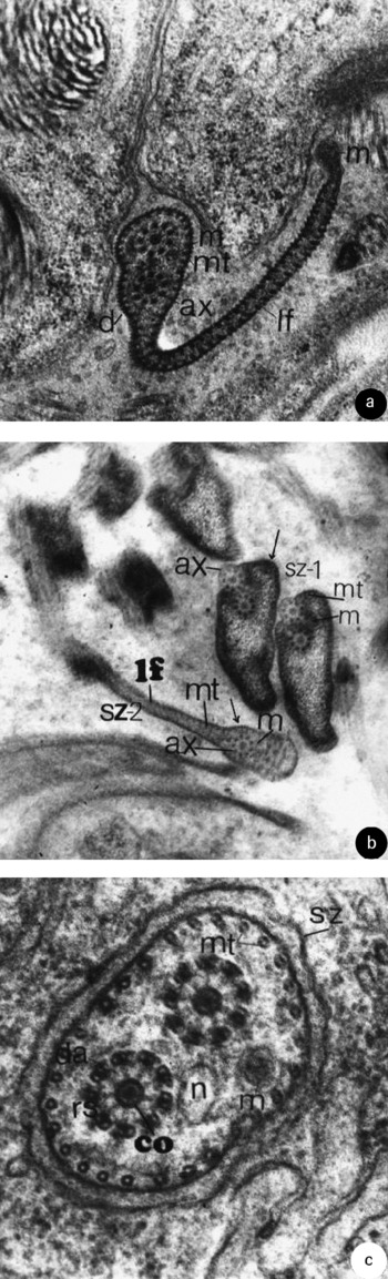
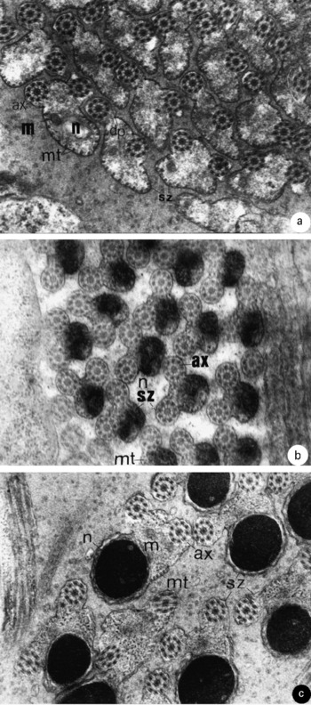
The sperm shaft of both paramphistome species exhibits a variety of profiles along its length during elongation. A cytoskeleton consisting of microtubules lies in close proximity to the plasma membrane. Two dense plaques attached to the inner surface of the plasma membrane are visible dorsally and ventrally between the microtubules; these plaques indicate the points of fusion of the two flagella with the median cytoplasmic processes (fig. 3b,d). In addition, several sections in the characteristic lateral flange of the spermatozoa are recorded in both species (fig. 4a,b). These lateral cytoplasmic extensions are of variable size with numerous longitudinal microtubules arranged at intervals and ending in a small protrusion, in addition to a dense external ornamentation on the cell membrane. Numerous transverse sections of spermatozoa with two axonemes consisting of the doublet 9+‘1’ arrangement are seen, as they extend from the basal bodies and comprise nine triplet tubules. These doublets are concentrically arranged around a single central complex (fig. 4c).
Transmission electron micrographs of the testis of: (a) Sandonia sudanensis to show a transverse section of a fully formed spermatozoon with two axonemes (ax), each with a 9+’1’ configuration and one small mitochondrion (m); note the formation of the lateral flange (lf) which ends in a small globular protrusion; the spermatozoon is surrounded internally by microtubules (mt) and externally by a dense external ornamentation except around the protrusion ( × 25,000); (b) Basidiodiscus ectorchis with a section through the spermatozoan shafts to show two stages in the formation of the lateral flange (lf); the first spermatozoa (sz-1) shows the incorporation of the second axoneme (ax) into the shafts with the presence of mitochondria (m), cortical microtubules (mt) and external dense material (arrowed); the second spermatozoon (sz-2) contains two axonemes (ax), one mitochondrion (m) and is surrounded internally by microtubules (mt) and externally by the dense material (arrowed) ( × 15,000); (c) S. sudanensis to show the spermatozoan shaft (sz) with two axonemes: each axoneme consists of nine doublets of microtubules arranged in a clockwise direction and provided with dynein arms (da) in addition to being connected to the central core (co) via radial spokes (rs); note the presence of the nucleus (n), mitochondria (m) completely surrounded by cortical microtubules (mt) ( × 60,000).

In S. sudanensis, the fully formed spermatozoon at the level of the lateral flange (fig. 4a) consists of two axonemes with the 9+’1’ configuration and a small mitochondrion. The lateral flange occasionally ends in a slight constriction and a small swelling. In B. ectorchis the developing spermatozoon fuses with the second axoneme, the first being completely encircled by a median cytoplasmic process (fig. 4b).
More distally, the nucleus becomes broader and not completely surrounded by microtubules (fig. 5a). Within the nucleus, the chromatin appears as coarse strands and closely-packed lamellae and the nuclear envelope is thickened and irregularly folded. The number of microtubules decreases towards the nuclear end of the spermatozoon with few microtubules in the nuclear region (fig. 5b,c).
Transmisison electron micrographs of the testis of: (a) Sandonia sudanensis to show various spermatozoan (sz) shafts sectioned at different levels; note the presence of two axonemes (ax) and the incompletely surrounding microtubules (mt) near the axonemes; note also the nucleus (n), mitochondria (m) and dense plaques (dp) marking the points of fusion of flagella with the median process ( × 25,000); (b) Basidiodiscus ectorchis at the distal part of the spermatozoan shaft (sz) showing two axonemes (ax), scarce microtubules (mt) and a large nucleus (n) ( × 25,000); (c) S. sudanensis to show the distal part of the spermatozoan shaft (sz) with a large nucleus (n), two axonemes (ax), scarce microtubules (mt) and two mitochondria (m) ( × 25,000).

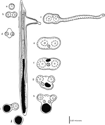
The longitudinal arrangement of elements in the sperms of each species is shown in fig. 6 which represents the schematic diagram of the ultrastructural organization of the mature spermatozoon of S. sudanensis. This shows a single axoneme (fig. 6a) and a second axoneme commencing a short distance below the anterior tip (fig. 6b). The fusion of the second flagellum with the median process is accompanied by the formation of the lateral flange (fig. 6c). At this level, the spermatozoon consists of two axonemes in the ’9+1’ configuration and one small mitochondrion, in addition, the sperm shaft ends in a small globular protrusion. The spermatozoon is completely surrounded internally by microtubules and externally by dense material representing the external ornamentation except around the protrusion. The first mitochondrion tapers off rapidly (fig. 6d) and is separated by a short intermediate region where only axonemes and the microtubules are seen. More distally, the second mitochondrion (fig. 6e, f) is followed by an elongated nucleus which becomes broader distally and the number of microtubules decreases towards the nuclear end (fig. 6g). Finally, a third mitochondrion appears (fig. 6i) before the tapering of the nucleus (fig. 6j).
Schematic diagram of the ultrastructural organization of the mature spermatozoon of Sandonia sudanensis. Transverse sections showing (a) one axoneme; (b) two axonemes; (c) the formation of the lateral flange; (d) axonemes and microtubules; (e) appearance of a second mitochondrion; (f) appearance of a nucleus; (g) decrease in the number of microtubules; (h) microtubules are arranged between the nucleus and one axial unit; (i) appearance of a third mitochondrion; (j) the nucleus occupying the entire sperm diameter.

Discussion
Although paramphistomes are considered as the most primitive digenetic trematodes (Kiger et al., Reference Kiger, Rashid, Griffon, Haque, Moens, Gibson, Poyart and Marden1998), they are highly successful intestinal parasites infecting a wide range of vertebrates. Rohde (Reference Rohde1980) and Justine et al. (Reference Justine, Lambert and Mattei1985) proposed that the ultrastructure of the spermatozoa of Platyhelminthes is suitable for establishing a phylogenetic relationship as they are not linked to parasitism or the influences that might be related to host specificity. Although morphologically paramphistomes are considered as primitive forms, spermiogenesis and karyotypes of the present two species may suggest that they are more advanced amphistomes compared to others previously studied.
In the present study, B. ectorchis and S. sudanensis follow the same pattern during spermatozoon formation at the level of the lateral flange. In previously studied amphistomes, neither the lateral flange nor the external ornamentation were recorded (Li & Wang, Reference Li and Wang1997; Taelab, Reference Taelab2000) and these structures had only been described in the Monogenea (Ashour et al., Reference Ashour, Soliman, Mezied and Mohammed2003), Aspidogastrea (Watson & Rohde, Reference Watson and Rohde1995) and in Fasciola gigantica (Ndiaye et al., Reference Ndiaye, Miquel, Ba and Marchand2004) suggesting that the structure might be of phylogenetic significance.
The karyotype of B. ectorchis and S. sudanensis previously described by Ashour et al. (Reference Ashour, Tohamy and Gamil2006) consisted mostly of metacentric chromosomes, although White (Reference White1973) showed that a primitive karyotype within any given phyletic group consists typically of acrocentric chromosomes. This view was confirmed by Grossman & Cain (Reference Grossman and Cain1981) and Ashour et al. (Reference Ashour, Hassan, Ibrahim and El-Kabbany1995) who recorded acrocentric chromosomes respectively in the amphistomes Megalodiscus temperatus, Calicophoron microbothrium and Carmyerius gregarius. These authors regarded the karyotype as showing a lesser degree of evolution in chromosomal configuration than any other member of the digenetic trematodes. However, in the present study, spermiogenesis together with the karyotype of S. sudanensis and B. ectorchis and the absence of any acrocentric chromosomes (Ashour et al., Reference Ashour, Tohamy and Gamil2006) suggest that the two species belong to more advanced amphistomes.
The axonemes of the spermatozoa of B. ectorchis and S. sudanensis show a 9+’1’ configuration, which matches the universal pattern in parasitic helminths (Rohde, Reference Rohde1980). Rohde et al. (Reference Rohde, Watson and Cannon1988) reported that the central axis of the axoneme has a double helical structure unique among sperms in the animal kingdom. In the present study, the two axonemes are enclosed entirely within the body of the spermatozoon and this is typical in digeneans. In addition, the axonemes are staggered along the length of the spermatozoon of these paramphistomes.
However, knowledge of amphistome spermiogenesis based on ultrastructural details is still fragmentary. Extensive application of ultrastructural data to phylogenetic problems must await further studies on additional species in various genera.