Introduction
In recent years, human embryonic stem (hES) cells have generated much interest over their potential applications in clinical therapy (Gerecht-Nir & Itskovitz-Eldor, Reference Gerecht-Nir and Itskovitz-Eldor2004), basic scientific research (Chen et al., Reference Chen, Lewis and Kaufman2003; Eisenberg et al., Reference Eisenberg, Kubalak and Eisenberg2004) and for pharmacological and cytotoxicity screening in vitro (Rohwedel et al., Reference Rohwedel, Guan, Hegert and Wobus2001; Gribaldo et al., Reference Gribaldo, Alison, Andrews, Bremer, Donovan, Knaan-Shanzer, Mertelsmann, Spielmann, Testa, Triffitt, Zipori and de Wynter2002). Nevertheless, a major bottleneck impeding the widespread application of these cells is the lack of efficient cryopreservation protocols.
Unlike ordinary somatic cells and murine embryonic stem cells, hES cells do not survive well under conventional cryopreservation protocols that utilize standard cryoprotectant concentrations, i.e. 10% (v/v) dimethyl sulfoxide (DMSO) and slow cooling in a −20 °C or −80 °C refrigerator (Reubinoff et al., Reference Reubinoff, Pera, Vajta and Trounson2001; Richards et al., Reference Richards, Tan, Tan, Chan and Bongso2004; Heng et al., Reference Heng, Kuleshova, Bested, Liu and Cao2005), with typically less than 5% of hES cells remaining viable, as reported by previous studies (Reubinoff et al., Reference Reubinoff, Pera, Vajta and Trounson2001; Richards et al., Reference Richards, Tan, Tan, Chan and Bongso2004). Although vitrification protocols (Reubinoff et al., Reference Reubinoff, Pera, Vajta and Trounson2001; Richards et al., Reference Richards, Tan, Tan, Chan and Bongso2004) utilizing high cryoprotectant concentrations together with flash freezing in liquid nitrogen have been reported to yield much higher survival rates; such protocols are tedious to perform manually and are clearly unsuited for handling bulk quantities of hES cells that would almost certainly be required for various clinical and non-clinical applications (Heng et al., Reference Heng, Kuleshova, Bested, Liu and Cao2005).
Hence, the pertinent question that arises is why hES cells do not adapt well to conventional cryo-preservation protocols? To answer this, we investigated cell death in hES colonies cryopreserved under standard conditions, i.e. with 10% (v/v) DMSO and slow cooling in a −80 °C refrigerator. It is hypothesized that cell death from cryopreservation could be induced by two major mechanisms. Firstly, cellular necrosis as a direct result of physical cryoinjury sustained through ice crystallization or osmotic shock. Secondly, a self-induced apoptotic mechanism may be triggered in hES cells as an indirect result of mild cryoinjury that is not directly lethal to the cell (i.e. damage to gap junctions, extracellular matrix) or by physical environmental stress encountered during cryopreservation (i.e. cold shock, osmotic stress).
If hES cell death during cryopreservation is the direct result of cryoinjury sustained through ice crystallization and osmotic shock, and then there should be an immediate loss of physical integrity of the cell membrane. This in turn is easily detected through trypan blue staining and we would expect a high proportion of non-viable cells to be stained blue immediately after post-thaw washing. By contrast, if a self-induced apoptotic mechanism is involved, then there should be a gradual loss of cell viability, as the apoptotic cascade would obviously need time to activate and exert its effect on the cell after freeze-thaw.
Hence, this study sought to determine whether there is an immediate or gradual loss of cell viability upon freeze-thawing of hES colonies cryopreserved with standard protocols. This in turn would be indicative of the major mechanism involved in hES cell death during cryopreservation, i.e. cellular necrosis or apoptosis? Additionally, this study also investigated whether cell death could somehow be abated by maintaining the freshly thawed hES cells at a low temperature (4 °C). If this were the case, then there would be stronger evidence for an apoptotic mechanism; as the various intracellular enzymes and proteins implicated in the apoptotic cascade should temporarily lose their activity at low temperature.
Materials and methods
hES cells, media, reagents and chemicals
hES cells were obtained from the Wicell Research Institute (Madison, WI, USA) and were from the H1 line listed on the National Institute of Health (NIH) registry, which had received Federal approval for US government-supported research funding (NIH, 2005). Unless otherwise stated, all liquid media, serum and serum replacements were purchased from Gibco BRL, while all other reagents and chemicals were purchased from Sigma–Aldrich.
Culture and propagation of hES cells in the undifferentiated state
Undifferentiated hES cells were maintained on a feeder layer of mitomycin C-inactivated murine embryonic fibroblast feeder (MEF) cells (Thomson et al., Reference Thomson, Itskovitz-Eldor, Shapiro, Waknitz, Swiergiel, Marshall and Jones1998; Reubinoff et al., Reference Reubinoff, Pera, Fong, Trounson and Bongso2000). These were harvested from the CF1 inbred mouse strain purchased from Charles River Laboratories. The culture medium was DMEM/F12 supplemented with 20% (v/v) Knockout (KO) serum replacement, 1 mM l-glutamine, 1% non-essential amino acid, 100 mM β-mercaptoethanol and 4 ng/ml basic fibroblast growth factor (bFGF). All cell cultures were carried out on 6-well culture dishes (Nunc) in a humidified 5% CO2 incubator set at 37 °C. The culture medium was changed daily with routine passage of hES cells on a fresh MEF layer being carried out once a week. Dissociation of hES colonies into cell clumps for serial passage was achieved through treatment with 1 mg/ml collagenase type IV, for between 3 to 5 min.
Cryopreservation of intact and adherent hES colonies
hES cells were cryopreserved by a modified protocol from that recommended by Wicell Inc (Wicell Research Institute, 2005). The cryopreservation solution was similar and consisted of DMEM/F12 medium supplemented with 10% (v/v) DMSO and 20% (v/v) defined fetal bovine serum. The major difference was that adherent hES colonies were cryopreserved intact on 6-well dishes, instead of dissociated cell clumps (through collagenase type IV treatment) in cryovials. This was similar to the protocol utilized by a number of previous studies (Chan & Evans, Reference Chan and Evans1991; Ure et al., Reference Ure, Fiering and Smith1992; Udy & Evans, Reference Udy and Evans1994; Ji et al., Reference Ji, de Pablo and Palecek2004). To achieve a gentler drop in temperature in the −80 °C refrigerator, the 6-well dishes were placed in an insulated Styrofoam box instead of an isopropanol container. A minimal volume of cryopreservation solution (0.3 ml) was added to each well of the dish, just enough to cover the adherent hES colonies. This was to facilitate rapid thawing. The adherent hES colonies cryopreserved on 6-well dishes were stored in the −80 °C refrigerator for at least 1 week prior to thawing.
Thawing and post-thaw wash of cryopreserved hES colonies
Cryopreserved hES colonies on 6-well dishes were thawed for 1 to 2 min in a 37 °C water bath. Special care was taken to ensure that no water from the bath seeped into the dish during thawing. To minimize osmotic shock to the cells, the thawed cryopreservation solution (0.3 ml) in each well was gradually diluted by dropwise addition of 2 ml of hES culture medium. After that, each well was consecutively washed twice with 2 ml of hES culture media. Depending on the particular experiment, the hES media used for post-thaw wash was either kept at 4 °C or 37 °C prior to use.
Effects of temperature on the viability and detachment of frozen-thawed hES colonies
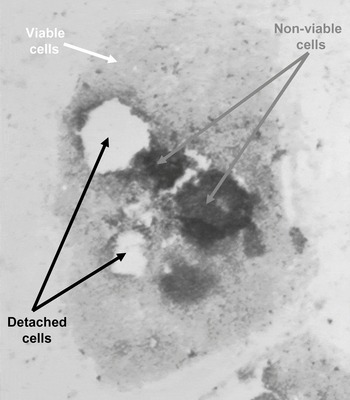
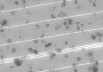
The freshly thawed hES colonies were placed either in a 37 °C incubator or the 4 °C chiller compartment of a refrigerator. At different time points (up to 90 min duration), the viability and detachment of the frozen-thawed hES colonies were assessed by trypan blue staining and observation under bright-field microscopy. All digital images were taken in 5 min of trypan blue staining. Viable cells in the hES colony remained unstained, while non-viable cells were clearly stained blue (Fig. 1). In some of the frozen-thawed hES colonies, some cells began to detach over time, leaving empty patches in the colony (Fig. 1). The detached cells were collected by centrifugation, stained with trypan blue and observed under a hemocytometer with bright-field microscopy (Fig. 2). It was found that virtually all detached cells were non-viable, as indicated by positive staining (Fig. 2). Hence, it can be reasonably assumed that detached areas in hES colonies represented only non-viable cells.
Non-viable cells in frozen-thawed hES colonies are positively stained by trypan blue (grey arrows), while viable cells remain unstained (white arrows). Some of the non-viable cells have detached from the hES colony (black arrows). The image was captured under bright-field microscopy at ×40 magnification.

Cells that have detached from hES colonies after freeze-thawing were collected by centrifugation, stained with trypan blue and observed using a hemocytometer with bright field microscopy at ×200 magnification. Virtually all detached cells were non-viable, as indicated by positive staining.

Assessment of viability and degree of detachment in trypan blue-stained frozen-thawed hES colonies through area analysis
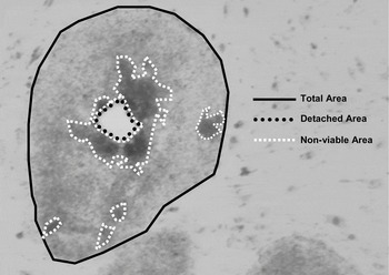
The viability and degree of detachment in trypan blue-stained frozen-thawed hES colonies were determined by area analysis, using ImageTool software (University of Texas Health Science Center). It was assumed that the hES cells were uniformly distributed throughout each colony. The total area occupied by each individual hES colony was first determined, followed by the blue-stained and detached areas in the same colony. This was achieved by manually tracing out the different areas (Fig. 3), using the ImageTool software. The total proportion of non-viable cells was taken to be the combined ratios of blue-stained and detached areas in each hES colony; while the proportion of viable cells was taken to be the ratio of adherent unstained areas in the colony. For each data set, between 20 to 30 trypan blue-stained frozen-thawed hES colonies were analysed in this manner.
hES colonies were stained with trypan blue and digital images were captured under bright-field microscopy at ×40 magnification. The total area of each individual hES colony was traced out (black solid line), together with the detached (black dotted line) and non-viable (white dotted line) areas.

Statistical analysis of data
The results from each data set were expressed as mean ± standard derivations (computed from area analysis of a total of 20 to 30 hES colonies). Differences between data sets were assessed by ANOVA analysis, with a p-value less than 0.05 being considered significantly different.
Results
Kinetics of cell death and detachment in frozen-thawed hES colonies incubated at 37 °C and 4 °C
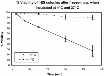
Just after the post-thaw wash at the 5 min time point, it was observed that 97–98% of hES cells were viable, as assessed by the trypan-blue exclusion test (Fig. 4), regardless of whether the cells were washed in medium kept at 4 °C or 37 °C. However, when the freshly thawed hES cells were placed in a 37 °C incubator, there was observed to be a steep decrease in cell viability over time. By the 90 min time point, only 27.3 ± 10.7% of cells remained viable (Fig. 4). By contrast, when the cells were kept at 4 °C, there was only a slight decrease in cell viability over the same duration, with 90.6 ± 4.1% of cells still remaining viable after 90 min (Fig. 4). At all corresponding time points, except the 5 min time point, there were significant differences in the cell viability of hES colonies maintained at 4 °C and 37 °C (p-value less than 0.01).
The percentage viability of hES colonies incubated at 4 °C and 37 °C was assessed by trypan blue staining and bright field microscopy, over a time course of 90 min.

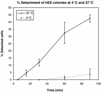
Additionally, a vast difference in the kinetics of cell detachment in hES colonies maintained at 4 °C and 37 °C was also observed (Fig. 5). Just after the post-thaw wash at the 5 min time point, virtually none of hES cells detached, regardless of whether the cells were washed in medium kept at 4 °C or 37 °C. However, when the freshly thawed hES cells were placed in a 37 °C incubator, there was a steep increase in cell detachment over time. By the 90 min time point, 42.5 ± 2.4% of cells in hES colonies have detached (Fig. 5). By contrast, when the cells were kept at 4 °C, there was only a slight increase in cell detachment over the same duration, with 4.1 ± 3.0% of cells being detached after 90 min (Fig. 5). At all corresponding time points except the 5 min time point, there were significant differences in cell detachment of hES colonies maintained at 4 °C and 37 °C (p-value less than 0.01).
The percentage detachment of hES colonies incubated at 4 °C and 37 °C was assessed by bright-field microscopy, over a time course of 90 min.

Kinetics of cell death and detachment in frozen-thawed hES colonies kept at 4 °C for 90 min, followed by incubation at 37 °C for a further 90 min
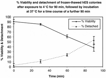
After incubation for 90 min at 4 °C, it was observed that 90.6 ± 4.1% of hES cells were viable, as assessed by the trypan-blue exclusion test (Fig. 6). However, when the low temperature-exposed hES cells were placed in a 37 °C incubator, there was a steep decrease in cell viability over time (Fig. 6). After a further 90 min incubation at 37 °C, only 38.1 ± 6.8% of cells retained their viability (Fig. 6). Additionally, there was also observed to be a steep increase in cell detachment, from 3.0 ± 3.8% to 53.3 ± 7.5% of cells being detached, after 90 min of further incubation at 37 °C.
The percentage viability and detachment of hES colonies initially exposed to 4 °C for 90 min, followed by a further 90 min of incubation at 37 °C, was assessed by trypan blue staining and bright-field microscopy.

Discussion
In the previous study of Ji et al. (Reference Ji, de Pablo and Palecek2004), adherent hES colonies were cryopreserved intact on 6-well dishes, utilizing conventional slow cooling with a standard cryoprotectant concentration of 10% (v/v) DMSO. In the absence of matrigel and trehalose loading, it was demonstrated that only a very small fraction of cells in intact adherent hES colonies survived freeze-thawing; typically less than 5%, which was comparable to the post-thaw recovery rate of enzymatically dispersed hES cell clumps cryopreserved in cryovials. Subsequently, Ji et al. (Reference Ji, de Pablo and Palecek2004) showed that matrigel embedment together with trehalose loading of the cells prior to cryopreservation, substantially enhanced post-thaw survival rates of adherent hES colonies. Nevertheless, the study of Ji et al. (Reference Ji, de Pablo and Palecek2004), did not attempt to characterize the mechanism responsible for the low post-thaw survival rates of adherent hES colonies cryopreserved without matrigel embedment and trehalose loading. It was somewhat presumed that the absence of matrigel and trehalose loading rendered the cells more vulnerable to cryoinjury induced by ice crystallization and osmotic shock. This would imply that the major mechanism of hES cell death during cryopreservation is cellular necrosis induced directly by cryoinjury. This study therefore attempted to investigate the validity of this presumption.
Surprisingly, the results clearly showed that immediately after the post-thaw wash (5 min time point), the overwhelming majority of hES cells in adherent colonies were viable (approximately 98%), as assessed by the trypan-blue exclusion test (Fig. 4). However, there was a steep decline in viability when the hES colonies were incubated at 37 °C, which was much less severe at 4 °C (Fig. 4). This slow down in the kinetics of cell death at 4 °C was somewhat reversible. Upon returning to physiological temperature 37 °C, there was again observed to be a steep decline of cell viability over time (Fig. 6). These observations strongly challenge the presumption that cellular necrosis induced directly by cryoinjury is the major mechanism of cell death during freeze-thaw of adherent hES colonies. Instead, the results are suggestive of a self-induced apoptotic mechanism (Raff et al., 2004), as the kinetics of cell death is so strongly dependent on physiological temperature (Fig. 4). This is made even more evident by the observation that the slow down in the kinetics of cell death at low temperature is reversible upon returning to physiological temperature (Fig. 6).
The pertinent question that arises, if a self-induced apoptotic mechanism is somehow implicated in hES cell death during cryopreservation, is why should this be abated by matrigel embedment and trehalose loading, as demonstrated by Ji et al. (Reference Ji, de Pablo and Palecek2004)? A possible conjecture is that in the absence of matrigel embedment and trehalose loading, cryodamage is predominantly localized on extracellular structural elements in hES colonies, i.e. gap junctions, intercellular adhesional contacts, rather than in the cell itself or its plasma membrane. Being ‘social’ cells that require co-operative interactions (Thomson et al., Reference Thomson, Itskovitz-Eldor, Shapiro, Waknitz, Swiergiel, Marshall and Jones1998; Reubinoff et al., Reference Reubinoff, Pera, Fong, Trounson and Bongso2000; Sathananthan et al., Reference Sathananthan, Pera and Trounson2002; Amit et al., Reference Amit, Shariki, Margulets and Itskovitz-Eldor2004; Cowan et al., Reference Cowan, Klimanskaya, McMahon, Atienza, Witmyer, Zucker, Wang, Morton, McMahon, Powers and Melton2004; Richards et al., Reference Richards, Fong, Tan, Chan and Bongso2004), hES cells are particularly vulnerable to any damage to gap junctional and other intercellular adhesional contacts. Nowhere is this better demonstrated than in the routine serial passage of hES colonies. In all culture protocols reported to date (Thomson et al., Reference Thomson, Itskovitz-Eldor, Shapiro, Waknitz, Swiergiel, Marshall and Jones1998; Reubinoff et al., Reference Reubinoff, Pera, Fong, Trounson and Bongso2000; Amit et al., Reference Amit, Shariki, Margulets and Itskovitz-Eldor2004; Cowan et al., Reference Cowan, Klimanskaya, McMahon, Atienza, Witmyer, Zucker, Wang, Morton, McMahon, Powers and Melton2004), special care is always taken to ensure that hES colonies are mildly dissociated into relatively large cell clumps during serial passage. Indeed, it was clearly demonstrated that extensive dissociation (enzymatic or mechanical) of hES colonies into single cells or clusters containing less than 10 cells during serial passage can severely compromise the survival and propagation of hES cell in vitro (Reubinoff et al., Reference Reubinoff, Pera, Fong, Trounson and Bongso2000). A cell clump size of around 100 cells has been reported to be optimal for hES colony growth and propagation (Reubinoff et al., Reference Reubinoff, Pera, Fong, Trounson and Bongso2000). The highly ‘co-operative’ nature of hES cells is further confirmed by electron-microscopic ultrastructural studies, which showed an abundance of tight and gap junctions in hES colonies (Sathananthan et al., Reference Sathananthan, Pera and Trounson2002), as well as by transcriptome profile analysis (Richards et al., Reference Richards, Fong, Tan, Chan and Bongso2004), which revealed high levels of expression of gap junction and adhesion proteins (i.e. connexin-43 and claudin-6).
Presumably, matrigel embedment and trehalose loading could somehow mitigate against cryodamage to these gap-junctional and adhesional elements in hES colonies. This in turn led to the enhanced post-thaw survival rates, as reported by Ji et al. (Reference Ji, de Pablo and Palecek2004). We hypothesize that cryodamage to gap-junctional and adhesional elements in hES colonies somehow triggers an apoptotic cascade in individual hES cells. As would be expected, the various intracellular proteins and enzymes implicated in this putative apoptotic cascade are reversibly and temporarily inhibited by low temperatures, as demonstrated by our results (Fig. 6). Future work would therefore attempt to investigate the molecular mechanisms of this putative apoptotic cascade.