INTRODUCTION
Trypanosoma equiperdum infects horses and other animals from the Equidae family under natural conditions, and causes a venereally transmitted disease called dourine or covering sickness. Trypanosoma equiperdum together with Trypanosoma brucei and Trypanosoma evansi make up the subgenus Trypanozoon of Salivarian trypanosomes (Stevens and Brisse, Reference Stevens, Brisse, Maudlin, Holmes and Miles2004). These parasites thwart the adaptive immune response of their mammalian host by maintaining a variant surface glycoprotein (VSG) coat which expresses one of more than thousand surface glycoprotein genes (Horn and McCulloch, Reference Horn and McCulloch2010). This ensures that once the host sets up an effective complementary immune response to the predominantly expressed VSG variant, the infection will persist into the next generation until the host mounts another complementary response. Mono-allelic expression of a single VSG gene is ensured because transcription is restricted to telomeric VSG expression sites. Several other expression site-associated genes (ESAG1–12) (Cully et al. Reference Cully, Ip and Cross1985) are located in those expression sites and are co-transcribed with the active VSG.
All VSGs are glycosylphosphatidylinositol (GPI)-anchored glycoproteins that are N-glycosylated at least in one site (Mehlert et al. Reference Mehlert, Zitzmann, Richardson, Treumann and Ferguson1998). Bloodstream forms of Trypanozoon parasites, and other Salivarian trypanosomes, also express a GPI-specific phospholipase C (GPI-PLC). The GPI-PLC enzyme cleaves the GPI anchor of the VSG and other GPI-anchored proteins, provided the two-position on the inositol is underivatized, forming free diacylglycerol in the membrane and a 1,2-cyclic phosphate on the inositol ring, which remains attached to the released VSG (Ferguson et al. Reference Ferguson, Haldar and Cross1985). This cleavage converts the membrane-bound form of the VSG (mVSG) to the soluble released form of the VSG (sVSG) (Cardoso de Almeida and Turner, Reference Cardoso de Almeida and Turner1983). The conversion can be detected immunologically because it exposes the cross-reacting determinant (CRD), which contains the inositol-1,2-cyclic phosphate (Holder and Cross, Reference Holder and Cross1981).
Interactions between different proteins are essential to their functions; therefore, identifying protein-binding partners is of major importance for characterizing the biological functions of a given protein. Since no specific ligands have been identified for VSGs, we initially used far-Western blots to search for VSG-interacting proteins in T. equiperdum. Anti-idiotypic antibodies were raised against anti-sVSG and anti-mVSG antibodies given that antibodies can be generated against the combining sites or idiotypes of other antibodies (Sege and Peterson, Reference Sege and Peterson1978). These anti-idiotypic antibodies that structurally mimic the VSG surface were also employed to identify protein candidates for VSG-binding partners by Western blot. By using resins coupled to the generated anti-idiotypic antibodies, we found that heat-shock protein 70 (Hsp70), tryparedoxin peroxidase, a protein homologous to the T. congolense glutamic acid/alanine-rich protein (GARP), VSG variants, the ESAG6 subunit of the transferrin receptor complex, and two hypothetical proteins are molecules that appeared to bind and interact with the VSG from T. equiperdum. Our data demonstrated that such anti-idiotypic antibodies appear to be useful tools in identifying potential VSG-interacting proteins.
MATERIALS AND METHODS
Materials
Reagents were purchased from the following sources: anti-mouse IgG alkaline phosphatase conjugate, anti-rabbit IgG alkaline phosphatase conjugate, Promega; 5-bromo-4-chloro-3 indolyl phosphate (BCIP), nitro blue tetrazolium (NBT), Thermo Scientific Pierce; p-chloromercuribenzenesulfonic acid (pCMBS), fibrous diethylaminoethyl (DEAE)-cellulose, Q-Sepharose, S-Sepharose, complete and incomplete Freund's adjuvants, N-acetyl-muramyl-L-alanyl-d-isoglutamine (muramyl dipeptide), n-octyl-β-glucopyranoside, benzamidine, iodoacetamide, phenyl methyl sulfonyl fluoride (PMSF), N-α-p-tosyl-L-lysine chloromethyl ketone (TLCK), Sigma; lipid A, Ribi Immunochemical Research; CNBr-activated Sepharose 4B, GE Healthcare Life Sciences; n-dodecyl β-d-maltoside, Anatrace. All other reagents were analytical grade.
Parasites
Cryopreserved parasites of T. equiperdum from the Venezuelan TeAp-N/D1 strain and of T. evansi from the Venezuelan TeAp-ElFrio01 and TeGu-Terecay323 isolates were passaged using Sprague-Dawley adult albino rats (Velásquez et al. Reference Velásquez, Camargo, Uzcanga and Bubis2014). When the number of parasites reached ⩾106 trypanosomes mL−1, the blood was extracted from the rats by cardiac puncture using 2% EDTA as anticoagulant. Trypanosomes were purified by anion-exchange chromatography using a fibrous DEAE-cellulose column (Lanham and Godfrey, Reference Lanham and Godfrey1970) equilibrated with PBS-G buffer [57 mm Na2HPO4, 3 mm NaH2PO4, 43·8 mm NaCl, 1% glucose (pH 8·0)]. Parasites eluting from the column were collected by centrifugation at 1475 g , for 20 min, and washed three times with the PBS-G buffer. In all the cases, the number of parasites was determined using a Neubauer chamber, and the final parasite pellet was kept frozen at − 80 °C until further use.
Purification of sVSG from T. equiperdum
sVSG was purified from frozen TeAp-N/D1 T. equiperdum parasites as previously described (Uzcanga et al. Reference Uzcanga, Perrone, Noda, Pérez-Pazos, Medina, Hoebeke and Bubis2004, Reference Uzcanga, Pérez-Rojas, Camargo, Izquier, Noda, Chacín, Parra, Ron, Rodríguez-Hidalgo and Bubis2016). Briefly, the parasite soluble fraction was loaded onto a Q-Sepharose column (40 mL) connected in tandem with an S-Sepharose column (15 mL). Under these conditions, sVSG was eluted in the non-adhering fraction.
Purification of mVSG from T. equiperdum
Two methods were applied to purify mVSG from freshly isolated TeAp-N/D1 T. equiperdum parasites. The first method was performed according to Jackson et al. (Reference Jackson, Owen and Voorheis1985) with few modifications. Briefly, parasites (~1010) were initially suspended in lysis buffer [5 mm Tris–HCl (pH 7·2), 1 mm benzamidine, 1 mm iodoacetamide, 1 mm PMSF, 5 mm EDTA], treated with an equal volume of 10% trichloroacetic acid, and homogenized at 4 °C by sonication. After centrifugation at 9000 g for 1 min, the loose pellet that was obtained at this stage was suspended in distilled water and rehomogenized by sonication. The suspension was extracted with 20 volumes of chloroform/methanol (2:1, v/v) with vigorous shaking for 5 min and then by continuous agitation for 12 h at 4 °C. Following addition of 0·2 volumes of 0·95% NaCl, and centrifugation at 12 000 g for 1 h, the resulting extract was separated into two phases. The purified mVSG was obtained in the aqueous upper phase, which was removed by aspiration and dialysed exhaustively against distilled water at 4 °C.
In the second method, a combination of the protocols described by Schell and Overath (Reference Schell and Overath1990) and Stijlemans et al. (Reference Stijlemans, Baral, Guilliams, Brys, Korf, Drennan, Van Den Abbeele, De Baetselier and Magez2007) was employed in the homogenization step. Briefly, parasites were extracted by sonication using 10 mm 4-(2-hydroxyethyl)-1-piperazineethanesulfonic acid (Hepes) (pH 6·9), 2 mm TLCK, 1 mm benzamidine, 1 mm iodoacetamide, 1 mm PMSF, 5 mm EDTA and 10 mm pCMBS, a known inhibitor of the parasite GPI-PLC (Bülow and Overath, Reference Bülow and Overath1985; Schell and Overath, Reference Schell and Overath1990). The homogenate was centrifuged at 100 000 g for 45 min at 4 °C in order to obtain the parasite soluble and particulate fractions, and the resulting pellet was re-extracted with the same buffer solution containing 2% n-octyl-β-glucopyranoside as a solubilizing non-denaturing detergent. Following centrifugation at 100 000 g for 60 min, the detergent-dissolved pellet was loaded onto a Q-Sepharose column connected in tandem with an S-Sepharose column (Uzcanga et al. Reference Uzcanga, Perrone, Noda, Pérez-Pazos, Medina, Hoebeke and Bubis2004), and the parasite mVSG eluted in the flow-throughout fraction.
Production and purification of mouse anti-sVSG polyclonal antibodies
Purified sVSG was used to raise polyclonal antibodies in ascitic liquid from Balb/C mice. Mice were pre-immunized twice, at 7 and 9 weeks of age, under conditions that did not involve ascites fluid formation, using lipid A and muramyl dipeptide as adjuvants. When the mice were 10 weeks old, the standard protocol described by Tung et al. (Reference Tung, Ju, Sato and Nisonoff1976) was followed. Mouse ascitic fluid was drained every week for a 4-month period.
To purify the antibodies, sVSG (0·56 mg) was adsorbed onto a nitrocellulose paper (11 × 7 cm2). The filter was then blocked using TBST [50 mm Tris (pH 8·0), 150 mm NaCl, 0·05% Tween-20] containing 5% non-fat milk powder, and subsequently incubated for 2 h at room temperature with mice ascitic liquid containing the anti-sVSG polyclonal antibodies (dilution 1:50 in TBST). Following three washes with TBST, the specific anti-sVSG antibodies were eluted employing 100 mm glycine-HCl (pH 2·8). Purified antibodies were immediately neutralized to pH 8·0 by the addition of solid Tris, dialysed against 20 mm sodium phosphate buffer (pH 8·0), and concentrated by dialysis against solid polyethylene glycol. The concentration of the purified antibodies was determined spectrophotometrically by using the molar extinction coefficient of mouse immunoglobulins at 280 nm, which is 11·8 m −1cm−1 (Harlow and Lane, Reference Harlow and Lane1988).
Production and purification of rabbit anti-sVSG and anti-mVSG polyclonal antibodies
Both sVSG and mVSG were employed as immunogens to generate polyclonal antibodies in individual New Zealand rabbits. Before performing the immunizations, the animals were bled to acquire the prebleed or pre-immune sera. Then, the proteins (0·5–1 mg) were emulsified with complete Freund's adjuvant, using a 1:1 (v/v) ratio, in a total volume of 1·5–2 mL, and the protein-adjuvant mixtures were inoculated intradermally into each rabbit using several injections of 100–200 µL. Twenty-one days after the priming immunization, rabbits were given booster injections at intervals of 3–4 weeks but using incomplete Freund's adjuvant in the emulsified mixtures. Seven to 10 days after each boost, rabbits were bled by ear puncture and sera were prepared.
Rabbit sera containing the anti-sVSG and anti-mVSG polyclonal antibodies were initially fractionated by using 50% ammonium sulphate. Following centrifugation at 10 000 g for 15 min, pellets were separated, resuspended and precipitated again using 37% ammonium sulphate. Following centrifugation at 10 000 g , for 15 min, the resulting pellets were dissolved in 20 mm sodium phosphate buffer (pH 8·0) and dialysed against the same buffer. Antibody-enriched samples were then loaded onto a DEAE-cellulose column (10 mL), and the elution of the purified antibodies was monitored at 280 nm. The concentration of the purified antibodies was determined by using the molar extinction coefficient of rabbit immunoglobulins, which is 13·5 m −1cm−1 (Harlow and Lane, Reference Harlow and Lane1988).
Far-Western blotting
Since mouse anti-sVSG antibodies were capable of recognizing the predominant VSG from the T. equiperdum TeAp-N/D1 isolate, but not the VSGs from the TeAp-ElFrio01 and TeGu-Terecay323 T. evansi isolates (Uzcanga et al. Reference Uzcanga, Perrone, Noda, Pérez-Pazos, Medina, Hoebeke and Bubis2004), far-Western blotting was employed to probe for parasite VSG-binding partners on the blot. Crude lysates of TeAp-ElFrio01 and TeGu-Terecay323 T. evansi (~107 parasites) containing prey proteins were firstly separated by sodium dodecyl sulphate polyacrylamide gel electrophoresis (SDS–PAGE) (Laemmli, Reference Laemmli1970), and electrotransferred to nitrocellulose membranes (Towbin et al. Reference Towbin, Staehelin and Gordon1979). The membranes were then blocked with TBST in the presence of 5% skimmed milk powder, and probed with the purified T. equiperdum mVSG (dilution 1:10) as a bait protein. After reaction for 12 h, the blots were washed three times with TBST, and incubated with purified mouse anti-sVSG polyclonal antibodies (dilution 1:2500 or 1:4000) for 1 h, at room temperature. The sheets were then incubated with alkaline phosphatase-conjugated antibodies against mouse IgG (dilution 1:4000) following the instructions of the supplier. Finally, the polypeptide bands were visualized by the addition of NBT and BCIP. The bait protein will be detected on the membranes where the prey proteins are located, only if the bait protein and the prey proteins together form a complex.
Production and purification of anti-idiotypic polyclonal antibodies
Purified rabbit anti-mVSG polyclonal antibodies were employed to generate anti-idiotypic polyclonal antibodies in ascites fluid from Balb/C mice. Mice ascitic fluid was precipitated with ammonium sulphate and separated by anion-exchange chromatography on a DEAE-cellulose column. Control anti-idiotypic antibodies were produced in mice ascites by inoculating purified antibodies from rabbit pre-immune sera. In addition, purified rabbit anti-mVSG polyclonal antibodies were also used to raise anti-idiotypic polyclonal antibodies in sera from New Zealand rabbits. By using the same animal species, we sought for antibodies that would specifically recognize immunoglobulin binding epitopes and, consequently, should share antigenic determinants with mVSG.
Western blot and dot blot analyses using anti-idiotypic antibodies
Freshly purified T. equiperdum TeAp-N/D1, T. evansi TeAp-ElFrio01, and T. evansi TeGu-Terecay323 parasites (106–107) were homogenized in the presence of SDS–PAGE sample buffer (Laemmli, Reference Laemmli1970). After boiling the samples for 5 min, the parasite extracts were separated by SDS–PAGE and electrotransferred to nitrocellulose sheets (Towbin et al. Reference Towbin, Staehelin and Gordon1979). Membranes were first incubated with either mouse anti-idiotypic antibodies (dilutions 1:10 or 1:100) or rabbit anti-idiotypic antibodies (dilution 1:500), and then with alkaline phosphatase-conjugated antibodies against mouse IgG (dilution 1:4000) or rabbit IgG (dilution 1:3000), respectively. Polypeptide bands were visualized by the addition of NBT and BCIP according to the supplier.
Dot blotting was employed to identify whether the proteins recognized by the anti-idiotypic antibodies were located in the parasite soluble or particulate fractions. For that, freshly isolated T. equiperdum parasites (~109) were homogenized by sonication with lysis buffer. After centrifugation at 100 000 g for 30 min at 4 °C, the whole-cell lysate was separated into a supernatant and a pellet containing the soluble and particulate fractions, respectively. The pellet was then solubilized using lysis buffer in the presence of 0·6 mm n-dodecyl β-d-maltoside and 0·5 mm Triton X-100. Aliquots of 25 µL per well of all samples were blotted onto nitrocellulose sheets using a dot blot apparatus connected to a vacuum pump. Membranes were developed using the purified mouse and rabbit anti-idiotypic antibodies as described above.
Purification of VSG-binding proteins by affinity chromatography on anti-idiotypic-Sepharose columns
Purified mouse anti-idiotypic antibodies (0·7 mg mL−1 of resin) and rabbit anti-idiotypic antibodies (1 mg mL−1 of resin) were coupled to CNBr-activated Sepharose 4B according to the procedure described by the manufacturer. Freshly purified T. equiperdum trypanosomes (7 × 10⁹) were homogenized with lysis buffer, and centrifuged at 100 000 g , at 4 °C, to separate the parasite soluble and particulate fractions. The soluble fraction was immediately incubated with either the mouse anti-idiotypic-Sepharose resin or the rabbit anti-idiotypic-Sepharose resin, by continuous agitation for 12 h, at 4 °C, in a batch-wise manner. The particulate fraction was solubilized in lysis buffer containing 0·6 mm n-dodecyl β-d-maltoside and 0·4 mm Triton X-100. After centrifugation at 10 000 g for 30 min, the resulting detergent-solubilized supernatant was batch-wise incubated with either the mouse anti-idiotypic-Sepharose resin or the rabbit anti-idiotypic-Sepharose resin as above. All resins were extensively washed with 0·1 m NaHCO3 (pH 8·5) containing 0·5 M NaCl, in order to eliminate non-bound proteins, which were separated in the flow-throughout material. Finally, the bound proteins were eluted using 0·1 M glycine–HCl (pH 2·5). After neutralization using 1 m Tris–HCl (pH 8·5), the affinity-purified proteins were dialysed against PBS (pH 7·4).
Mass spectrometry (MS) analyses
Lyophilized samples of purified sVSG and mVSG (~10–20 µg) were reduced with 2 mm dithiothreitol, alkylated with 20 mm iodoacetamide, and digested at 37 °C, overnight, with 1:50 (w/w) trypsin to protein, in 50 mm NH4HC03 (pH 8·3). The resulting peptides were analysed by liquid chromatography/electrospray ionization–tandem mass spectrometry (LC/ESI–MS/MS) using the Ultimate3000 (Dionex Inc.) nanoLC system coupled to a 4000 QTRAP (Applied Biosystems) (McMahon et al. Reference McMahon, Lamont, Beattie and Hayes2010), at the University of Dundee FingerPrints Proteomics Facility (http://proteomics.lifesci.dundee.ac.uk). MASCOT was employed for the identification of the tryptic peptides after searching the NCBInr and the T. brucei brucei genome database as there is not yet a T. equiperdum genomic database available.
The GPI anchor of the purified mVSG from T. equiperdum was deaminated with nitrous acid (Field and Menon, Reference Field, Menon, Hooper and Turner1992; Bütikofer et al. Reference Bütikofer, Greganova, Liu, Edwards, Lehane and Acosta-Serrano2010) in order to break the bond between the glucosamine sugar and the inositol moiety. Following an extraction with 9% (v/v) butan-1-ol in water, the released phosphatidylinositol (PI) species was recovered in the butan-1-ol phase and analysed by ESI-MS and ESI-collision-induced dissociation (CID) MS/MS on an ABI Q-Star XL (Bütikofer et al. Reference Bütikofer, Greganova, Liu, Edwards, Lehane and Acosta-Serrano2010).
Immunoaffinity purified proteins obtained by using the anti-idiotypic antibodies-Sepharose columns were separated by SDS–PAGE. Coomassie blue or silver stained-bands were cut and digested with trypsin. The resulting peptides were loaded on a reverse-phase C18 column using an Agilent 1100 HPLC system (Agilent Technologies, Santa Clara, CA). The peptides were eluted with acetonitrile to an LTQ-Orbitrap XL mass spectrometer (Thermo Fisher Scientific, San Jose, CA). Results were compared with the available data stored in TriTrypDB (http://tritrypdb.org) using the Blastp program.
RESULTS
Purification of sVSG and mVSG
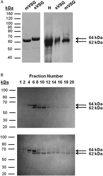
SDS–PAGE and immunoblotting showed that sVSG from the T. equiperdum TeAp-N/D1 strain was purified to homogeneity, and corresponded to a polypeptide band with an apparent molecular mass of 64 kDa (Fig. 1A). A yield of 10·2 mg of sVSG was obtained from ~109 trypanosomes. A 62 kDa polypeptide band corresponding to mVSG was obtained from freshly isolated T. equiperdum parasites by extraction of the acid-treated protein into chloroform/methanol, followed by selective repartition into aqueous salt solution (Fig. 1A). A yield of 2 mg of mVSG was acquired from ~1011 trypanosomes. Parasites were also homogenized in the presence of n-octyl-β-glucopyranoside and pCMBS. Following centrifugation, the resulting detergent soluble fraction was separated by ion-exchange chromatography on a Q-Sepharose column connected in tandem with an S-Sepharose column, and mVSG eluted in the non-adhering fraction. Analyses by SDS–PAGE and Western blot showed that both VSG forms, sVSG and mVSG, were acquired even in the presence of the GPI-PLC inhibitor (Fig. 1B), demonstrating that 10 mm pCMBS was not sufficient to completely inactivate the T. equiperdum GPI-PLC enzymatic activity. Nevertheless, mVSG was enriched in some of the fractions (Fig. 1B). Following concentration of the purified protein by freeze-drying, we obtained 5·5 mg of mVSG from ~1010 parasites.
Purification of T. equiperdum sVSG and mVSG. (A) sVSG was purified as previously described (Uzcanga et al. Reference Uzcanga, Perrone, Noda, Pérez-Pazos, Medina, Hoebeke and Bubis2004, Reference Uzcanga, Pérez-Rojas, Camargo, Izquier, Noda, Chacín, Parra, Ron, Rodríguez-Hidalgo and Bubis2016), and mVSG was purified from freshly isolated T. equiperdum parasites by organic solvent extraction. Aliquots of both purified proteins were separated by SDS–PAGE and stained with Coomassie Brilliant Blue (left), or electrotransferred to a nitrocellulose filter and detected using mouse anti-sVSG polyclonal antibodies (right). H = whole T. equiperdum homogenate. (B) Freshly prepared T. equiperdum parasites were homogenized in the presence of n-octyl-β-glucopyranoside and pCMBS, and the detergent-solubilized fraction was loaded onto a Q-Sepharose column connected in tandem with an S-Sepharose column. Fractions were acquired while the flow-through eluted from both columns, and analysed by SDS–PAGE using Coomassie blue staining (top), or by immunoblotting using mouse anti-sVSG polyclonal antibodies (bottom). Both sVSG and mVSG eluted in the non-adhering fraction.

Cardoso de Almeida and Turner (Reference Cardoso de Almeida and Turner1983) have demonstrated by SDS–PAGE that a molecular weight change is associated with the release of sVSGs from T. brucei. Similarly, the purified T. equiperdum sVSG possesses a higher apparent molecular mass than purified mVSG.
Identification of sVSG and mVSG by mass spectrometry
In order to identify the purified proteins, samples of sVSG and mVSGs were trypsinized in solution and the peptides were analysed by LC/ESI–MS/MS. Analysis of the tryptic peptides from sVSG and the mVSGs obtained by both methods retrieved the following peptides sequences: (i) KGDKLVTNILRN, (ii) KEIFDTPLDSRQ, (iii) KFKEIFDTPLDSRQ and (iv) KALTALATASERN. According to the examined databases, the obtained sequences yielded only one hit to a putative VSG from T. b. brucei TREU927 (Tb927.4.5460), which possesses a molecular mass of 54 331 Da based on the primary structure of the translated gene product.
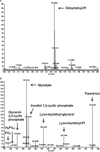
To confirm that both purified mVSGs from T. equiperdum possessed the GPI anchor, the GPI lipid composition was analysed by negative-ion ESI–MS and ESI–MS/MS following organic solvent extraction (Fig. 2A). An abundant [M–H]–ion (m/z 753·54) that corresponds to dimyristoyl-PI was clearly seen (Fig. 2A). To confirm assignment, the dimyristoyl-PI molecule was subjected to ESI–MS/MS analysis. Besides the parental ion at m/z 753·54, Fig. 2B illustrates the occurrence of signature [M–H]– product ions consistent with the presence of myristate (m/z 227·24), inositol 1,2-cyclic phosphate (m/z 241·05), glycerol-2,3-cyclic phosphate (m/z 153·03), lyso-myristoyl glycerol (m/z 363·25), lyso-myristoyl PI (m/z 625·32), H2PO4 – (m/z 97), and PO3 – (m/z 78·98). Analogous results were obtained for the mVSG sample purified by column chromatography (data not shown). Furthermore, before nitrous acid treatment, the sample was washed five times with butanol and water to eliminate PI and other phospholipid contaminants. The fifth wash was re-analysed by ESI–MS. No ion of m/z 753·54 was obtained in this case, demonstrating that PI was liberated from the mVSG sample and was not a contamination product (data not shown). Collectivelly, the lipid moiety of the T. equiperdum mVSG consists exclusively of dimyristoyl-PI as reported for T. brucei bloodstream-forms GPI anchors (Ferguson et al. Reference Ferguson, Homans, Dwek and Rademacher1988).
ESI–MS and ESI–CID MS/MS analyses of the phosphatidylinositol species released from the purified T. equiperdum mVSG. The lipid composition of the mVSG GPI anchor was analysed by ESI–MS and ESI–MS/MS following deamination and extraction with 9% butan-1-ol in water. Top, ESI–MS spectrum showing an ion peak for dimyristoyl-phosphatidylinositol (PI). Bottom, the dimyristoyl-PI molecule was fragmented and analysed by ESI–CID MS/MS. Key signature ions for myristate, inositol 1,2-cyclic phosphate, glycerol-2,3-cyclic phosphate, lyso-myristoyl glycerol, lyso-myristoyl PI, H2PO4 −, and PO3 − were obtained.

Production of polyclonal anti-sVSG and anti-mVSG antibodies
Purified T. equiperdum sVSG was employed to raise anti-sVSG polyclonal antibodies in mouse ascites. Western blot analyses show that these antibodies specifically recognized VSG from parasite whole-cell homogenates (Fig. 1A). As illustrated in Fig. 1A and B, both purified sVSG and mVSG were recognized by these ascitic fluids. Additionally, no recognition of either VSG in whole parasite extracts or purified sVSG or mVSG was obtained by anti-sVSG antibodies (dilution 1:5000) that were pre-incubated with ⩾5 µg of purified sVSG (data not shown). These results confirmed the specificity of the ascites anti-sVSG polyclonal antibodies.
Purified T. equiperdum sVSG and mVSG were also employed to raise anti-sVSG and anti-mVSG polyclonal antibodies in rabbit sera. Rabbit anti-sVSG and anti-mVSG polyclonal antibodies were purified by ammonium sulphate precipitation followed by ion-exchange chromatography throughout a DEAE-cellulose column. As expected, SDS–PAGE analysis showed two polypeptide bands of 55 and 25 kDa that corresponded to the heavy and light chains of the purified IgGs, respectively (data not shown). The generated antibodies were titrated by Western blot using either the whole-cell T. equiperdum lysate or the purified sVSG or mVSG, and polypeptide bands of 64 and 62 kDa, respectively, were specifically recognized even at the maximal dilution used (1:20 000) (data not shown).
Use of far-Western blotting to search for potential VSG-binding partners
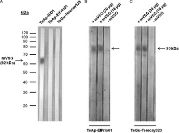
As illustrated in Fig. 3A, the generated mouse anti-sVSG antibodies were capable of recognizing the VSG from the T. equiperdum TeAp-N/D1 isolate, but not the VSGs from two different T. evansi isolates, TeAp-ElFrio01 and TeGu-Terecay323. Similar results were previously reported by Uzcanga et al. (Reference Uzcanga, Perrone, Noda, Pérez-Pazos, Medina, Hoebeke and Bubis2004). In order to identify VSG-interacting proteins, nitrocellulose membranes containing whole-cell homogenates of T. evansi TeAp-ElFrio01 and T. evansi TeGu-Terecay323 were incubated first with the purified mVSG from T. equiperdum and then with the mouse anti-sVSG antibodies. Control experiments were carried out by incubating directly with anti-sVSG antibodies in the absence of mVSG. As shown in Fig. 3 (panels B and C), a polypeptide band with an apparent molecular mass of about 80 kDa was detected only in the membranes that were pre-incubated with the purified mVSG.
Far-Western blot assays. (A) Western blot experiments using parasite crude extracts (25 µg of total protein) showed that mouse anti-sVSG polyclonal antibodies specifically recognized the VSG variant from the T. equiperdum TeAp-N/D1 isolate, but not the VSGs from the T. evansi TeAp-ElFrio01 or the T. evansi TeGu-Terecay323 isolates. Aliquots of whole-cell homogenates (25 µg of total protein) from the T. evansi TeAp-ElFrio01 isolate (B) or the T. evansi TeGu-Terecay323 isolate (C), were electrotransferred to nitrocellulose membranes and incubated in the absence or presence of various amounts of mVSG (16 or 30 µg). Blots were then incubated with specific mouse anti-sVSG polyclonal antibodies, and revealed using alkaline phosphatase-conjugated anti-mouse IgG as secondary antibodies. Arrows indicate the migration of mVSG and the apparent molecular mass of the polypeptide band that was detected.

Western blotting using anti-idiotypic antibodies to detect prospective VSG-interacting proteins
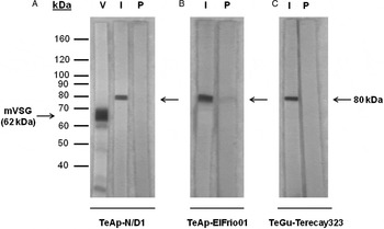
As illustrated in Fig. 4A by Western blot, the generated mouse anti-idiotypic antibodies recognized a band of ~80 kDa in T. equiperdum TeAp-N/D1, and in both T. evansi isolates, TeAp-ElFrio01 and TeGu-Terecay323 (Fig. 4B and C), supporting the results obtained by far-Western blot and suggesting again that the ~80-kDa polypeptide band probably contains VSG-interacting proteins. To discard unspecific reactions, we also immunized mice with purified IgG from rabbit pre-immune sera. As shown in Fig. 4, these control anti-idiotypic antibodies were not capable of recognizing proteins in crude extracts from T. equiperdum TeAp-N/D1 (panel A) or T. evansi TeGu-Terecay323 (panel C). However, a very faint band of ~80 kDa was detected on blots containing T. evansi TeAp-ElFrio01 homogenates that were incubated with mouse anti-idiotypic antibodies directed against rabbit pre-immune sera (panel B). This band was negligible when compared with the band obtained when the mouse anti-idiotypic antibodies directed against rabbit anti-mVSG polyclonal antibodies were employed.
Identification of potential VSG-interacting proteins by immunoblotting using anti-idiotypic antibodies generated in mice ascitic fluids. Aliquots of whole-cell homogenates (25 µg of total protein) from T. equiperdum TeAp-N/D1 (A) T. evansi TeAp-ElFrio01 (B) and T. evansi TeGu-Terecay323 (C) were separated by SDS–PAGE and electrotransferred to nitrocellulose membranes. The filters were incubated either with mouse anti-idiotypic antibodies directed against rabbit anti-mVSG polyclonal antibodies (I) or with mouse anti-idiotypic antibodies directed against rabbit pre-immune sera (P). In A, a nitrocellulose membrane containing the T. equiperdum TeAp-N/D1 crude extract was incubated with rabbit anti-mVSG polyclonal antibodies (V) as a control. Arrows indicate the migration of mVSG and the apparent molecular mass of the polypeptide band that was detected.

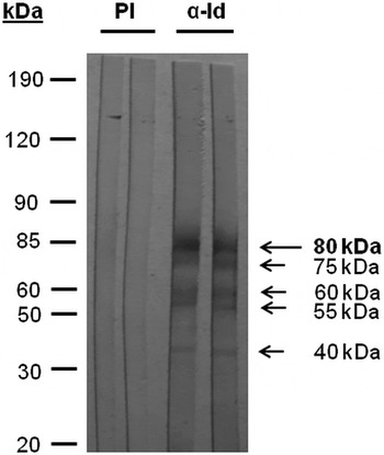
Anti-idiotypic antibodies were also prepared in rabbit sera by inoculating rabbit anti-mVSG antibodies. Similar to the results obtained with mouse anti-idiotypic antibodies, rabbit sera also detected a polypeptide band of ~80 kDa (Fig. 5). However, other minor bands of about 75, 60, 55 and 40 kDa were also recognized (Fig. 5). No recognition of polypeptides was seen when rabbit pre-immune sera were used as controls.
Identification of prospective VSG-binding protein partners by immunoblotting using anti-idiotypic antibodies raised in rabbit sera. Nitrocellulose membranes containing aliquots of the parasite crude extracts (25 µg of total protein) from T. equiperdum TeAp-N/D1 were incubated with anti-idiotypic antibodies generated in rabbits inoculated with rabbit anti-mVSG antibodies (α-Id). Control blots were incubated with pre-immune sera (PI). Arrows indicate the apparent molecular masses of the polypeptide bands that were detected.

Purification of VSG-interacting proteins by affinity chromatography
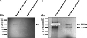
In order to generate matrixes capable of purifying VSG-interacting proteins by affinity column chromatography, either purified mouse anti-idiotypic antibodies or purified rabbit anti-idiotypic antibodies were coupled to CNBr-Sepharose activated resins. Freshly purified T. equiperdum parasites (~109) were homogenized and the corresponding soluble and particulate fractions were separated by centrifugation. Following application of the soluble and particulate parasite portions to each of the generated anti-idiotypic antibodies-affinity matrixes, the separated fractions were tracked by dot blot analyses using the corresponding anti-idiotypic antibodies. Then, the fractions that were immunologically recognized were pooled and subjected to SDS–PAGE. Essentially, the anti-idiotypic antibodies-binding proteins were found in the T. equiperdum soluble fraction (data not shown). After staining the gel with Coomassie blue, a polypeptide band with an apparent molecular mass of ~80 kDa (M2) was obtained when the resin coupled to mouse anti-idiotypic antibodies was employed as bait (Fig. 6A). Once this band was sliced, the gel was completely distained and restained with silver (Fig. 6B). As shown in Fig. 6B, an additional polypeptide band of ~55 kDa (M1) was obtained using the resin coupled to mouse anti-idiotypic antibodies. Bands of about 80 and 55 kDa (R2 and R1, respectively) were also obtained when the resin coupled to rabbit anti-idiotypic antibodies was employed (Fig. 6B). Since the ~55-kDa polypeptide band was not detected by far-Western blot (Fig. 3), no additional evidences were obtained indicating that this band contained proteins capable of interacting with VSG.
Purification of VSG-interacting proteins by immunoaffinity chromatography. Proteins purified by affinity chromatography through rabbit or mouse anti-idiotypic antibodies-Sepharose columns were separated by SDS–PAGE. The gel was initially stained with Coomassie Brilliant Blue (A) After slicing the Coomassie blue-coloured band, the gel was carefully destained and restained with silver (B) α-Id = anti-idiotypic antibodies. Arrows indicate the apparent molecular masses of the polypeptide bands that were detected. Bands obtained from the mouse α-Id-Sepharose and the rabbit α-Id-Sepharose were designed M and R, respectively.

Identification of VSG-interacting proteins by mass spectrometry
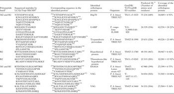
Gel slices containing the purified VSG-interacting proteins were trypsinized and the resulting peptides were analysed by LC/Q-Trap-MS/MS. Since the separation was carried out by one-dimensional SDS–gel electrophoresis, it is likely to obtain various proteins in each sliced band. Analysis of the peptides obtained from the gel bands retrieved a series of amino acid sequences that are summarized in Table 1. Unfortunately, proteomic analysis showed that both M1 and M2 were contaminated with human keratin and serum proteins. We believe that these contaminating proteins, mostly keratin, probably masked the sequencing of the purified candidates for VSG-interacting proteins, thus hindering their identification. In contrast, we obtained clearer results from R1 and R2. Therefore, in the specific cases of M1 and M2, we only reported those sequences that matched with the findings obtained after sequencing R1 and R2.
Identification of potential candidates for VSG-interacting proteins in T. equiperdum

a Polypeptide bands obtained from the mouse anti-idiotypic antibodies-Sepharose and the rabbit anti-idiotypic antibodies-Sepharose (Fig. 6) were designed M and R, respectively.
b Peptide sequences are reported using one-letter codes for the amino acid residues. M* = methionine sulfoxide produced by oxidation.
c Residues are numbered according to the primary structure of the translated gene products stored in TriTrypDB (http://tritrypdb.org). All sequenced peptides from the T. equiperdum potential VSG-binding partners gave an exact 100% match with the corresponding amino acid sequences of the orthologous proteins.
d Mr, Molecular weight of the identified orthologous protein.
According to the examined databases, the obtained sequences from M2 and R2 yielded a hit with a Hsp70 from T. b. brucei TREU927 (Tb11.v5·1035). The molecular weight of the T. brucei Hsp70 was predicted to be 75 234 on the basis of the primary structure of the translated gene product (Table 1). This size is consistent with the migration of M2 and R2 on SDS–PAGE (~80 kDa).
R2 also retrieved peptide sequences that yielded a hit with a sequence highly homologous to Trypanosoma congolense GARP. GARP is a highly glycosylated GPI-anchored surface glycoprotein expressed in the epimastigote stage of T. congolense (Thomson et al. Reference Thomson, Lamont, Mehlert, Barry and Ferguson2002) that is homologous to T. brucei alanine-rich protein (BARP) (Urwyler et al. Reference Urwyler, Studer, Renggli and Roditi2007). Although the predicted size for T. congolense GARP is 26 539 on the basis of its amino acid sequence (Table 1), it is known that GARP migrates on gels as a heterogeneous series of bands with a much higher apparent molecular mass due in part to the attachment to complex GPI anchors (Bayne et al. Reference Bayne, Kilbride, Lainson, Tetley and Barry1993; Beecroft et al. Reference Beecroft, Roditi and Pearson1993). Hence, it is not unreasonable to find a GARP-like T. equiperdum orthologous protein that migrates with an apparent molecular mass of ~80 kDa. However, the expression of GARP in T. congolense coincides with the loss and gain of VSG molecules in the tsetse vector. Since Urwyler et al. (Reference Urwyler, Studer, Renggli and Roditi2007) have shown that T. brucei BARPs could not be detected in the parasite bloodstream states and T. equiperdum only occurs as bloodstream forms, it is unusual for T. equiperdum to express GARP-like factors that are, to date, considered to be only expressed in the tsetse fly.
Another protein contained in R2 appeared to be the putative T. equiperdum tryparedoxin peroxidase, on the basis of peptide sequences that yielded a hit with the tryparedoxin peroxidase from T. b. brucei TREU927 (Tb927.8.1990, predicted molecular weight = 25 631) (Table 1). In addition, various peptides acquired in R2 yielded a hit with a hypothetical protein from T. b. brucei TREU927 (Tb 927.5.1780, predicted molecular weight = 48 191), and another hypothetical protein from T. b. brucei TREU927 (Tb11.v5·0265, predicted molecular weight = 22 315) that corresponds to a putative thioredoxin-like protein (Table 1). Since these proteins migrate with an apparent molecular mass of ~80 kDa, which is significantly higher than predicted from their amino acid sequences, it is plausible that these proteins are larger in T. equiperdum and/or undergo complex and extensive covalent modfications that cause a substantial increase on their sizes.
Analysis of the tryptic peptides from R1 retrieved a series of amino acid sequences that yielded a hit with a VSG from T. b. brucei TREU927 (Tb927.11.17120) that has a predicted molecular weight of 54 054 based on its primary structure (Table 1). R1 also retrieved various peptides that yielded a hit with another VSG from T. b. brucei TREU927 (Tb927.4.5460) (Table 1), which represents the predominant VSG from the T. equiperdum TeAp-N/D1 isolate (Camargo et al. Reference Camargo, Izquier, Uzcanga, Perrone, Acosta-Serrano, Carrasquel, Arias, Escalona, Cardozo and Bubis2015), and possesses a predicted molecular mass of 54 331 Da. Both VSG variants are expected to migrate at about the mass of the band in which they were found (~55 kDa).
Another protein contained in R1 appeared to be the ESAG6 subunit of the parasite transferrin receptor complex (Steverding et al. Reference Steverding, Stierhof, Chaudhri, Ligtenberg, Schell, Beck-Sickinger and Overath1994), as seen by three peptides that yielded a hit with the ESAG6 protein from T. b. brucei Lister strain 427 (Tb427.BES134·3, predicted molecular weight = 43 980) (Table 1). Interestingly, the peptides that yielded a hit with the T. brucei ESAG6 were also retrieved from M1 (Table 1). In T. brucei, ESAG6 is a heterogeneously glycosylated GPI-anchored protein of 50–60 kDa (Schell et al. Reference Schell, Evers, Preis, Ziegelbauer, Kiefer, Lottspeich, Cornelissen and Overath1991). As other GPI-anchored molecules, the T. equiperdum ESAG6 protein probably undergoes extensive N-glycosylation, and as such, it is expected to migrate at about the mass of R1 and M1 (~55 kDa).
As shown in Table 1, all the peptides that were sequenced by LC/Q-Trap-MS/MS were 100% identical to the corresponding hits in each of the orthologous target proteins, strenghtening the conclusion that these proteins are candidates for VSG-interacting partners in T. equiperdum. The extent of coverage with the proposed orthologous proteins ranged between 4·93% and 24·22% (Table 1).
All potential VSG protein partners were categorized by Gene Ontology (GO) terms according to the biological processes in which they might participate. The identified proteins belong to the following GO classes: (i) gene products involved in protein folding such as Hsp70; (ii) proteins involved in antigenic variation such as VSG variants; (iii) proteins involved in evasion or tolerance of the host immune response such as the ESAG6 subunit of the transferrin receptor complex; (iv) proteins involved in oxidation–reduction reactions such as tryparedoxin peroxidase; and (v) proteins with no function yet reported such as the GARP-like protein and the two hypothetical proteins that were also identified.
DISCUSSION
We purified here the T. equiperdum sVSG and mVSG forms. The presence of the GPI anchor on the purified mVSG was clearly established by analysing the GPI lipid composition by ESI–MS and ESI–MS/MS. Like the T. brucei bloodstream-forms GPI anchors, the lipid moiety of the T. equiperdum mVSG consisted exclusively of dimyristoyl-PI.
Affinity chromatography separations of the T. equiperdum soluble fraction using anti-idiotypic antibodies-Sepharose yielded polypeptide bands of approximately 80 and 55 kDa by SDS–PAGE. Gel slices containing the isolated polypeptide bands were trypsinized, and the resulting peptides were analysed by LC/Q-Trap-MS/MS. On the basis of the retrieved sequences, a series of potential VSG protein partners were acknowledged that included Hsp70, a GARP-like protein, VSG variants, the transferrin receptor ESAG6 subunit, tryparedoxin peroxidase, and two hypothetical proteins. Some of the identified partners such as the GARP-like protein, the VSG variants and ESAG6 represent GPI-anchored glycoproteins, which must colocalize with VSG in the surface glycocalyx of T. equiperdum (Acosta-Serrano et al. Reference Acosta-Serrano, Hutchinson, Nakayasu, Almeida, Carrington, Barry, McCulloch, Mottram and Acosta-Serrano2007). The release of these GPI-modified proteins into the parasite soluble fraction is certainly caused by the parasite GPI-PLC, which is responsible of cleaving their GPI anchor. Although our observation that VSG is capable of interacting with a GARP-like protein in T. equiperdum is appealing, it is rather unusual because Urwyler et al. (Reference Urwyler, Studer, Renggli and Roditi2007) reported that T. brucei BARPs could not be detected in the parasite bloodstream states. Thus, the process in which the interaction between VSG and the homologous T. equiperdum GARP-like protein takes place remains elusive.
Hsp70 appears to be the most prominent candidate likely to be a true in vivo VSG-interacting partner. Members of this family of proteins play key housekeeping roles in protein folding, synthesis, assembly, transportation across biological membranes and degradation. Since Hsp70 proteins are expressed in subcellular compartments, their function during stress conditions is regularly confined to the intracellular environment (Mayer and Bukau, Reference Mayer and Bukau2005). Consistently, Hsp70 has been found alongside other trypanosome endoplasmic reticulum and cytosolic proteins. Given that VSG is the most abundant protein in bloodstream forms of Salivarian trypanosomes, Hsp70 is probably required to interact with VSG during its synthesis and folding in order to correctly generate the closely packed protective coat that covers the parasite. However, in higher eukaryotes Hsp70 proteins can also be found in the outer leaflet of the plasma membrane where they appear to act as an alert stress signal (De Maio and Vazquez, Reference De Maio and Vazquez2013). Although there is no evidence suggesting that Hsp70 is localized on the surface of African trypanosomes, it is feasible that T. equiperdum Hsp70 may also be found extracellularly merely by analogy with higher eukaryotes. In that case, Hsp70 might interact with the trypanosome GPI-anchored VSG.
Finding that a particular T. equiperdum VSG is capable of interacting with other molecules of the same or a different VSG variant is not uncommon, considering that VSGs from Salivarian trypanosomes comprise a family of proteins holding a homodimeric quaternary structure that adopts similar shapes. The trypanosome transferrin receptor, which is a VSG-related protein, is a heterodimeric or higher order heteroligomeric complex that is encoded by the ESAG6 and ESAG7 genes (Steverding et al. Reference Steverding, Stierhof, Chaudhri, Ligtenberg, Schell, Beck-Sickinger and Overath1994). Structurally, the heterodimeric receptor protein resembles a classical VSG dimer, and its subunits ESAG6 and ESAG7 fold like the N-terminal domain of the VSG. Since homodimeric and heterodimeric quaternary structures have been reported for VSGs and VSG-related gene products, VSG variants and ESAG6 are protein candidates that might really interact with VSG in vivo. In T. brucei, ESAG6 and 7 are restricted to the flagellar pocket and are not widely distributed on the parasite surface like the VSG molecules. Since the density of the VSG coat in the flagellar pocket is similar to that on the rest of the plasma membrane (Grünfelder et al. Reference Grünfelder, Engstler, Weise, Schwarz, Stierhof, Boshart and Overath2002), it is plausible to postulate that only VSGs localized in or near the T. equiperdum flagellar pocket are capable of interacting wih ESAG6.
Trypanosoma brucei encodes three nearly identical genes for cysteine containing glutathione peroxidase-type tryparedoxin peroxidases (Hillebrand et al. Reference Hillebrand, Schmidt and Krauth-Siegel2003), which are reported to occur in the cytosol and the mitochondrion of the parasites (Schlecker et al. Reference Schlecker, Schmidt, Dirdjaja, Voncken, Clayton and Krauth-Siegel2005). Recently, various Hsp70 variants were identified as T. cruzi mitochondrial tryparedoxin peroxidases partners under both physiological and oxidative stress conditions (Peloso et al. Reference Peloso, Dias, Queiroz, Leme, Pereira, Carnielli, Werneck, Sousa, Ricart and Gadelha2016). Thus, it is interesting to find that both tryparedoxin peroxidase and Hsp70 are potential candidates for VSG-interacting partners in T. equiperdum. Moreover, a hypothetical protein that in T. b. brucei correspond to a putative thioredoxin-like protein was also identified as a prospective VSG protein partner in T. equiperdum.
It is important to acknowledge that the interactions reported here may not be related to the cell surface. In T. brucei, Grünfelder et al. (Reference Grünfelder, Engstler, Weise, Schwarz, Stierhof, Boshart and Overath2002) have quantified the steady-state distribution of VSG and estimated that ~89% is located on the cell surface and ~11% is intracellular. About 75% of the intracellular VSG is predominantly located in the endosomal compartment, while the rest is associated with the endoplasmic reticulum, Golgi apparatus and lysosomes, suggesting sorting between several membrane compartments (Grünfelder et al. Reference Grünfelder, Engstler, Weise, Schwarz, Stierhof, Boshart and Overath2002). Since the localization of ESAG6 is thought to be limited to the flagellar pocket, which is the only site for endocytosis and exocytosis in these organisms, and Hsp70 and enzymes of thiol metabolism are reported to be cytosolic, our findings may reflect interactions during trafficking of the VSG molecules to the surface or during recycling via the flagellar pocket. Moreover, the findings reported here are highly preliminary given that the proposed interactions have not been further tested. More experiments using, for example, specific antibodies against the putative VSG-interacting partners, need to be performed in order to validate our results. Likewise, we cannot state unequivocally that the potential VSG-binding proteins identified here are genuine partners that interact with VSG in live cells under physiological conditions, because some of the proposed interaction may perhaps be artifacts. For instance, Hsp70 and tryparedoxin peroxidase are abundant parasite proteins and the binding to the anti-idiotypic antibodies may simply reflect non-specific binding to proteins, especially for the case of Hsp70 that binds to misfolded proteins. The recognition of the various GPI-anchored proteins by the anti-idiotypic antibodies might also be merely a cross-reaction. Nonetheless, we demonstrated here that anti-idiotypic antibodies can be used as tools to recognize, localize and purify potential VSG-interacting proteins from T. equiperdum, and to the best of our knowledge, this seems to be the first report that recognizes plausible protein–protein interactions with a trypanosome VSG.
ACKNOWLEDGEMENTS
We want to thank Antonio Gutiérrez (Instituto de Biología Experimental, Universidad Central de Venezuela, Caracas, Venezuela) for kindly donating the p-chloromercuribenzenesulfonic acid (pCMBS) that was used in this work. We also thank Mike Ferguson and Douglas Lamont (University of Dundee, Scotland) for generous use of the Q-Star XL.
FINANCIAL SUPPORT
This research was supported by Grant no. 2009000681 from FONACIT, Caracas, Venezuela.
ETHICAL STANDARDS
We declare that all the experiments in this paper were carried out in accordance with the legal and ethical standards of Venezuela.
CONFLICT OF INTERESTS
The authors declare that there is no conflict of interests regarding the publication of this paper.