Melatonin is a hormone that is secreted primarily by the pineal gland in response to darkness. As such, melatonin levels rise and fall throughout the night and day, respectively. The circadian synthesis and release of melatonin is modulated by the suprachiasmatic nucleus of the hypothalamus (the ‘master biological clock’) (Refs Reference Reiter1, Reference Pandi-Perumal2). The functions of melatonin in the body are extensive, but its role in entraining circadian rhythms to the light–dark cycle is the most well known. People have used melatonin for many years to reset the body's internal clock during travel and to reduce sleep-onset latency in sleep disorders.
Melatonin is a complicated hormone to study because of its chronobiotic nature and its ability to produce diverse responses in the body through both receptor-dependent and receptor-independent actions. Complicating things further is the fact that melatonin receptor density and sensitivity fluctuate throughout the 24 h cycle in both a diurnal and a circadian manner (Refs Reference Witt-Enderby3, Reference Poirel4, Reference Gauer5, Reference Gauer6). Therefore, for receptor-dependent effects, a melatonin dose given at a time when melatonin receptor expression and sensitivity are lowest may show no effect compared with this same dose given when melatonin receptor expression and sensitivity are highest, which may occur at a different time of the day.
Besides melatonin and its receptors, many other bodily functions oscillate in response to their own endogenous pacemaker and/or in response to the light–dark cycle, including gene expression (Refs Reference Ostrowska7, Reference Bailey8, Reference Bailey9). In more recent work, certain pathological states such as tumour activity have been found to be influenced by the light–dark cycle via melatonin (Ref. Reference Blask, Sauer and Dauchy10) or via the clock genes (Ref. Reference Rutter, Reick and McKnight11).
Over the past 20 years, there has been an explosion of research redefining the role of melatonin in the body, especially in the area of cancer. Melatonin ‘disruption’ by exposure to light at night has been hypothesised to play a role in breast cancer (Refs Reference Ravindra, Lakshmi and Ahuja12, Reference Jasser, Blask and Brainard13, Reference Reiter14). Similar effects on the pattern and levels of melatonin production have also been observed in patients with sleep disorders, jetlag, depression, stress and immunological disorders, and it has been hypothesised that such disruption in the circadian rhythm may be associated with genomic instability and subsequent cancer susceptibility (Ref. Reference Shadan15). Melatonin supplementation has been successful at restoring sleep quality and circadian rhythmicity in the elderly (Refs Reference Valtonen16, Reference Gubin17), suggesting that such supplementation could also ameliorate the effects of melatonin disruption, including increased cancer incidence, in younger subjects with genetic-, disease- or lifestyle-induced low levels of melatonin secretion. Melatonin may be effective as a chemopreventative agent if it inhibits the initial growth or subsequent progression of a tumour (Ref. Reference Marks, Furstenberger and Muller-Decker18).
This article does not repeat the summary of in vitro studies of melatonin as reviewed by Panzer and Viljoen (Ref. Reference Panzer and Viljoen19). Rather, this review focuses on the potential of melatonin as a chemopreventative agent or chemotherapeutic for breast cancer. In recent years, many studies have examined the mechanisms underlying the response of cells to melatonin, and many support a protective role in breast cancer. It is important to ascertain whether melatonin shows similar protection against breast cancer in the clinic, controlling for the variables that may influence melatonin's efficacy. Thus, this review also highlights the issues or variables that one must consider when conducting melatonin research on breast cancer to assess its efficacy as a cancer-protective agent.
Implication of melatonin as an anticancer agent
Cellular and molecular actions
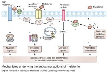
Melatonin acts to counteract tumour formation by reducing cell proliferation (Refs Reference Vijayalaxmi20, Reference Cos and Sanchez-Barcelo21, Reference Cos and Sanchez-Barcelo22, Reference Lenoir23, Reference Blask24). In addition, it has antimetastatic properties in cultured MCF-7 cells (a breast-cancer-derived cell line) (Ref. Reference Cos and Sanchez-Barcelo22), which may be due to a reduced chemotactic response and increased expression of cell-surface adhesion molecules (Ref. Reference Cos and Sanchez-Barcelo22). Physiological levels of melatonin have been shown to reduce the invasion of MCF-7 cells, and counteract the increase in oestrogen-induced invasiveness (Ref. Reference Cos and Blask25). Melatonin downregulates oestrogen (estrogen) receptor (ER) levels in breast cancer cells and blocks ER binding to DNA and transactivation functions (Refs Reference Baturin26, Reference Rato27, Reference Kiefer28, Reference Sanchez-Barcelo29, Reference del Rio30). In addition, the induction of differentiation by melatonin has been demonstrated in numerous studies (Refs Reference Satomura31, Reference Sanchez-Hidalgo32, Reference Schuster33, Reference Johnston34, Reference Radio, Doctor and Witt-Enderby35, Reference Sainz36, Reference Bellon37, Reference Moriya38, Reference Jimenez-Jorge39). Highly differentiated cells have a diminished proliferative potential (Ref. Reference Radio, Doctor and Witt-Enderby35). The myriad of intracellular mechanisms underlying the actions of melatonin are discussed in the following sections and summarised in Figure 1.
Mechanisms underlying the anticancer actions of melatonin. Melatonin can act through membrane-bound G-protein-coupled receptors (MT1, MT2), or in a receptor-independent manner. (a) Melatonin binding to its cell-surface receptors can activate Gi proteins (Giαβγ) to cause dissociation into Giα-GTP and Giβγ. Increases in Giα-GTP or Giβγ can affect the mitogen-activated protein kinase (MAPK) pathway. (b) In certain cell types, Giβγ may act via matrix metalloproteinases (MPs) to liberate heparin-bound EGF to activate EGFRs in an autocrine loop (Ref. Reference Radio, Doctor and Witt-Enderby35). Subsequent activation of the downstream effectors MEKs and ERKs may drive cell proliferation or differentiation (hence dashed arrow) through nuclear events. (c) Also, activation of Gi may result in the internalisation of melatonin receptors and binding of β-arrestin-2 (probably to phosphorylated melatonin receptors) to activate MEKs and ERKs to target cytosolic proteins leading to differentiation. (d) Microtubules (purple rectangle) can directly bind to MEK/ERK and modulate their activity and translocation patterns in a cell. (e) Besides effects on the MAPK pathway, activated Gi leads to the inhibition of adenylate cyclase (AC) via Giα-GTP to decrease cAMP levels within the cell. Decreases in cAMP leads to decreases in the transcription of genes responsive to oestrogen receptors (ERs). Gi can also be activated directly by microtubules to increase Giα-GTP through α–β tubulin heterodimers (not shown). (f) Melatonin can also act independently of melatonin receptors to drive cellular events, as it can easily traverse membranes. (g) It can inhibit the cytosolic protein calmodulin, which, in turn, can decrease adenylate cyclases sensitive to calmodulin (types I, III and VIII). Decreases in adenylate cyclase activity reduce cAMP levels to attenuate the transcription of ER-responsive genes. (h) Melatonin can also inhibit telomerase and aromatase to reduce cellular proliferation, and (i) can scavenge free radicals, thereby reducing oxidative damage. Other abbreviations: EGF, epidermal growth factor; EGFR, epidermal growth factor receptor; ERK, extracellular-signal-regulated kinase (also known as MAPK1); MEK, MAPK/ERK kinase (also known as MAP2K1).

The multiple modes of action of melatonin (antiproliferative, antimetastatic and prodifferentiation) within the cell make it an excellent candidate hormone for the prevention of cancer in those individuals at risk or for the treatment of cancer in combination with other conventional therapies.
Receptor-dependent actions of melatonin
Melatonin can act through G-protein-coupled receptors – MT1 (encoded by MTNR1A) and MT2 (encoded by MTNR1B) – located at the plasma membrane (Ref. Reference Witt-Enderby40). One pathway for melatonin-receptor-induced cellular differentiation may be via the mitogen-activated protein kinase (MAPK) cascade, by transactivation of epidermal growth factor receptors (EGFRs) (Ref. Reference Radio, Doctor and Witt-Enderby35) or through the formation of β-arrestin scaffolds (Ref. Reference Bondi41) (Fig. 1). In addition to MAPK signalling, melatonin receptors can activate the G protein Gi, leading to inhibition of adenylate cyclase and hence the cAMP-dependent signalling pathway (Refs Reference Jarzynka42, Reference Witt-Enderby and Dubocovich43, Reference Brydon44) (Fig. 1). Reduced cAMP leads to decreased ER binding to oestrogen-response elements of genes (Refs Reference Rato27, Reference Kiefer28, Reference Sanchez-Barcelo29, Reference del Rio30, Reference Witt-Enderby40) such as cyclin D1, through its actions on JUN and ATF2 (transcription factors known to bind to minimal oestrogen-sensitive cyclin D1 promoter elements) (Ref. Reference Cini45). In general, an attenuation of ER-mediated signalling in a cell should show protection against ER-positive breast cancers, similar to that occurring in response to the ER blockers tamoxifen, raloxifene and fulvestrant.
Recently, microtubules have been implicated in melatonin receptor signalling through a direct effect on Gi activation and also through receptor trafficking and sequestration of MAPK signalling components (Refs Reference Bondi41, Reference Jarzynka42).
Receptor-independent actions of melatonin
Melatonin can also act independently of melatonin receptors to drive cell differentiation events, reduce tumour cell proliferation and reduce invasive properties. Melatonin can easily traverse membranes, and can bind to cytosolic proteins such as calmodulin (Ref. Reference Dai46), which in turn can decrease adenylate cyclases sensitive to calmodulin (AC types I, III and VIII). As indicated above, reductions in adenylate cyclase activity and cAMP levels within a cell can attenuate the transcription of ER- and AP-1-induced genes (Refs Reference Sanchez-Barcelo29, Reference Martinez-Campa47) and thus decrease oestrogen-regulated factors (Ref. Reference Cini45). The action of melatonin on such genes appears to be through ERα and not ERβ (Refs Reference del Rio30, Reference Martinez-Campa47). Besides modulating ER-responsive genes, melatonin can act to reduce oestrogen biosynthesis by decreasing the release of gonadotropins (Ref. Reference Roy48) or in breast cancer cells via its inhibitory actions on aromatases (Refs Reference Cos49, Reference Cos50).
Melatonin also has several other cellular effects that may be protective against breast cancer. It reduces oxidative damage through its direct free-radical-scavenging properties (Ref. Reference Reiter51), its actions on quinone reductases (Refs Reference Tan52, Reference Nosjean53, Reference Nosjean54), and its induction of antioxidant enzymes (Ref. Reference Dziegiel, Podhorska-Okolow and Zabel55). In addition, it modulates the immune system (Refs Reference Pawlikowski, Winczyk and Karasek56, Reference Moss57, Reference Lissoni58, Reference Carrillo-Vico59) and is implicated in tumour surveillance (Ref. Reference Cos and Sanchez-Barcelo22). Melatonin can also decrease telomerase activity in the tumours of nude mice by decreasing mRNA expression of essential telomerase subunits (Ref. Reference Leon-Blanco60).
Animal model studies
Multiple studies that manipulate circulating melatonin levels – through melatonin administration, changes in light–dark cycles, or pinealectomy – demonstrate that melatonin has antitumour activities. Melatonin reduces the incidence of cancer and the time of developing mammary cancer (latency) in carcinogen-induced and spontaneous mammary cancer animal models (Refs Reference Vijayalaxmi20, Reference Cos and Sanchez-Barcelo21, Reference Cos and Sanchez-Barcelo22, Reference Lenoir23, Reference Blask24). Melatonin-deficient (pinealectomised) rats appear to suffer increased oxidative damage over their lifetime (Ref. Reference Reiter61), whereas life-long melatonin administration decreases DNA damage in the brain (Ref. Reference Morioka, Okatani and Wakatsuki62). Long-term melatonin supplementation increases lifespan in animal model systems (mice and fruit flies) (Refs Reference Anisimov63, Reference Vinogradova64) (although it should be borne in mind that the biology of ageing may be specific to the lifespan of each organism; Ref. Reference Braeckman, Demetrius and Vanfleteren65), and reduces the spontaneous incidence of tumours in mice (Refs Reference Anisimov63, Reference Vinogradova64) perhaps by a reduction in oxidative damage.
Light-induced effects on melatonin and breast cancer
Light exposure at night (e.g. night-shift work) may possess carcinogenic potential including that for breast cancer (Refs Reference Reiter14, Reference Bartsch and Bartsch66, Reference Davis and Mirick67). Since light inhibits the synthesis and secretion of melatonin from the pineal gland (Refs Reference Reiter1, Reference Pandi-Perumal2), and given the anticancer properties of melatonin discussed above, the diminished production of melatonin may explain the increase in breast cancer risk. In an elegant set of experiments, it was shown in rat hepatoma and human breast xenografts that linoleic acid uptake, tumour proliferation, cAMP levels and activity of MEKs/ERKs (downstream effectors of the MAPK pathway) are reduced when these xenografts are bathed in blood taken from human subjects at night (melatonin-rich) when compared with blood taken during the night with interrupted light exposure (low melatonin levels) (Ref. Reference Blask24). Furthermore, these effects were shown to be mediated through membrane melatonin receptors.
Even though light-induced decreases in melatonin production probably play very significant roles in increasing breast cancer risk, light effects on clock genes located in the breast tissue may also be involved. Light may directly affect tumour growth through the key clock genes PER1- and PER2, which may regulate cell-cycle- and apoptosis-related genes (Refs Reference Moriya68, Reference Gery69). PER2 inhibits the proliferation of several cancers that are epithelial in nature, including breast, prostate and lung (Ref. Reference Gery69), and its expression is downregulated in several human B-ALL and lymphoma cell lines. Thus PER inactivation, perhaps via phosphorylation by casein kinase 1ɛ (Ref. Reference Gery69) or by the methylation of PER gene promoters (Ref. Reference Chen70) might induce the development of these B-cell leukaemias and lymphomas (Ref. Reference Gery69) or breast cancer (Ref. Reference Chen70) in humans.
Melatonin and human breast cancer
Melatonin as a biomarker of breast cancer
Risk
Biomarkers of cancer risk are usually indicative of two conditions: individuals with an inherent genetic susceptibility to a disease, and individuals who have sustained exposures associated with the initiation or promotion of the disease process. There are at least three reasons why a deficiency of melatonin could be used as a biomarker in public health screening to identify individuals who would benefit from increased surveillance and/or behavioural or chemical disease prevention strategies. First, other than the rather specific group of early-onset ‘childhood’ cancers, age is by far the most significant and biologically important risk factor in human cancer (Ref. Reference Migliore and Coppede71); since melatonin production declines with age, constitutive or premature deficit of melatonin production could thus be indicative of a physiological ageing process that also impacts upon cancer incidence. Second, given that melatonin has been found to be a powerful antioxidant (both in vitro and in vivo) (Ref. Reference Nogues72) and oxidation of DNA is a known mechanism for the production of mutations that play a fundamental role in carcinogenesis (Ref. Reference Loft and Poulsen73), lower melatonin levels could allow for a constitutive increase in radical-induced mutagenesis or render a cell hypersensitive to physiological or induced oxidative stress. Third, as indicated above, melatonin secretion can be downmodulated by sleep deprivation, nightshift work or other exposure to light at night (Ref. Reference Schernhammer74), and several types of cancer, particularly breast cancer, seem to occur at higher incidence in such populations (Ref. Reference Megdal75).
Several retrospective studies have demonstrated significantly reduced melatonin levels in patients with breast cancer (Refs Reference Davis and Mirick67, Reference Schernhammer74, Reference Schernhammer and Hankinson76). However, no such association was observed in a large prospective study (Ref. Reference Travis77). Also, low plasma melatonin levels were not associated with breast cancer risk in high-risk individuals (Ref. Reference Blask, Yu and Reiter78), perhaps due to melatonin's antioxidant properties. Circulating melatonin levels were actually increased almost two-fold in smokers relative to controls (Ref. Reference Tarquini79), perhaps as a mechanism of detoxification.
One way to rationalise the observation of decreased melatonin production in cancer patients with no such association in high-risk patients and patients in the prospective cancer study is to hypothesise that the presence of a tumour can affect melatonin levels (Ref. Reference Bartsch and Bartsch66). The regulation of the circadian rhythm of melatonin production has been reported to be affected in patients with breast, prostate, colorectal or uterine cancer, with the magnitude of the decrease in nocturnal melatonin levels correlated with the size and proliferation index of the tumour (Refs Reference Bartsch80, Reference Bartsch81, Reference Blask, Shafii and Shafii82). Tumours are known to affect both local and systemic regulatory mechanisms, especially with regard to growth stimulation and immune function (Ref. Reference Kusmartsev and Gabrilovich83), and this may be dependent upon tumour type (epithelial verus sarcoma) and ER status (Ref. Reference Bartsch and Bartsch66). Inflammation is often associated with tumour growth (Refs Reference Kawanishi and Hiraku84, Reference Chen85), which could stimulate the production of melatonin as an antioxidant. Therefore, size, type and grade of tumour may influence melatonin levels (increase or decrease) and, as such, caution should be used when using melatonin as a biomarker of cancer, although most studies support a role for melatonin deficiencies in increasing cancer.
Prognosis
Differences in melatonin production, particularly the magnitude of the nocturnal peak in the serum or the accumulation in breast cyst fluids (Ref. Reference Burch86) in cancer patients may differentiate between different aspects of the disease, thus affecting treatment or ultimate outcome. Low levels of melatonin are associated with poor prognosis in breast cancer, based on measures of tumour aggression (proliferating cell nuclear antigen) (Ref. Reference Bartsch81), differentiation (nuclear grade) (Ref. Reference Maestroni and Conti87) and progression (loss of ER or progesterone receptor) (Ref. Reference Blask, Shafii and Shafii82). Such parameters are potentially useful in staging a tumour and selecting a course of treatment, as they can provide information without the need for tumour biopsy.
Melatonin as a breast cancer chemopreventative agent
Melatonin may be effective as a chemopreventative agent if it inhibits the growth or progression of a premalignant lesion, inhibits the growth of the initial transformed cell into a palpable tumour, or inhibits the progression of the initial tumour into a more aggressive (e.g. metastatic) form (Ref. Reference Marks, Furstenberger and Muller-Decker18). Given the evidence suggesting various anticancer effects of melatonin, melatonin supplementation may thus act as a chemopreventative treatment (Ref. Reference Poeggeler88). Melatonin administration has little toxicity in vivo, so any beneficial effects come with low risk. A particular effect of melatonin that impacts on both aging and carcinogenesis is its antioxidative property (Ref. Reference Poeggeler89), which should act to reduce the mutagenesis and therefore carcinogenesis of environmental genotoxic exposures (Ref. Reference Brozmanova, Dudas and Henriques90). Indeed, melatonin is protective against the genotoxic effects of ionising radiation, which has been associated with almost every type of cancer (Ref. Reference Becr VII Phase 291), as well as chemical mimics of the oxidative effects of radiation, such as those in tobacco smoke (Ref. Reference Vesnushkin92). Because melatonin supplementation has been successful at restoring sleep quality and circadian rhythmicity in the elderly (Refs Reference Valtonen16, Reference Gubin17), such supplementation could also ameliorate the effects of melatonin disruption (through genetics, disease or lifestyles) in younger individuals.
Melatonin as a cancer therapeutic agent
Based on its potential oncostatic effects, melatonin has also long been considered as a possible cancer chemotherapeutic agent, most often in combination with established genotoxic or immunological regimens. Human data come mainly from a large series of studies by Lissoni and colleagues (Ref. Reference Lissoni93) that used melatonin on relatively small numbers of patients, with many different types of cancer. Based on these studies (Ref. Reference Lissoni93), there are several grounds for invoking melatonin in cancer treatment, but most have been poorly investigated. For example, melatonin supplementation seems to promote general well-being in cancer patients (Ref. Reference Norsa and Martino94), which might allow them to tolerate higher doses of toxic compounds or be more likely to complete standardised regimens of such compounds. Also, melatonin seems to be effective at preventing or reducing treatment-associated hypotension (Ref. Reference Lissoni93), thrombocytopaenia (Refs Reference Lissoni95, Reference Lissoni96) and cachexia (Ref. Reference Lissoni97); however, it is not clear whether the greater tolerance for treatment elicited by melatonin administration has been exploited for greater overall treatment in any study.
In endocrine-dependent tumours, particularly breast cancer, the anti-oestrogenic effects of melatonin may interfere with hormone-induced cell proliferation, susceptibility to carcinogenesis and tumour progression (Ref. Reference Cos98). Such tumour cells often overexpress hormone receptors (Ref. Reference Treilleux99), express altered ratios of receptors (Ref. Reference Cunat, Hoffmann and Pujol100), overexpress receptor accessory proteins (Ref. Reference Naderi101) or are hormone-dependent for survival, making them hyper-responsive to modulation of exogenous hormone levels. This differentiates them from normal hormone-responsive cells and provides the basis of a therapeutic index for melatonin treatment. The need for such a therapeutic index restricts discussion of other potential mechanisms of melatonin efficacy in chemotherapy. For example, the antioxidative effect of melatonin may well protect normal cells and the entire organism from the systemic effects of chemotherapy (Ref. Reference Tiligada102), but, since most of the in vitro work on melatonin has been performed only on cancer cells, and not concurrently on normal cells, there is no reason to believe that, in general, such protective effects will not be conferred equally on tumour cells.
Human trials of melatonin as a cancer chemotherapy agent, particularly those of Lissoni et al., have been reviewed in detail (Ref. Reference Panzer and Viljoen19). It is significant that when attempting a comprehensive review and meta-analysis of studies examining melatonin as a chemotherapeutic agent, Mills et al. (Ref. Reference Mills103) were able to find only ten reports, all from the Lissoni group, that analysed melatonin definitively alone or in combination with standard therapies. All studies were performed on advanced cancer patients, were unblinded and did not include placebos. These reports included: three studies on lung cancer (one alone and two in conjunction with genotoxic chemotherapy, including cisplatin); one study each on breast cancer, renal cell cancer, glioblastoma and malignant melanoma; two studies of mixed populations of patients with either advanced or metastatic solid tumours; and one study specifically of brain metastases from assorted solid tumours. The average number of subjects in these studies was 75. The meta-analysis concludes that melatonin significantly reduced the risk of death of these patients at one year.
Expert opinion on the status of melatonin on breast cancer
Melatonin and cancer treatment
In a systematic review of randomised controlled trials of melatonin in solid-tumour cancer patients and on one-year survival, Mills et al. (Ref. Reference Mills103) have shown that there is a positive benefit from the use of melatonin. These trials were all performed by the same group in the same hospital and are thus without independent confirmation from any other group; the results, although intriguing, are therefore difficult to assess. In addition, one-year survival is very short term in the context of many cancers such as breast (with a mean recurrence time of seven years) and a much longer trial would need to be conducted. The most significant finding is that no adverse outcomes were reported from the use of melatonin. There seems to be very limited toxicity of this chemical in the context of what can be very toxic exposures during chemotherapy. This characteristic would make larger clinical trials of melatonin feasible.
Although it is a proven antioxidant, it is not clear that melatonin supplementation would have a significant protective effect in disease-free individuals, or a selective protective effect in cancer patients undergoing radio- or chemotherapy (Ref. Reference Vijayalaxmi104). Melatonin supplementation has proven efficacy in mitigating the effects of shift work or sleep deprivation. Although the connection between disruption of the circadian rhythm and carcinogenesis has not been established mechanistically, studies suggest that it is probably the result of low circulating levels of melatonin. This chronobiotic hormone needs to be given at appropriate times of the day to achieve maximum efficacy on treating or preventing breast cancer. The possibility that its connection with regulation of the circadian rhythm would result in different effects of melatonin when administered at different times during the day has generally reduced the enthusiasm to develop it as a clinical treatment, and, if true, has also probably contributed to the ambiguity in clinical data.
Probably the most important property of melatonin administration in humans, and the one that may eventually allow it to proceed to treatment, is its lack of toxicity. Thus, melatonin may be added to large clinical trials with a theoretical possibility of its improving the efficacy of treatment and with little concern that it will interfere with the action of the primary agent (genotoxic or immunological). Although recent studies by the Lissoni group have involved larger experimental populations (Ref. Reference Lissoni105), ultimately, clinically beneficial effects of melatonin will be demonstrated only through its addition to large multicentre studies targeting specific tumour types in conjunction with specific types of therapy.
Use of MCF-7 cells as a cancer cell model
It is worth adding a comment on the widespread use of the established breast-cancer-derived cell line MCF-7. This cell line represents the product of a stage IV breast tumour metastasised to the pleural sack around the lung of a patient who eventually died of breast cancer. The cell line has also been in culture for over 20 years and has an amplification of the ERα gene of approximately 140 times the normal copy number (Ref. Reference Jonsson, Staaf, Olssen, Heidenblad, Vallon-Christersson, Osoegawa, de Jong, Oredsson, Ringr, Hoglund and Borg106). The putative oncostatic/antiproliferative properties of melatonin have been demonstrated primarily in these MCF-7 cells, as opposed to other cell lines from other tumour types (Refs Reference Crespo107, Reference Hill and Blask108). The theory that melatonin may exert its effects through its effect on ER may be affected by the fact that this cell line overexpresses the ERα gene. However, there are discrepancies within the MCF-7 melatonin literature, which may be explained by the chromosomal instability of MCF-7 (Refs Reference Wenger109, Reference Klein110).
Use of other in vivo model systems to study melatonin and cancer
Fewer studies using melatonin have utilised xenograft models or animal models of tumour growth as opposed to in vitro studies. These in vivo studies must be performed because the in vitro studies cannot deal with the possibility that tumours themselves may have circadian rhythms and may manifest peak times for treatment regimens. Tumours can also alter local and systemic regulatory systems. In addition, in vitro systems cannot simulate the complex interactions between host responses to the melatonin (natural and exogenous) and to the tumour itself.
Modification of the design of future clinical trials
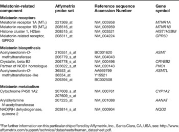
If melatonin levels in urine or serum are indicative of cancer risk or prognosis, they would constitute a relatively simple and noninvasive method of population biomonitoring. However, based on the preliminary studies summarised above, it is likely that melatonin measurements would be more useful as part of a larger screening strategy that included a number of markers (Ref. Reference Chatterjee and Zetter111). One way to develop such a screening panel would be through high-content, high-throughput analyses of tumours themselves (Ref. Reference Wang112). For example, probes to measure the expression levels of several genes related to melatonin biosynthesis, metabolism and signal transduction are present in commercially available chips (Table 1), and the data from such experiments are required to be deposited in open on-line databases upon publication. Thus, using such modern techniques, screening experiments targeting other markers concurrently provide data on the relevance of melatonin-related genes in cancer. Even if not ‘hypothesis-driven’, analysis of such studies is usually confined to only the few genes showing the greatest differences in expression, and such markers may not prove to be the most consistent and reproducible. However, even if changes in melatonin-related gene expression are directly and causally implicated in carcinogenesis, they themselves may not provide the best biomarker of the process. Similarly, measurements of melatonin itself and its metabolites are present in proteomic screens of blood and urine from cancer patients (Ref. Reference Stemke-Hale113), but here it may be more difficult to identify melatonin-related fragments unless they are specifically detected immunologically or by sequencing and taken at peak times of melatonin secretion.
Melatonin-related probes on the Affymetrix U133 + 2 gene chipa

a For further information on this particular chip offered by Affymetrix, Inc., Santa Clara, CA, USA, see: http://www.affymetrix.com/support/technical/datasheets/human_datasheet.pdf.
Future considerations for using melatonin as a therapy for breast cancer
More fundamental research in the area of melatonin is imperative. It still needs to be determined what constitutes a physiological dose of melatonin, as discussed in Ref. Reference Reiter and Tan114. Melatonin activates its membrane receptors and its lipophilic nature also allows it to traverse plasma membranes and accumulate within the cell. Even though circulating concentrations of melatonin at night range between 50 and 100 pg/ml (Ref. Reference Reiter1), nanomolar to micromolar concentrations of melatonin may be reached within or near the cell dependent upon the concentrating abilities of the tissue. Melatonin shows efficacy as an oncostatic agent using concentrations that range between high picomolar to low micromolar. Whether these levels are reached physiologically in tissue still needs to be ascertained, but for now these levels should be the therapeutic targets when designing future studies. More fundamental research with respect to determining the signalling mechanisms modulated by melatonin in tumours needs to be pursued, considering that the efficacy of melatonin to retard or prevent breast cancer is variable and may depend on the type of cancer (Ref. Reference Bartsch and Bartsch66). Additionally, other models, both cellular and in vivo, should be used to verify the mechanisms underlying melatonin action as it relates to cellular differentiation.
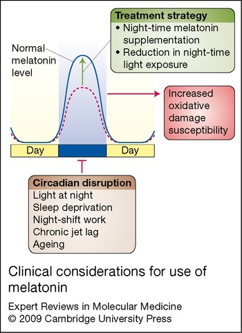
More translational research is warranted seeing that the World Health Organization (WHO) and International Agency for Research on Cancer (IARC) classify night-shift work as a potential carcinogen. Elucidating how light at night affects breast cancer risk, and whether or not melatonin supplementation at night shows any efficacy to prevent and/or treat breast cancer, should be given top priority when conducting clinical trials to direct future research. The clinical trials should be expanded to include a more heterogeneous population of individuals from around the world and be hypothesis driven. Factors like dosage and timing of dose need to be ascertained and genomic endpoints determined with respect to the use of melatonin as a treatment for breast cancer (Fig. 2; Table 1). The findings that melatonin inhibits the oestrogen-dependent signalling cascades should lead to more studies in this area (both basic and clinical) to determine if melatonin shows a synergistic interaction with mainstay therapies using tamoxifen, raloxifene or fulvestrant (Refs Reference Sanchez-Barcelo29, Reference Witt-Enderby40, Reference Aust115). The outcome(s) of these endeavours could lead to the novel use of melatonin in the clinic for the prevention and/or treatment of breast cancer. The results of such studies could benefit many.
Clinical considerations for use of melatonin. As shown in this schematic, factors that disrupt the circadian rhythm (e.g. light at night, sleep deprivation, shift work, chronic jet lag and ageing) may increase susceptibility to oxidative damage through a suppression of the nocturnal melatonin surge (dotted line). The goal of therapy, then, would be to restore one's nocturnal melatonin levels back to normal (solid line) by supplementing with melatonin at night (dosage still needs to be determined) and by reducing night-time light exposure.

Acknowledgements and funding
The authors thank the peer reviewers for their thoughtful insight and expert comments and suggestions.