Introduction
Ensign or hatchet wasps (family Evaniidae) are solitary predators on cockroach eggs in oothecae, and are easily diagnosed by a uniquely ensign- or hatchet-shaped metasoma with a tubular petiole and a shortened, laterally compressed metasoma. The fauna comprises ~650 extant species in 22 extant and 13 fossil genera (Deans, Reference Deans2005; Deans et al., Reference Deans, Yoder and Dole2013; Engel et al., Reference Engel, Huang., Alqarni and Cao2016). This relatively rich record of fossil taxa compared with some other parasitoid Hymenoptera is undoubtedly due to their searching for host cockroaches on tree trunks, and thus an increased chance of being trapped in tree resin.
The genus Parevania Kieffer, Reference Kieffer1907 is known from 26 extant species and one subspecies from the Old World (Deans and Huben, Reference Deans and Huben2003; Deans et al., Reference Deans, Yoder and Dole2013), and three fossil species, P. brevis (Brues, Reference Brues1933), P. producta (Brues, Reference Brues1933), and P. remanea (Brues, Reference Brues1933) (Nel et al., Reference Nel, Waller, Hodebert and De Ploëg2002; Deans, Reference Deans2005). Although Bradley (Reference Bradley1908) treated Parevania as a subgenus of Zeuxevania Kieffer, Reference Kieffer1903, Deans and Huben (Reference Deans and Huben2003) suggested that the genus is distinct but similar to Evaniella Bradley, Reference Bradley1905. However, Deans et al. (Reference Deans, Gillespie and Yoder2006) found that Parevania is not monophyletic, and suggested it should be synonymized with Zeuxevania and Papatuka Deans, Reference Deans2002 although this has not been formalized.
Twenty-eight evaniid species have been described from Mesozoic (Early to Late Cretaceous) or Tertiary ambers (Eocene to Miocene) and seven species from non-amber deposits, mostly Early to Late Cretaceous (see Jennings et al., Reference Jennings, Krogmann and Mew2012, Reference Jennings, Krogmann and Mew2013a, Reference Jennings, Krogmann and Priyab; Peréz-de la Fuente et al., Reference Peréz-de la Fuente, Peñalver and Ortega-Blanco2012; Engel et al., Reference Engel, Huang., Alqarni and Cao2016). Five evaniid species are known from Eocene Baltic amber: Evaniella eocenica (Sawoniewicz and Kupryjanowicz, Reference Sawoniewicz and Kupryjanowicz2003), Hyptia hennigi Jennings, Krogmann, and Priya, Reference Jennings, Krogmann and Priya2013b, and three species of Parevania; P. brevis, P. producta, and P. remanea (Brues, Reference Brues1933; Nel et al., Reference Nel, Waller, Hodebert and De Ploëg2002; Deans, Reference Deans2005). Unfortunately the holotypes of the latter three species were deposited in Gdansk and destroyed during World War II (Nel et al., Reference Nel, Waller, Hodebert and De Ploëg2002).
Recently, a large quantity of Eocene Baltic amber with hymenopteran inclusions was obtained by the State Museum of Natural History, Stuttgart. Examination of this material has revealed a new species of Parevania that has the most highly modified eyes of any known member of the order, and possibly of all insects, in that the eye surface has a prominent acute ridge running across and dividing the eye into two distinct regions. Here we describe this new species, present a brief analysis of the optical consequences of the eye-surface discontinuity, and discuss the likely function of the eye ridge compared with the types of compound eye modifications that occur in other Hymenoptera and other insects.
Materials and methods
Imaging and terminology
The amber piece containing the fossil specimen, which had previously been polished, was immersed in 50% glycerol, and images taken using a Visionary Digital BK plus imaging system with a Canon EOS 7D 18 megapixel camera. Images were produced using Zerene Stacker, Zerene Systems LLC, software and were cropped and resized in Photoshop CS6. Anatomical terminology follows the Hymenoptera Anatomy Consortium (http://glossary.hymao.org); wing venation terminology follows Deans and Huben (Reference Deans and Huben2003).
Optical calculations
Ommatidial facet diameters were estimated from the inter-facet center spacing, with facet centers identified in images that had been pre-processed by high-pass filtering with the Photoshop CS6 unsharp-mask filter, and then segmented using custom software written in Matlab R2015a (Mathworks).
Repository and institutional abbreviation
The holotype specimen in polished amber (figured here) is deposited in the State Museum of Natural History, Stuttgart (SMNS).
Systematic paleontology
Class Insecta Linnaeus, Reference Linnaeus1758
Order Hymenoptera Fabricius, Reference Fabricius1804
Family Evaniidae Latreille, Reference Latreille1802
Genus Parevania Kieffer, Reference Kieffer1907
Type species
Parevania semirufa Kieffer, Reference Kieffer1907, by monotypy.
Synonyms
See Deans (Reference Deans2005) for a complete taxonomic history.
Diagnosis
Eyes large; antennae with 11 flagellomeres; notauli present; fore wing with seven complete cells; fore wing vein 1RS attached to Sc+R basal to pterostigma, curved slightly towards wing base; M+CU often as long as hind jugal lobe; distance between mid- and hind-coxae 0.6 x or less than distance between fore- and mid-coxae; hind tarsomeres not elongated into spines.
Parevania oculiseparata Jennings, Krogmann, and Austin new species
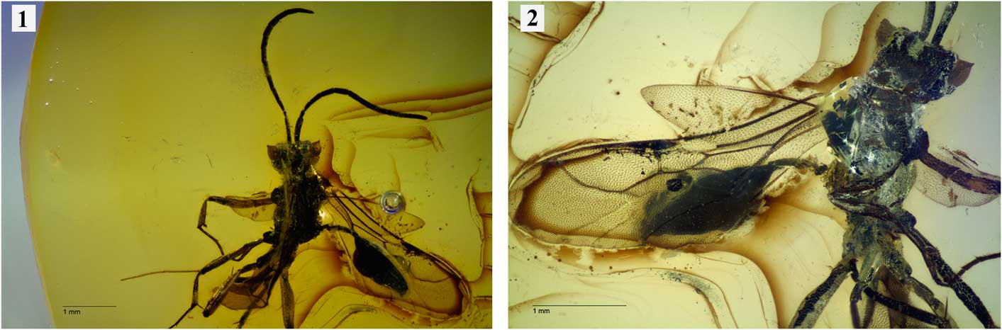
Parevania oculiseparata Jennings, Krogmann, and Austin n. sp., holotype ♂ (SMNS BB-2494). (1) Dorsal habitus; (2) ventral habitus. Scale lines=1 mm.

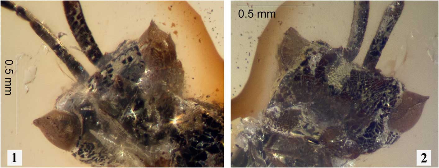
Parevania oculiseparata Jennings, Krogmann, and Austin n. sp., holotype ♂ (SMNS BB-2494). (1) Dorsal head; (2) ventral head. Scale lines as indicated.

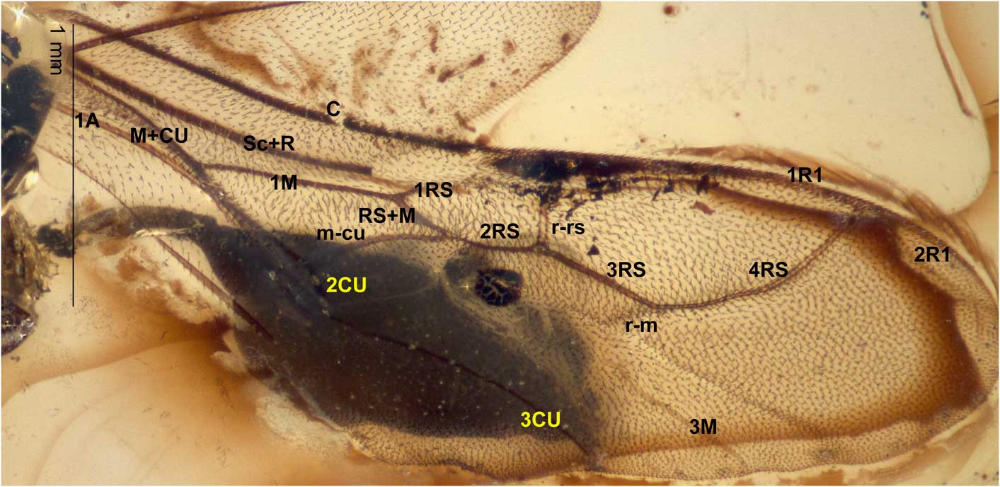
Parevania oculiseparata Jennings, Krogmann, and Austin n. sp., holotype ♂ (SMNS BB-2494), fore wing. Abbreviations for wing veins: A=Anal, C=Costa, CU=Cubitus, M=Media, R=Radius, RS=Radial Sector, Sc=Subcosta, m-cu=medial-cubital cross-vein, r-m=radial-medial cross-vein, r-rs=radial-radial sector cross-vein. Scale line as indicated.

Holotype
Male, specimen within a polished piece of amber. Deposited in SMNS (registration SMNS BB-2494).
Occurrence
An extremely well-preserved specimen as a single inclusion within Eocene Baltic amber, 56–34 Ma, according to Szwedo and Sontag (Reference Szwedo and Sontag2009). Purchased from the collection of Jürgen Velten, Idstein, Germany, by the State Museum of Natural History, Stuttgart.
Diagnosis
The new species can be diagnosed as a member of the genus Parevania based primarily on the putative synapomorphy for the genus, fore wing vein 1RS being attached to Sc+R basal to the pterostigma (Fig. 3). Parevania oculiseparata n. sp. differs from all extant species, and from the descriptions of the three (lost) Baltic amber fossil species of Brues (Reference Brues1933), in that the compound eye has two angled surfaces that gives the appearance of having a prominent acute ridge (Fig. 2).
Description (male)
Relatively small compared with most other members of the genus; total body length 4.3 mm; head length 0.59 mm, mesosoma 1.50 mm, metasoma including petiole 2.29 mm. Body and metasoma apparently dark brown.
Head width 1.07 mm; vertex of head 0.5×head width; compound eye height 0.47 mm; eye with prominent acute ridge extending across the entire dorso-ventral elongation of the eye surface (Fig. 2); shortest distance between eyes 0.73 mm; distance of lateral ocellus to compound eye 0.17 mm; occipital carina obscured; malar space 0.29 mm; antennae inserted slightly above mid-line of eye, distance between toruli 0.85 mm; distance from torulus to eye 0.24 mm; antenna with 11 flagellomeres; scape 3.97×as long as pedicel; first flagellomere 0.21 mm in length; all flagellomeres longer than wide.
Mesosoma with surface of mesoscutum and mesoscutellum rugose-punctate; notauli apparently absent (obscured); propodeum rugose-punctate; legs elongate; hind leg slightly more robust; hind tibia slightly widened apically; distance between mid- and hind-coxa 0.25×distance between fore- and mid-coxae; pretarsal claws short, slender, simple, arolium present.
Fore wing venation relatively complete (Fig. 3); first marginal cell elongate; second submarginal cell separated from first discal cell; 1RS attached to Sc+R basal to pterostigma, slightly curved; r-rs straight and arising near middle of pterostigma (Fig. 3); pterostigma narrow, ~4×as long as wide; 3M and 2CU+3CU nebulous, extending to wing margin; 4RS curved, obtuse; r-m apparently nebulous; first discal cell ~1.8×as long as wide; hind wing with 5 hamuli.
Metasomal petiole thin, subcylindrical, 0.48×length of remaining metasoma (Fig. 1); metasoma smooth, elliptical.
Description (female)
Unknown.
Etymology
The species epithet refers to the acute ridge of the compound eyes having two separate faces or surfaces.
Eye structure of Parevania oculiseparata
There seems little doubt that the acute ridge of the compound eye is a real morphological structure and not just a distortion artefact resulting from shrinkage during fossilization. We base this on: (1) the uniformity of the ridge between the eyes (i.e., they are exactly the same shape and of the same dimensions); (2) the clearly undistorted structure of the rest of the body; and (3) the relatively normal appearance of compound eyes in all other species preserved in a similar manner. This begs the question as to the functional role of this ridge.
As in most Hymenoptera, the compound eyes are vertically elongated in P. oculiseparata n. sp. They have a diameter of ~300 µm at the equator (defined with respect to the presumed normal orientation of the head in living evaniids) and a dorso-ventral extent of around double this. The eye morphology is highly unusual, given the prominent acute ridge extending across the entire dorso-ventral elongation of the eye, being most prominent at the equator (Figs. 1, 2). Ommatidial facets are relatively uniform in diameter across the eye (10–13 µm), including along this acute ridge. At the equator of the eye, the ommatidia immediately medial to the acute ridge are directed forwards, with ~12–14 facet rows between the ridge and the frontal margin of the eye. Along this dimension, the ommatidial orientation changes only gradually. Ommatidia immediately lateral to the acute ridge are directed predominantly sideways, with approximately 25 facet rows, again curving only gradually towards the posterior margin of the eye where they are directed rearwards.
Discussion
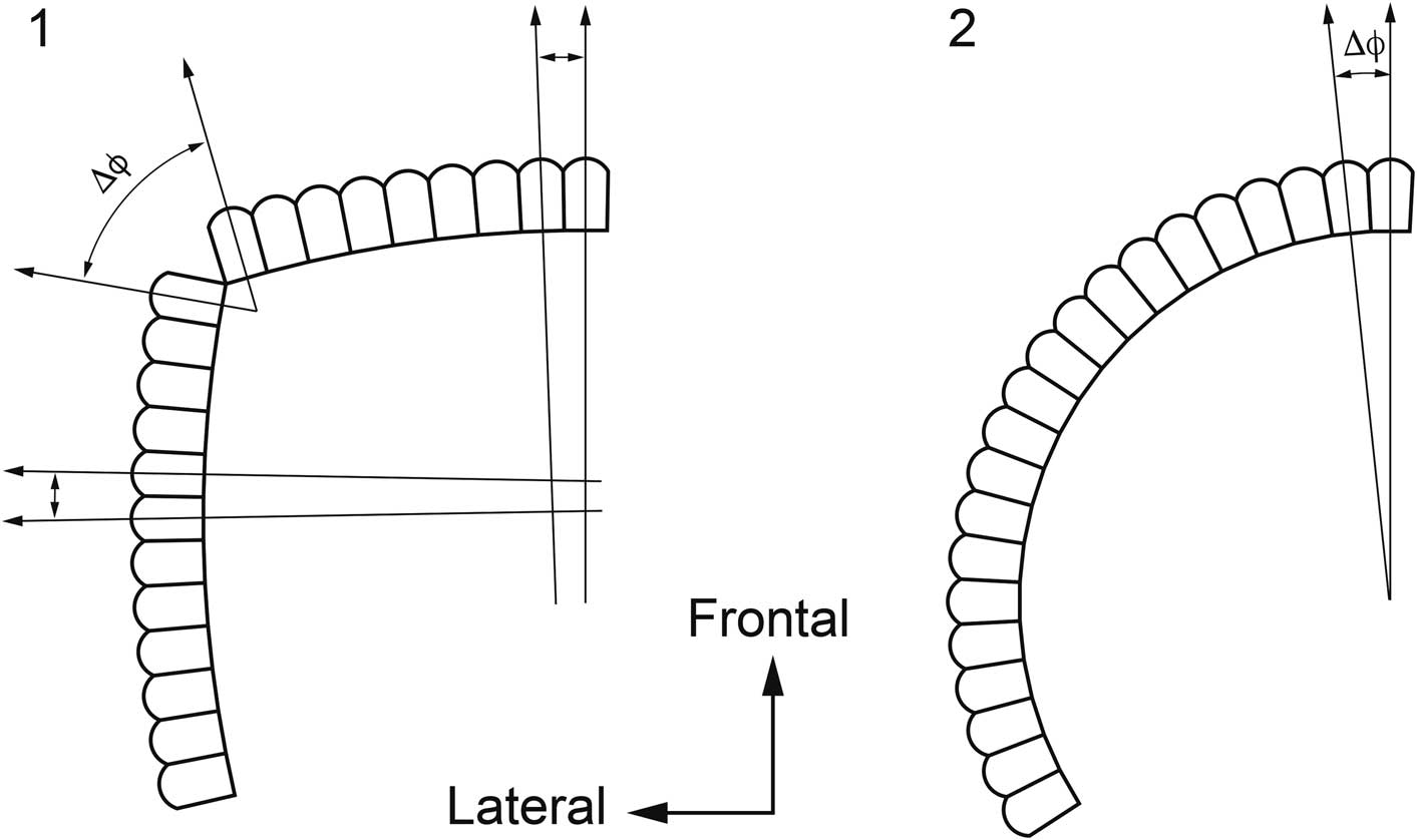
The acute ridge in P. oculiseparata n. sp. clearly divides the eye into two regions, each with a locally large radius of curvature (in the horizontal dimension). Local radius of curvature in apposition compound eyes is generally correlated with the angle subtended by adjacent ommatidial axes (the inter-ommatidial angle, ∆ϕ), which itself limits the sampling resolution of the eye (Land, Reference Land1981). Although we cannot be sure that the photoreceptors under each facet lens are exactly aligned with their ommatidial axis (it is common for some distortion in this relationship, particularly at the margins of the eye), this arrangement nevertheless suggests that the horizontal inter-ommatidial angles in the frontal and lateral field of view would be smaller than those of ommatidia along the crest of the acute ridge (Fig. 4.1). Although this would lead to a region of low resolution in the transition zone between the frontal and lateral eye fields, it would permit potentially higher resolution than would normally be expected from a normal eye with the same total horizontal (equatorial) diameter (Fig. 4.2).
Optical consequences of a vertical discontinuity in apposition eye morphology. (1) Cross-sectional view through a row of ommatidia in an eye as observed in Parevania oculiseparata Jennings, Krogmann, and Austin n. sp. The angular separation of adjacent ommatidia (∆ϕ, inversely proportional to resolution) is large in the transition between the frontal and lateral eye fields, but small within these subregions; (2) a similar cross-section for a similar-sized eye with identical facets, but a uniform local radius of curvature. ∆ϕ is constant across the eye but is always 3×larger than in the frontal and regions in (1).

Eye modifications in other Hymenoptera
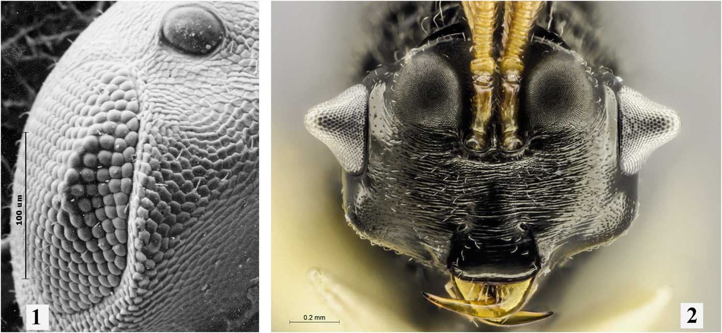
Tubercules and eye ridges are known to occur on the compound eyes of other Hymenoptera, but they are extremely rare within the order. A small ridge is present on the eyes of both sexes of two platygastrid species, Inostemma oculare Austin, Reference Austin1984 (Fig. 5.1) and I. apsyllae Austin, Reference Austin1984 from India (Austin, Reference Austin1984), which are parasitoids of cecidomyiids (gall midges). However, in these species the eye surface is curved normally and does not have opposing surfaces, and the ridge is only 2–3 ommatidia wide and does not extend across the full width of the eye (Fig. 5.1). The only other hymenopterans known to have modified eyes are the three species that comprise the eucharitid genus Isomerala Shipp, Reference Shipp1894, from Central and South America (Heraty, Reference Heraty2005) which, like all members of the family, are ectoparasitoids of ant larvae in their nests. In this case the species have a large, cone-shaped tubercle in the center of the eye, which is approximately one-third the height of the eye in diameter at its base (Heraty, Reference Heraty2005; Fig. 5.2). This structure must significantly distort the visual field of these species and it is difficult to speculate on its function. In these species there is a huge variation in facet diameter from the base to the tip of the eye cone, around a doubling in facet diameter to ~23 µm at the tip of the cone. While not unusually large in absolute terms, these are indeed large facets for such a small total eye diameter. Similar sized lenses in the miniature dipteran predator Coenosia attenuata Stein, Reference Stein1903 support resolution well below 3°, so it is conceivable that these large facets are associated with a small region of high acuity (Gonzalez-Bellido et al., Reference Gonzalez-Bellido, Wardill and Juusola2011). That being the case, it is surprising that the largest facets are in this case found at a position that is almost the inverse of what one would expect, corresponding to a small radius of curvature (on the cone). Also perplexing is that the smaller facets in the transition area from the cone to the margin of the eye have a large radius of curvature. Normally in an acute zone the larger radius of curvature would be associated with larger facets. However, some caution is required in interpreting this arrangement because only the radius of curvature can be used to infer the direction that individual ommatidia view. This cannot take into account that, in many insect species, the underlying photoreceptors are displaced. Indeed, the bizarre position of a possible ‘acute zone’ is supported by a large pseudopupil visible from the frontal view of both eyes in Figure 5.2. In the cone-shaped eye structure of these species, it seems unlikely that this is not a region where the inter-ommatidial angle would be very large, yet the point-source resolution of individual facets of 23 µm is potentially quite high, thus being a somewhat paradoxical design representing a likely extreme form of what has been considered to be optical under-sampling. However, extreme under-sampling is seen in other taxa, including some so-called ‘bright-zone’ eyes of Diptera (van Hateren et al., Reference van Hateren, Hardie, Rudolph, Laughlin and Stavenga1989) and in the peripheral eyes of jumping spiders, where it appears to be an interesting adaptation to detect small contrasting features (Land, Reference Land1985). Whether this is the case for Isomerala is unknown given little if anything is known about its biology.
Hymenoptera with modified eyes. (1) Inostemma oculare Austin (Platygastridae) paratype ♀, from India, dorso-lateral view showing small horizontal ledge in the upper quadrant of the eye. Scale line=100 µm. (2) Isomerala azteca Girault, Reference Girault1920 (Eucharitidae) ♀, from central America, anterior head showing cone-like structure of the eye. Scale line=200 µm.

Eye modifications in other insects
Among other extant insects, the flower-mimicking hymenopodid mantids have strikingly modified eyes (Wipfler et al., Reference Wipfler, Wieland, Decarlo and Hörnschemeyer2012; O’Hanlon and Norma-Rashid, Reference O’Hanlon and Norma-Rashid2013). In these species, however, the vertical tubercular extension of the dorsal eye is a chitinous structure that lacks functional ommatidia, rather than part of the eye per se. This eye modification is therefore not an adaptation that changes or enhances visual acuity, but rather is more likely associated with behavioral recognition among conspecifics or associated with procrypsis.
A similar functional division as in P. oculiseparata n. sp. between the upper (dorsal) and lower (ventral) eye is seen in a number of extant bibionid flies (Zeil, Reference Zeil1983), usually with some discontinuity in the transition zone between these eye regions. However, a distinct and abrupt division between the frontal and lateral eye fields, as in P. oculiseparata n. sp., is not present in these flies.
A more abrupt division between the frontal and lateral eye fields (although still not ridge-like) is exhibited by some extant hypercephalic drosophilid flies, such as Zygothrica dispar Wiedemann, Reference Wiedemann1830, Z. latipanops Grimaldi, Reference Grimaldi1987, and Z. exuberans Wheeler, Reference Wheeler1968 (see Grimaldi and Fenster, Reference Grimaldi and Fenster1989, fig. 4), resulting in eye shapes that strongly resemble the condition found in P. oculiseparata n. sp. Interestingly, the eye modifications are only present in male Zygothrica Wiedemann, Reference Wiedemann1830. Unfortunately, the female of P. oculiseparata n. sp. is so far unknown, so we do not know whether the eye is sexually dimorphic in this fossil species. However, there is a very important difference between the eye modification in male Zygothrica and P. oculiseparata n. sp., which could be indicative of different biological functions. In these Zygothrica species the eyes are pushed antero-laterad due to the expansion of the dorsal portion of the fronto-orbital plate and the occipital sclerite (Grimaldi and Fenster, Reference Grimaldi and Fenster1989), while the eyes are in the normal position in P. oculiseparata n. sp. Thus, while the eyes of these drosophilids are very similar to P. oculiseparata n. sp., and may provide a similar optical solution, they may also result from completely different selective pressures since there is no sign of hypercephaly in P. oculiseparata n. sp., which seems to be associated with sexual dimorphism in drosophilids and aggressive behavior among male flies. Extremely aggressive behavior among males has been observed in various parasitoid wasp species (e.g., Abe et al., Reference Abe, Kamimura, Kondo and Shimada2003), but to our knowledge has not been reported for any members of the superfamily Evanioidea.
Local regions of higher frontal and lateral acuity are seen in the eyes of some dragonflies (Horridge, Reference Horridge1978), but with a relatively smooth transition (and accompanying functional vision) in the transition between these eye regions. Because of wave optical limitations to resolution (Stavenga, Reference Stavenga2003), compound eyes do not simply scale geometrically as facet diameter is reduced. In practice, many small to tiny insect species have facet diameters in the range 10–14 µm, as seen in P. oculiseparata n. sp. Instead, as the demands of a smaller body plan require smaller eyes, ommatidial facet number appears to be sacrificed (Fischer et al., Reference Fischer, Meyer-Rochow and Müller2013), and the resulting smaller radius of curvature leads to reduction in the overall resolution of the eye, if the spherical geometry of the eye is maintained. It is possible that in P. oculiseparata n. sp. the aspheric organization of the shelf-eye represents an alternative compromise between the conflicting demands imposed by miniaturization (and thus a small eye) and the need to preserve moderate acuity vision (requiring a large radius of curvature) in the frontal and lateral eye fields, at the cost of a loss of functional spatial vision in the intermediate region viewed by the ridge.
Acknowledgments
We wish to thank G. Bechly (State Museum of Natural History Stuttgart) for providing access to the amber specimen, J. Heraty and R. Burks for providing high-resolution images of Isomerala, and E. Fagan-Jeffries for formatting the plates. We also thank the School of Biological Sciences, The University of Adelaide for a Summer Scholarship awarded to Priya through the Faculty of Sciences. Figure 5 is reproduced with permission of the Bulletin of Entomological Research.