Introduction
Betta splendens, widely cultivated as an ornamental fish, is a very important species for fish farming. The betta fish has an auxiliary breathing organ known as the labyrinth, which allows them to breathe atmospheric oxygen and tolerate low levels of dissolved oxygen in the water (Damazio, Reference Damazio1992; Faria et al., Reference Faria, Crepaldi, Teixeira, Ribeiro, Souza, Carvalho, Melo and Saliba2006). Certain features of B. splendens, such as colour, fin length and temperament, have been selected by breeders for centuries for ornamental purposes and fighting. Therefore, in the wild, bettas are less aggressive with less colouring and shorter fins (Monvises et al., Reference Monvises, Nuangsaeng, Sriwattanarothai and Panijpan2009).
Among the several Betta species, the best known is Betta splendens (Faria et al., Reference Faria, Crepaldi, Teixeira, Ribeiro, Souza, Carvalho, Melo and Saliba2006; Monvises et al., Reference Monvises, Nuangsaeng, Sriwattanarothai and Panijpan2009). However, despite its commercial importance, little information is known about its ontogeny and it is during the early developmental stages, such as the onset of exogenous feeding, that high mortality rates are observed (Yúfera & Darias, Reference Yúfera and Darias2007). Therefore, study of the early developmental stages is necessary to establish good production methods (Maciel et al., Reference Maciel, Lanna, Junior, Donzele, Neves and Menin2010).
The embryonic developmental stage lasts from fertilization of the oocyte by the sperm until hatching of the larvae (Matkovic et al., Reference Matkovic, Cussac, Cukier, Guerrero and Maggese1985; Solnica-Krezel, Reference Solnica-Krezel2005; da Rocha Perini et al., Reference da Rocha Perini, Sato, Rizzo and Bazzoli2010). The larval development stage begins with hatching and lasts until metamorphosis is complete, when the larvae acquire morphological characteristics similar to those of the adults and are known as juveniles (Kendall et al., Reference Kendall, Ahlstrom, Moser, Moser, Richards, Cohen, Fahay, Kendall and Richardson1984). Meanwhile, a series of morphological changes essential to survival are observed, such as the development of fins, breathing and feeding (Osse, Reference Osse1989; da Rocha Perini et al., Reference da Rocha Perini, Sato, Rizzo and Bazzoli2010).
Stereomicroscopy and scanning electron microscopy show structural differences during the development of eggs and larvae (Paes et al., Reference Paes, Makino, Vasquez, Fernandez Kochenborger and Nakaghi2011), which provide important information about betta biology. Therefore, due to the importance and the lack of knowledge about the ontogeny of this species, the current study analyses the embryonic and larval development of B. splendens using stereomicroscopy and scanning electron microscopy.
Materials and methods
Site, animals and sampling
The experiment was carried out at the Ornamental Fish Laboratory of the Aquaculture Center of UNESP (CAUNESP), in Jaboticabal, São Paulo, Brazil. The average physico-chemical water parameters monitored in the tanks were as follows: average temperature 28 ± 0.2°C; dissolved oxygen 5.3 mg/L and conductivity 44 μS/cm2. Four animals (two males and two females) were kept in individual 2-litre tanks equipped with a water recirculation system for 10 days. After this period, the females with visible oviducts were transferred to the tanks with the males, where they remained inside plastic cups with holes and were released after a day to initiate reproductive behaviour.
Gamete release started after 3 days. This process was slow and lasted approximately 3 h (the two couples spawned and the eggs were pooled). The sampling started at pre-established times: initial collection (IC; as soon as eggs were observed in the nests), hourly until 6 h post-initial collection (hpIC), every 3 h until hatching, 1 h post-hatching (hPH), every 2 h until 19 hPH, every 4 h until 43 hPH, every 6 h until 91 hPH and every 7 days until 936 hPH. Sampling consisted of collecting 10 eggs and larvae at a time. From 72 hPH, the larvae were fed with Artemia sp. twice daily. The samples were fixed in 4% formaldehyde and 0.1 M phosphate buffer, pH 7.4 and modified Karnovsky (2.5% glutaraldehyde and 1.0% paraformaldehyde, with 1.0 M cacodylate buffer and pH 7.2).
Microscopy
Eggs and larvae were examined under a LEICA MZ8 stereomicroscope equipped with a LEICA DFC 280 camera, using the IM 50-LEICA software. Egg diameter and total length of the larvae (n = 10) were measured in each sample. For scanning electronic microscopy analysis, the samples were post-fixed in 1% osmium tetroxide for 2 h and washed in sodium phosphate buffer. Subsequently, they were dehydrated in graded series of ethanol at 30, 50, 70, 80, 90 and 95% concentrations plus three washes at 100% (10 min each). Soon after, the samples were dried to the critical point in a liquid CO2 drier, mounted on a copper grid, coated with gold–palladium ions, observed and photographed under a scanning electron microscope (JEOL-JSM 5410).
Results
Embryonic development
Betta splendens exhibited external fertilization and partitioned spawning, with gamete release that extended up to 3 h. Once spawning had finished, the females were removed due to the aggressive behaviour of the males.
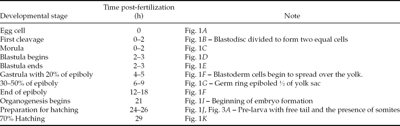
Eggs from a single collection exhibit different development stages due to partitioned spawning and the pooling of eggs from two different spawning. Therefore, eggs from the initial collection were light yellow, spherical, translucent, telolecithal and demersal. At this time, the eggs were at the egg cell, first cleavage and morula stages, with an average diameter of 1.08 ± 0.038 mm (Fig. 1A–C). The early and late blastula stages were identified between 2–3 hpIC while the cells continued to divide mitotically (Fig. 1D, E). The early gastrula was observed between 3–4 hpIC with 20% epiboly (Fig. 1F). Gastrula presented between 30 and 50% epiboly at 5–6 hpIC (Fig. 1G). In B. splendens, the blastoderm does not completely surround the yolk; therefore, no yolk plug is formed and approximately 50% of the yolk is covered. At 11–17 hpIC, thickening of the dorsal epiblast was observed, which would give rise to the head of the embryo (Fig. 1H).
Main stages of the embryonic development of Betta splendens. (A) Egg cell; (B) 0–2 h post-initial collection (hpIC): first cleavage; (C) 0–2 hpCI: morula; (D) 2–3 hpIC: blastula begins; (E) 2–3 hpIC: blastula ends; (F) 4–5 hpIC: beginning of the gastrula with 20% of epiboly; (G) 6–9 hpIC: gastrula with 50% of epiboly; (H) 12–18 hpIC: gastrula at the end of epiboly; (I) 21 hpIC: beginning of the formation of embryo and yolk pigmentation (arrow); (J) 24–26 hpIC: pre-larva, ready to hatch, showing the cephalic and tail regions (arrows) and the presence of somites (arrowheads); (K) 29 hpIC: hatching of the larva and eye pigmentation.

At 21 hpIC, the head and tail were differentiated and the first melanophores were present in the yolk, found mainly near the ventral region of the embryo (Fig. 1I). Over time, between 23–25 hpIC, the embryos became pre-larvae. During this period, pairs of somites occupied the entire notochord from the occipital to the caudal region. The pre-larva was ready to hatch and presented a free tail with strong and continuous movements. This period, propagation and increase of melanophores in the yolk toward the dorsal region of the pre-larva was also observed (Fig. 1J). The hatching began at 28 hpIC and the larvae presented poorly pigmented eyes (Fig. 1K). After 32 hpIC, 90% of the larvae had already hatched and after 38 hpIC, 100% of the larvae had hatched. The total average length of newly hatched larvae was 2.47 ± 0.04 mm. Table 1 and Fig. 1 show the main embryonic development stages of B. splendens.
Embryonic development of the Betta splendens at 28.4 ± 0.2 °C
Larval development
Hatching
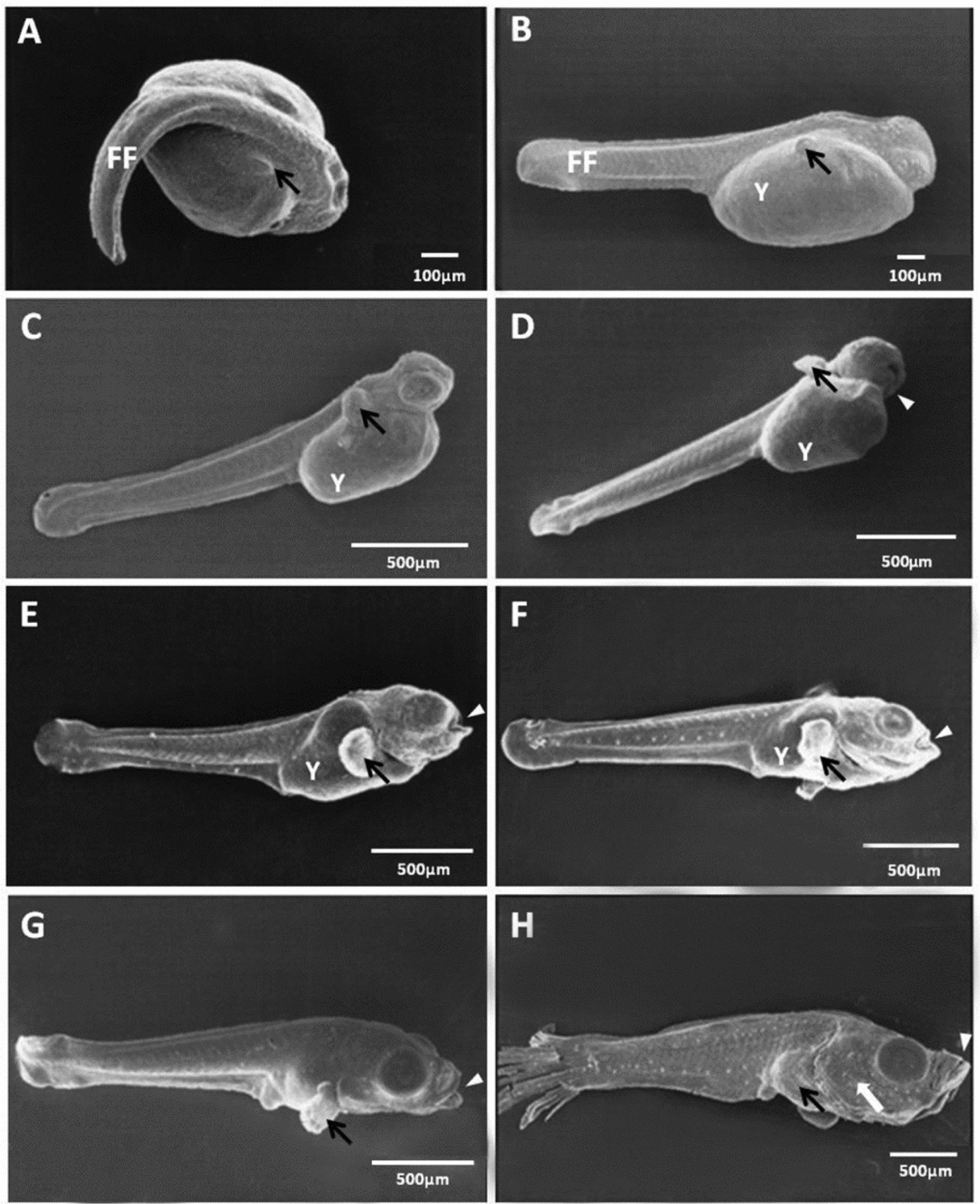
At the beginning of hatching, B. splendens measured 2.47 ± 0.044 mm total length (TL) and presented melanophores in the yolk at the antero-ventral axis. At this moment, the newly hatched larvae showed a closed mouth, large yolk sac, non-differentiated and slightly pigmented eyes (Fig. 2A), a pectoral fin bud and a well developed caudal fin (Fig. 3B).
Larval development of Betta splendens. (A) newly hatched larva (29 h post-fertilization); (B) 11 h post-hatching (hPH); (C) 23 hPH; (D) 49 hPH; (E) 73 hPH; (F) 432 hPH; (G) 768 hPH.
Electron micrographs of Betta splendens. (A) 25 h post-fertilization (hPF): Pre-larva ready to hatch; (B) 29 hPF: newly hatched larva; (C) 15 h post-hatching (hPH); (D) 17 hPH; (E) 43 hPH; (F) 61 hPH; (G) 85 hPH; (H) 264 hPH. FF: finfold. Y: yolk. Arrowheads: mouth. Black arrows: pectoral fin. White arrows: opercle.
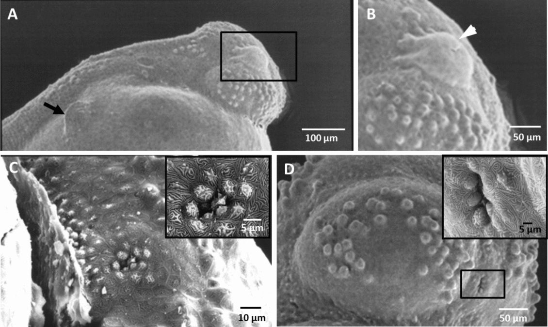
At this stage, the larvae had low swimming capacity and remained attached to the bubble nest under the parental care of the male. For this reason, they had adhesive glands that were identified in the dorsal region of the head, just above the eye (Fig. 4A, B).
Electron micrographs of Betta splendens showing some structures during development. (A,B) Newly hatch larvae, adhesive glands. (C) 1 h post-hatching (hPH): olfactory cavity surrounded by mucus-producing cells. (D) 7 hPH: olfactory cavity. Arrow: pectoral fin. Arrowhead: adhesive glands.
1–11 hPH
The olfactory cavity was formed after 1 hPH, surrounded by mucus-producing cells (Fig. 4C), and after 7 hPH it was deeper (Fig. 4D). It was observed that the eyes were more pigmented; the notochord more visible and the yolk volume reduced after 11 hPH (Fig. 2B). The melanophores were more evident in the entire yolk and concentrated mainly on the larval antero-ventral axis (Fig. 2B). At this stage, the disappearance of the adhesive gland was also observed.
11–43 hPH
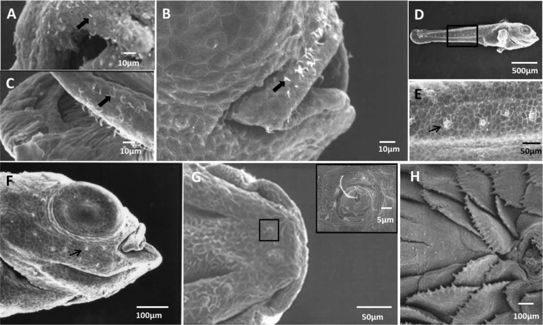
Cilia were identified on the upper lip and the mouth opening at 17 hPH (Fig. 5A), when the larva TL was 2.66 ± 0.068 mm. At 43 hPH, the opercle covered the gills (Fig. 3E). Despite the mouth opening was present, we did not observe larvae feeding at this stage.
Electron micrographs of Betta splendens showing some structures during development. (A) 17 h post-hatching (hPH); (B) 23 hPH; (C) 85 hPH; (D–F) 65 hPH: neuromasts on the lateral line region and around the eye; (G) 86 hPH: neuromasts on the lower jaw region; (H) 936 hPH: detail showing scales in juveniles. Thick arrows: ciliated cells. Thin arrows: neuromasts.
43–65 hPH
At 49 hPH the eyes and body were more pigmented (Fig. 2D). At 65 hPH, a large amount of neuromasts was observed in the lateral line and around the eye (Fig. 5D–F). At this stage, a greater swimming ability was observed, with the larvae displaying morphological and sensory structures that enabled greater mobility and perception of the surroundings.
65–264 hPH
At this stage, although the larvae still had yolk, exogenous feeding had begun, as Artemia sp. nauplii could be seen inside their bodies at 72 hPH (Fig. 6A). The yolk was completely absorbed at 73 hPH, when the larvae measured 3.20 ± 0.176 mm in length. From this phase onwards, the larvae exclusively obtained food via exogenous feeding, by actively chasing the nauplii (Artemia sp.) (Fig. 6B). After 86 hPH, neuromasts were observed in the lower jaw (Fig. 5G). At 156 hPH, the heart was pumping blood throughout the entire larval extension (Fig. 6D). At 264 hPH, the caudal fin was fully formed, with opercle and eyes well developed (Fig. 3H).
Photomicrographs of Betta splendens. (A) 72 h post-hatching (hPH). (B) 96 hPH. (C) 156 hPH. (D) 156 hPH. Circle: Heart. Black thick arrow: Artemia sp. ingested by the larva. Black thin arrow: mouth opened for respiration.
264–768 hPH
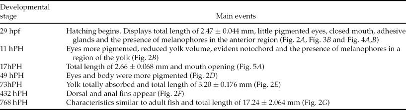
At 432 hPH, the dorsal and anal fins were being formed and the caudal fin rays were clearly evident (Fig. 2F). Characteristics similar to the adults were observed when larval TL was 17.24 ± 2.064 mm, which characterized the end of the larval stage (Fig. 2G), after 768 hPH. From this point on, the animals are considered juveniles. Table 2 describes the main stages of larval development.
Main events during larval development of B. splendens
hpf: hours post-fertilization; hPH: hours post-hatching.
Discussion
Betta splendens are sedentary fish with parental care, whose eggs adhere to the bubble nest. Betta eggs are non-adhesive as, morphologically, they do not present any of the structures that define this characteristic such as zona radiata with hexagonal pore canals, filaments, villous blood cells or gelatin covers (Godinho & Godinho, Reference Godinho and Godinho2003). However, the eggs remain and develop in bubble nests constructed by the male, who produces in his mouth a thick mucus that consists of glycoproteins that help to maintain the permanence of the bubbles in the nest (Kang & Lee, Reference Kang and Lee2010); thus, it is believed that this mucus may also be linked to egg permanence in the bubble nest. However, adhesive eggs are described for sedentary fish such as Acestrorhynchus britskii, Acestrorhynchus lacustris, Serrasalmus spilopleura (Rizzo et al., Reference Rizzo, Sato, Barreto and Godinho2002), Astronotus ocellatus (Paes et al., Reference Paes, Makino, Vasquez, Fernandez Kochenborger and Nakaghi2011) and Franciscodoras marmoratus (Alberto Weber et al., Reference Alberto Weber, Sato, Enemir Santos, Rizzo and Bazzoli2012).
Betta splendens eggs do not have oil droplets and are telolecithal. The yolk is most concentrated at the vegetal pole while the organelles and cytoplasm are concentrated in the animal pole (Kunz, Reference Kunz2004; Ninhaus-Silveira et al., Reference Ninhaus-Silveira, Foresti and de Azevedo2006). The eggs are demersal because their specific gravity is greater than water (Godinho & Godinho, Reference Godinho and Godinho2003) and the animal pole is oriented upward. This event occurs because the yolk sac has a greater relative gravity than the blastodisc (Kunz, Reference Kunz2004).
Egg average diameter in the current study was approximately 1.08 ± 0.038 mm. This value is close to the 0.8 mm reported by Watson & Chapman (Reference Watson and Chapman2002) as the average for ornamental species. The egg diameter and quality are related to factors such as parental care, breeder nutrition, ecological strategy, water quality, photoperiod, animal welfare and genetic influence (Brooks et al., Reference Brooks, Tyler and Sumpter1997; Kolm & Ahnesjo, Reference Kolm and Ahnesjo2005). Parental care is directly related to egg size: the larger the egg, the greater the parental care (Kolm & Ahnesjo, Reference Kolm and Ahnesjo2005). Usually, migratory fish exhibit another strategy, which does not present parental care and produce a large number of small eggs (Godinho et al., Reference Godinho, Lamas and Godinho2010).
The size and shape of the eggs may be important for systematic and phylogenetic studies, as observed in the identification of species of the genus Gobius (Borges et al., Reference Borges, Faria, Gil, Goncalves and Almada2003). Furthermore, it is also relevant to identify spawning areas and to implement programmes to protect and preserve the species (Nakatani et al., Reference Nakatani, Agostinho, Baumgartner, Bialetzki, Sanches and Cavicchioli2001).
The cleavage process, which consists of the division of the egg into smaller cells named blastomeres, starts after fertilization and zygote formation (egg cell). This process varies greatly among vertebrates and depends on the amount of egg yolk (Gilbert, Reference Gilbert2003). The eggs of B. splendens undergo meroblastic or partial cleavage because the mitotic divisions occur only in the animal pole of the egg. This type of cleavage is typical of fish that accumulate a large amount of yolk (Leme dos Santos & Azoubel, Reference Leme dos Santos and Azoubel1996; Gilbert, Reference Gilbert2003; Takeuchi et al., Reference Takeuchi, Okabe, Aizawa, Gann and Crotty2008), such as Gymnocorymbus ternetzi (Celik et al., Reference Celik, Celik, Cirik, Gurkan and Hayretdag2012), F. marmoratus (Alberto Weber et al., Reference Alberto Weber, Sato, Enemir Santos, Rizzo and Bazzoli2012) and Brycon gouldingi (Faustino et al., Reference Faustino, Nakaghi and Neumann2010b).
The morula stage is reached once the zygote has divided into 64 blastomeres. Through these divisions, the number of cells increases while the volume of each individual cell decreases (Wolpert et al., Reference Wolpert, Beddington, Brockes, Jessell, Lawrence and Meyerowitz2000; Gilbert, Reference Gilbert2003), as observed for B. splendens. Immediately after the morula stage, the blastula, which is characterized by the blastoderm, is formed (Marques et al., Reference Marques, Okada Nakaghi, Faustino, Ganeco and Senhorini2008).
The gastrulation process is characterized by epiboly and involution movements (Wolpert et al., Reference Wolpert, Beddington, Brockes, Jessell, Lawrence and Meyerowitz2000; Gilbert, Reference Gilbert2003; Kunz, Reference Kunz2004). The epiboly movement consists of the spreading of the blastoderm toward the vegetal pole (Gilbert, Reference Gilbert2003; Faustino et al., Reference Faustino, Nakaghi, Marques, Ganeco and Makino2010a). After the blastoderm has engulfed at least the half of the yolk, the involution movement initiates (Gilbert, Reference Gilbert2003; Kunz, Reference Kunz2004). Wolpert et al. (Reference Wolpert, Beddington, Brockes, Jessell, Lawrence and Meyerowitz2000) and Kunz (Reference Kunz2004) show that this process leads to the formation of two layers: the epiblast, which gives rise to the ectoderm, and the hypoblast, which gives rise to mesoderm and endoderm.
Temperature strongly influences the duration of embryonic and larval development (Morrison et al., Reference Morrison, Miyake and Wright2001; Martell et al., Reference Martell, Kieffer and Trippel2005): the higher the temperature, the shorter developmental time and vice versa (Leme dos Santos & Azoubel, Reference Leme dos Santos and Azoubel1996; Martell et al., Reference Martell, Kieffer and Trippel2005).
The embryonic development of B. splendens was slower compared with other ornamental species. The cardinal tetra, Paracheirodon axelrodi, hatches 19–20 h post- fertilization (hpf) at an average temperature of 26 ± 1ºC (Anjos & Anjos, Reference Anjos and Anjos2006); Gymnocorymbus ternetzi hatches 20–21 hpf at 24 ± 0.5ºC (Celik et al., Reference Celik, Celik, Cirik, Gurkan and Hayretdag2012); however, A. ocellatus development was even slower, as it hatched after 46–58 hpf at a temperature of 27.5ºC (Paes et al., Reference Paes, Makino, Vasquez, Fernandez Kochenborger and Nakaghi2011). This variability can be explained by factors such as temperature and interspecific variation.
Fish species can be classified either as precocial or altricial depending upon the strategy adopted. The larvae of precocial species hatch from the eggs in the juvenile stage, while the altricial species hatch before this stage and the larvae exhibits an undifferentiated developmental stage (Bejarano-Escobar et al., Reference Bejarano-Escobar, Blasco, Degrip, Oyola-Velasco, Martín-Partido and Francisco-Morcillo2010). Betta splendens is an altricial species, as it hatches with several organs and systems in differentiation.
The newly hatched B. splendens larvae had an average TL of 2.47 ± 0.044 mm, higher than other sedentary species such as F. marmoratus (Alberto Weber et al., Reference Alberto Weber, Sato, Enemir Santos, Rizzo and Bazzoli2012) that hatches at 1.27 ± 0.4 mm and G. ternetzi (Celik et al., Reference Celik, Celik, Cirik, Gurkan and Hayretdag2012) at 1.44 mm, but smaller than P. axelrodi (Anjos & Anjos, Reference Anjos and Anjos2006) which hatches at 2.9 ± 0.2 mm. Coleman & Galvani (Reference Coleman and Galvani1998) stated that there is a relationship between egg size and the length of newly hatched larvae and analysed it in a wide range of tropical species, concluding that the larger the egg size, the longer would be the newly hatched larvae. However, there are few studies available on this topic.
After hatching, B. splendens larvae have adhesive glands that consist of mucous cells present in the head. These glands enable the larvae to remain attached to the nest, to increase parental efficiency (Araújo-Lima & Bittencourt, Reference Araújo-Lima and Bittencourt2001). These glands are similar to those described for A. ocellatus (Paes et al., Reference Paes, Makino, Vasquez, Fernandez Kochenborger and Nakaghi2011), Cichlasoma dimerus (Meijide & Guerrero, Reference Meijide and Guerrero2000) and Hoplias malabaricus (Araújo-Lima & Bittencourt, Reference Araújo-Lima and Bittencourt2002).
In the current study, the mouth opened very quickly, while depletion of the yolk sac happened later. According to Yúfera & Darias (Reference Yúfera and Darias2007), when the larvae initiates exogenous feeding it is important that all structures related to ingestion, digestion and assimilation are ready. Furthermore, it is necessary to highlight the importance of sensory structures such as neuromasts and eyes (Bilotta & Saszik, Reference Bilotta and Saszik2001). Along with the development of fins, these structures are essential for sensing and chasing food. Therefore, describing these ontogenetic events will help improve husbandry practices by making it possible to determine the real needs of animals at different developmental stages.
The larval stage was completed when the larvae reached an average TL of 17.20 mm, when their body features are similar to adults and become juveniles (Kendall et al., Reference Kendall, Ahlstrom, Moser, Moser, Richards, Cohen, Fahay, Kendall and Richardson1984). The results of the initial developmental stages of B. splendens provide important information for the biology, breeding and rearing of the species as well as a basis for further studies.
Acknowledgements
We would like to thank Drs Maria do Carmo Faria Paes and Sheryll Corchuelo for revising the manuscript and the ornamental fish laboratory of CAUNESP for supplying the animals.