Heart transplantation in paediatric patients was first reported in 1968. Reference Kantrowitz, Haller, Joos, Cerruti and Carstensen1 The number of paediatric heart transplantations has been increasing over the past two decades with marked improvements in survival rates. Data from the International Society for Heart and Lung Transplantation (ISHLT 2016 Pediatric Report) showed survival rates of > 80% at 5 years compared to < 60% a decade prior. Reference Rossano, Dipchand and Edwards2 Complex CHD is the most common indication for heart transplant in children below the age of 1 year, while dilated cardiomyopath is the most common indication in older children. Reference Rossano, Dipchand and Edwards2 Following transplantation, patients experience numerous acute and chronic complications. Diastolic dysfunction is a recognised complication of adult heart transplantation in both the early and late post-transplant periods. Reference Tallaj, Kirklin and Brown3,Reference Vejpongsa, Torre-Amione and Marcos-Abdala4 Prolonged graft ischaemic time and increased donor age are both associated with a higher risk of ventricular diastolic dysfunction. Reference Rustad, Nytrøen and Andreassen5 In the paediatric age group, cardiovascular MRI data showed a significantly reduced early diastolic strain rate compared to healthy controls long-term following transplantation. Reference Latus, Hachmann and Voges6
The echocardiographic assessment of diastolic dysfunction in children is challenging. There is no single parameter to evaluate diastolic function using echocardiography, but evaluation relies on multiple parameters. The current guidelines for evaluating left ventricular diastolic dysfunction by echocardiography in children are based on adult guidelines. In the adult heart transplant population, using Doppler echocardiographic parameters for estimating left ventricular filling pressures is controversial. There are conflicting data in both the adults and the paediatric population in this regard. Reference Bech-Hanssen, Al-Habeeb and Ahmed7–Reference Buddhe, Richmond, Gilbreth and Lai9
Monitoring the ventricular diastolic function is essential for the health and longevity of graft recipients. Invasive haemodynamic data are the gold standard for evaluating the left ventricular filling pressure in both paediatric and adult heart transplant patients. In this study, we aimed to evaluate the correlation between traditional echocardiographic indices for diastolic function and invasive pulmonary capillary wedge pressure along with invasive left ventricular end-diastolic pressure in paediatric patients in the long-term follow-up after heart transplantation.
Methods
Patients
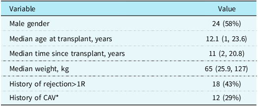
Chart review of 52 heart transplant recipients who received their transplanted hearts before the age of 24 years was performed. Patients underwent transplantation between 2001 and 2018. Data in this analysis included the most recent cardiac catheterisation (within two years from the time of this study) during a routine annual follow-up. Echocardiographic data included in this cohort were obtained on the day of cardiac catheterisation. Of the 52 patients, 41 were included in the analysis. Eleven patients were excluded from the study due to incomplete echocardiographic data needed for the purpose of this study. Demographic data, age at transplant, age since transplant, gender, weight, vital signs, laboratory data, any previous history of biopsy-proven rejection that is more than mild rejection (1R), and evidence of coronary artery vasculopathy were included in this analysis (Table 1).
Demographics of the patient cohort

* Cardiac allograft vasculopathy.
Echocardiographic examination
All patients included in this study underwent echocardiographic examination within 24 hours following cardiac catheterisation. The examinations were performed using Philips EPIQ CVx and Philips Epiq7c. Standard ultrasound protocols were used in these studies. Two Dimensional views, colour Doppler, and Doppler interrogation were applied using the currently available American Society of Echocardiography guidelines for both adult and paediatric patients. All echocardiograms in this study were performed after the cardiac catheterisation. The following echocardiographic parameters were evaluated in this study: left ventricular ejection fraction measured using Simpson’s biplane method, left ventricular end-diastolic volume, mitral inflow E and A velocities measured using pulsed-wave Doppler, and the E/A ratio and mitral valve deceleration time. Tissue Doppler velocities were obtained in the lateral and septal positions of the mitral valve annulus. The E/e’ average value was also calculated. Isovolumic relaxation time was measured using pulse tissue Doppler from the end of the systolic waveform to the early diastolic velocity.
Cardiac catheterisation
All patients underwent left and right cardiac catheterisation with coronary angiography as part of annual surveillance. Older patients were consciously sedated, but some younger patients were intubated and placed under general anaesthesia for the procedure. Pulmonary capillary wedge pressure was measured simultaneously with the left ventricular end-diastolic pressure. In most cases, cardiac output was measured using thermodilution. Following haemodynamic measurements, an endomyocardial biopsy was obtained from the septal surface of the right ventricle. Coronary artery angiography was performed to examine for any evidence of coronary artery vasculopathy.
Statistical analysis
Two software, SPSS for windows version 21 (SPSS Inc., Chicago, Illinois, USA) and GraphPad Prism version 8.4.2 (GraphPad Software, San Diego, CA, USA) were used to perform the statistical analysis. Kolmogorov Smirnov test was performed for normality checks of variables. Descriptive statistics were expressed into mean values and standard deviation. Independent sample t test was conducted for parametric data and Mann-Whitney t test was conducted for non-parametric data. Simple linear regression was also performed to find the relationship between echocardiographic data and pulmonary capillary wedge pressure (which was defined as mean pressure ≥ 15 mmHg). Similar echocardiographic data were also compared between patients with normal left ventricular end-diastolic pressure to patients with elevated left ventricular end-diastolic function (which was defined as left ventricular end-diastolic pressure ≥ 15 mmHg). P-value < 0.05 was considered statistically significant.
Results
This cohort included 41 patients, of whom 24 (58%) were male. The median age at transplant was 12.1 years (two patients were under 1 year of age), and the median time after transplantation was 11 years. Eighteen patients (43%) had a previous history of one or more episodes of rejection greater than mild in severity (>1R), as defined by the International Society for Heart and Lung Transplantation. Twelve patients (29%) had a history of cardiac allograft vasculopathy, including three patients who had evidence of severe allograft vasculopathy requiring stent placement.
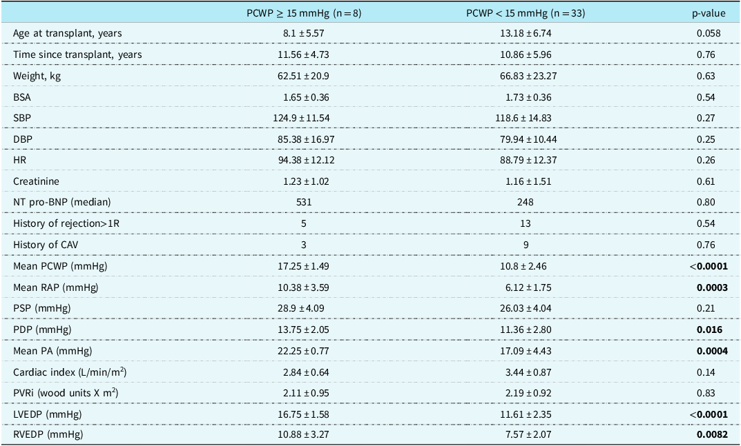
Eight patients (20%) had evidence of elevated pulmonary capillary wedge pressure and 33 patients had normal pulmonary capillary wedge pressure (Table 2). There was no significant difference in age at transplant or time since transplant between patients with elevated pulmonary capillary wedge pressure and those with normal pulmonary capillary wedge pressure. Elevated NTproB-type natriuretic peptide, history of allograft rejection, or the presence of cardiac allograft vasculopathy were not significantly correlated with elevated pulmonary capillary wedge pressure (Table 2).
Comparison of patient’s characteristics and catheterisation data based on PCWP
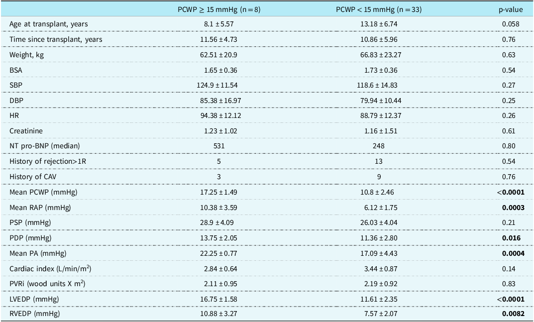
SBP = systolic blood pressure; DBP = diastolic blood pressure; HR = heart rate; NT pro-BNP: NTproB-type natriuretic peptide; CAV = cardiac allograft vasculopathy; RAP = right atrial pressure; PSP- pulmonary artery systolic pressure; PDP = pulmonary artery diastolic pressure; PA = pulmonary artery; PVRi= pulmonary vascular resistance index; LVEDP = left ventricular end-diastolic pressure; RVEDP = right ventricular end-diastolic pressure; PCWP = pulmonary capillary wedge pressure.
Data are presented in mean ± SD.
The haemodynamic data are presented in Table 2. The mean right atrial and mean pulmonary artery pressures were significantly elevated in patients with elevated pulmonary capillary wedge pressure. Cardiac index was lower in patients with elevated pulmonary capillary wedge pressure, but the difference was not significant (p = 0.14). Pulmonary vascular resistance indexed to body surface area was similar in patients with elevated pulmonary capillary wedge pressure (mean of 2.11 ± 0.95 compared to a mean of 2.19 ± 0.92 in patients with normal pulmonary capillary wedge pressure). Left and right ventricular end-diastolic pressures were significantly elevated in patients with elevated pulmonary capillary wedge pressure (p-value < 0.0001 and = 0.0082, respectively).
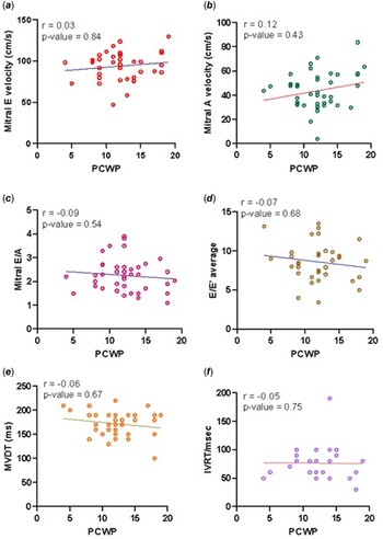
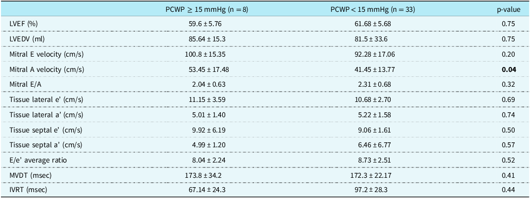
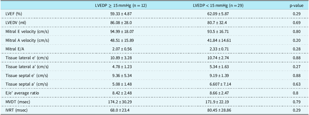
The echocardiographic findings are presented in Table 3. The mean left ventricular ejection fraction was normal and similar in both the groups (59.6 ± 5.76 and 61.68 ± 5.68). The mean mitral E velocity was also similar in both groups (mean of 100.8 ± 15.35 cm/s in patients with elevated pulmonary capillary wedge pressure compared to 92.28 ± 17.06 cm/s in patients with normal pulmonary capillary wedge pressure) (Fig. 1a). The mean mitral A velocity was higher in patients with elevated pulmonary capillary wedge pressure than in patients with normal pulmonary capillary wedge pressure (53.45 ± 17.48 cm/s and 41.45 ± 13.77 cm/s), but no significant correlation was found when applying simple linear regression (Fig. 1b). The mean mitral E/A ratio was slightly higher in patients with normal pulmonary capillary wedge pressure (2.31 ± 0.68 compared to 2.04 ± 0.63 in patients with elevated pulmonary capillary wedge pressure, Fig. 1c) but was not statistically significant. Averaged tissue Doppler myocardial velocities obtained at the lateral mitral annulus (e’ and a’ lateral) were similar in both groups with no statistical difference (p-value = 0.69 for e’ velocities and p-value = 0.74 for a’ velocities). Tissue Doppler myocardial velocities obtained at the septal mitral annulus (e’ and a’ septal) were also similar in both groups with no statistical difference (p-value = 0.50 for e’ velocities and p-value = 0.57 for a’ velocities). The E/e’ ratio averaged over three consecutive beats was similar in both groups (8.04 ± 2.24 and 8.73 ± 2.51, Fig. 1d). The mean mitral valve deceleration time was normal and similar in both groups (173.8 ± 34.2 msec and 172.3 ± 22.17 msec, Fig. 1e). The mean isovolumic relaxation time was normal and similar in both groups (67.14 ± 24.3 msec and 97.2 ± 28.3 msec, Fig. 1f). Similar echocardiographic indices were also compared between patients with elevated left ventricular end-diastolic pressure (12 patients) to patients with normal left ventricular end-diastolic pressure (29 patients), and no correlations were found (Table 4).
Scatter plots show the relationship between pulmonary capillary wedge pressure and echocardiographic data. ( a ) mitral E velocity, ( b ) mitral A velocity, ( c ) mitral E/A, ( d ) E/e’ average ratio. ( e ) mitral valve deceleration time, ( f ) isovolumic relaxation time.

Comparison of echocardiographic data with invasive PCWP

PCWP = pulmonary capillary wedge pressure; LVEF = left ventricular ejection fraction; LVEDV = left ventricular end-diastolic volume; MVDT = mitral valve deceleration time; IVRT = isovolumic relaxation time.
Data are presented in mean ± SD.
Comparison of echocardiographic data with invasive LVEDP

LVEDP = left ventricular end-diastolic pressure; LVEF = left ventricular ejection fraction; LVEDV = left ventricular end-diastolic volume; MVDT = mitral valve deceleration time; IVRT = isovolumic relaxation time.
Data are presented in mean ± SD.
Discussion
Left ventricular diastolic dysfunction is a recognised complication of heart transplantation in both paediatric and adult populations. Invasive haemodynamic data remain the gold standard for evaluating filling pressure. To date, there has been no single echocardiographic parameter that can accurately predict the presence or severity of left ventricular diastolic dysfunction. The most recent update from the American Society of Echocardiography and the European Association of Cardiovascular Imaging recommends evaluating both the clinical status and echocardiographic data to assess for ventricular diastolic dysfunction. Reference Nagueh, Smiseth and Appleton10 These reports also recommend using more than one parameter (average E/e’, lateral and septal e’ velocities, tricuspid regurgitation velocity, and left atrial volume index), and if one or more of these parameters are abnormal, the likelihood of left ventricular diastolic dysfunction increases.
Acute right ventricular diastolic dysfunction is a well-known entity in the early period following cardiac transplantation and is a strong predictor of early mortality, but it also tends to improve within the first year after transplantation. Reference Tallaj, Kirklin and Brown3 The development of left ventricular diastolic dysfunction is not common in the first few years following transplantation in the paediatric group and is often not detected within the first five years of cardiac transplant in children. Reference Fyfe, Mahle, Kanter, Wu, Vincent and Ketchum11 This study aimed to evaluate the long-term evolution of left ventricular diastolic dysfunction post-transplantation. The mean “time since transplant” in our study was 11 years, and 29% of our cohort had elevated left ventricular filling pressures. We found that none of the traditional echocardiographic parameters we used correlated with invasive pulmonary capillary wedge pressure or left ventricular end-diastolic pressure measurements, which is similar to other studies published on the paediatric population that investigated paediatric patients a few years following transplant. Reference Buddhe, Richmond, Gilbreth and Lai9,Reference Loar, Pignatelli and Morris12 This is different from available adult data where echocardiographic parameters, including mitral E/A, average E/e’, DT, and isovolumic relaxation time, correlated with pulmonary capillary wedge pressure. Reference Bech-Hanssen, Al-Habeeb and Ahmed7,Reference Broch, Al-Ani, Gude, Gullestad and Aakhus8 There are possible explanations for why the paediatric data are inconsistent with the adult data. One of the most likely explanations is age, while the normal values of the commonly used parameters are available; they are age-dependent, and the values vary widely within the paediatric age group. Children and young adults present a challenging haemodynamic profile, and these echocardiographic parameters depend heavily on heart rate, preload/afterload, and regional wall motion abnormalities. In addition, the paediatric donor heart tends to develop tachycardia, which adds to the inherent fast heart rates that usually young adults experience, which may lead to fusion of mitral E and A velocities and makes measuring deceleration time and E/A ratio difficult and inaccurate. Regional wall motions are abnormal in transplanted children even in the absence of rejection episodes, Reference Pauliks, Pietra and Kirby13 leading to significantly lower tissue Doppler velocities and potential differences in measurements of these velocities along with the E/e’ ratio when compared to normal values published for healthy children. In addition, tissue Doppler measurements are preload dependent, and subjects included in this study fasted for more than eight hours, which creates different haemodynamic conditions, especially for young adults. Isovolumic relaxation time is a measurement of the left ventricular relaxation and is prolonged with impaired relaxation even with normal left ventricular filling pressure; when the left ventricular pressure increases, isovolumic relaxation time is reduced. Isovolumic relaxation in our study was normal and similar in both groups, and it is also important to note that isovolumic relaxation is largely affected by heart rate.
An important observation in this study was that the mean pulmonary capillary wedge pressure in patients with elevated pulmonary capillary wedge pressure was 17.25 ± 1.49 mmHg. Echocardiography might be limited in detecting subtle elevations in filling pressures, and this could change if the cohort is larger with more significant elevations in filling pressure. It is also important to take into account that these patients were sedated (and some were under general anaesthesia), and myocardial demand was minimal, but if those patients were challenged with pharmacological or exercise tests, their cardiac systolic and diastolic dysfunction may become more profound and might be more evident if there was an associated coronary artery disease. Reference Arnold, Goebel, Ulmer, Gorenflo and Poerner14 Our findings are consistent with those in the literature, Reference Goldberg, Quartermain and Glatz15 and traditional echocardiographic parameters might not detect elevations in the filling pressures. These echocardiographic limitations apply not only to the paediatric transplant group but also to paediatric patients with other forms of cardiomyopathies. Reference Dragulescu, Mertens and Friedberg16,Reference Masutani, Saiki, Kurishima, Kuwata, Tamura and Senzaki17 New imaging techniques were found to have a better correlation with invasive pulmonary capillary wedge pressure in paediatric patients after cardiac transplantation. These include left atrial strain. Reference Loar, Pignatelli and Morris12,Reference Yeh, Aiyagari, Gajarski, Zamberlan and Lu18 Impaired left ventricular diastolic function and filling pressure affect the geometry and function of the left atrium, making left atrial strain measurements a useful tool to gauge left ventricular diastolic function by avoiding the limitations of traditional methods. Advanced imaging modalities, such as cardiac MRI, have superior capabilities over echocardiography. Standard cardiac MRI techniques, such as volumetric analysis and native T1 relaxation time and strain measurements, have been found to correlate with pulmonary capillary wedge pressure in paediatric heart transplants. Reference Belay, Godown and Chan19
There are some important limitations to this study. The retrospective nature of this review has inherent limitations owing to its design. The cardiac catheterisation and echocardiography were non simultaneous; thus, they were performed under different haemodynamic conditions, particularly for the few intubated patients whose intracardiac pressure may have been impacted. A larger number of patients with a more severe form of diastolic function may have increased the likelihood of detecting a meaningful link with echocardiographic data. However, only a small number of patients with evidence of diastolic dysfunction by invasive methods were included in this investigation.
Monitoring diastolic function is important for the health and longevity of grafts in paediatric heart transplants. Invasive cardiac catheterisation is still the standard method to monitor haemodynamics, the presence of coronary vascular disease, and for obtaining biopsy to monitor for rejection. New emerging techniques and modalities are promising and have the potential to replace invasive studies in selected populations.
Conclusion
Many paediatric patients tend to develop left ventricular diastolic dysfunction long term after heart transplantation. Traditional echocardiography indices did not correlate well with left ventricular filling pressure in paediatric heart transplant patients, which is consistent with other published paediatric short-term follow-up studies. This study shows that even long term after heart transplant in children, echocardiography has no correlation with invasive measurements. Other parameters should be considered when non-invasive assessment of diastolic function is required. Larger cohorts with more significant diastolic disease are needed to confirm the current findings.
Acknowledgements
None.
Financial support
None.
Competing interests
None.