INTRODUCTION
Mild cognitive impairment (MCI) is a state of cognitive dysfunction that lies between normal cognitive ageing and dementia. Published literature on MCI has been dominated by one subtype of MCI in which memory impairment is the salient feature, the so-called amnestic MCI (aMCI), and which is considered to progress to Alzheimer’s disease (AD) (Petersen, Reference Petersen and Petersen2003), albeit in a proportion of cases. MCI is, however, a broader concept, and arguably all patients with a dementia of gradual onset will pass through a phase of MCI in the predementia stage (Winblad et al., Reference Winblad, Palmer, Kivipelto, Jelic, Fratiglioni and Wahlund2004). Consistent with the fact that vascular impairment may be caused by vascular factors, a vascular sub-type of MCI (vaMCI) is diagnosable in many patients (O’Brien et al., Reference O’Brien, Erkinjuntti, Reisberg, Roman, Sawada and Pantoni2003).
As the concept of vascular cognitive impairment (VCI) has broadened to include cognitive impairment caused by a range of cerebrovascular disorders, the importance of the early recognition of VCI at the vaMCI stage is being appreciated. While several studies have examined vascular dementia (VaD) in comparison with AD, the study of vaMCI has been limited, and there is inadequate information on the neuropsychological profile of vaMCI and its rate of progression toward dementia. This is partly because a cohort of individuals with a well-characterized diagnosis of vaMCI is difficult to study. One approach is to examine post-stroke cognitive impairment that falls short of dementia diagnosis.
MCI is common in post-stroke patients. In the Sydney Stroke Study, nearly two in five patients were diagnosed to have vaMCI 3–6 months after a stroke (Sachdev et al., Reference Sachdev, Brodaty, Valenzuela, Lorentz, Looi and Berman2006), and their cognitive impairment was judged not to be explained by specific brain infarcts. This was in addition to approximately one in five stroke patients who met criteria for dementia diagnosis. Similar figures have been reported in other studies (Qui, Skoog, & Fratiglioni, Reference Qui, Skoog, Fratiglioni, Erkinjuntti and Gauthier2000; Serrano, Domingo, Rodriguez-Garcia, Castro, & Del Ser, Reference Serrano, Domingo, Rodriguez-Garcia, Castro and del Ser2007). The progression of vascular cognitive impairment without dementia is not well known and pattern of cognitive changes is rarely reported. In the clinical setting, it is generally considered that if cognitive impairment occurs after a stroke, it will show initial improvement followed by persistence of deficits, but without decline. In a study from Singapore, 31% of post-stroke patients diagnosed with cognitive impairment—no dementia (CIND) at 6 months were judged to be cognitively normal at 1 year (Tham et al., Reference Tham, Auchus, Thong, Goh, Chang and Wong2002). Similarly, improvement in CIND was reported in 44% of post-stroke patients over 2 years in a Spanish study (del Ser et al., Reference del Ser, Barba, Morin, Domingo, Cemillan and Pondal2005). Kokmen, Whisnant, O’Fallon, Chu, and Beard (Reference Kokmen, Whisnant, O’Fallon, Chu and Beard1996) suggested that progressive decline following a stroke that cannot be attributed to another stroke should be considered to be due to Alzheimer’s disease. Follow-up over 1 year in the Sydney Stroke Study showed only a mild decline in the post-stroke patients which on a composite neuropsychological index was not different from the control group, although the stroke patients showed slightly greater decline in verbal memory and visuoconstructive function (Sachdev, Brodaty, Valenzuela, Lorentz, & Koschera, Reference Sachdev, Brodaty, Valenzuela, Lorentz and Koschera2004). Other studies have reported greater progression in post-stroke patients who demonstrate some impairment at index assessment (Srikanth, Quinn, Donnan, Saling, & Thrift, Reference Srikanth, Quinn, Donnan, Saling and Thrift2006; Wentzel et al., Reference Wentzel, Rockwood, MacKnight, Hachinski, Hogan and Feldman2001). In fact, the Canadian Study of Health and Aging (Wentzel et al., Reference Wentzel, Rockwood, MacKnight, Hachinski, Hogan and Feldman2001) reported similar rates of progression to dementia in vaMCI over 5 years as in nonvascular MCI. Rate of progression in VCI varies, however, with lowest rates of progression in those with mild impairment and highest in those with VaD (Rockwood et al., Reference Rockwood, King Moorhouse, Song, MacKnight, Gauthier and Kertesz2007).
The literature on progression of vaMCI, therefore, remains inconclusive. This is complicated by the overlap of VCI with AD, which makes the etiology of progression difficult to ascertain. Vascular factors affect the progression of AD (1), and specifically, hypertension and diabetes was shown to increase the risk of Alzheimer’s disease (1, 2). Additionally, neuropathological lesions such as neuritic plaques and neurofibrillary tangles, typically suggestive of AD, are often detected in patients with VaD (3). The dividing line between VaD and AD is becoming blurred; therefore, these two types of dementia were believed by some to be two extremes in a broad spectrum of cognitive changes (4). In this study, we investigate neuropsychological features of the vaMCI and its progression over 3 years after their stroke in a cohort previously free of dementia. We also investigate risk factors of conversion to dementia in this group and magnetic resonance imaging (MRI) correlates to see if further accumulative vascular pathology can explain the progression of impairment. We expected gradual minor decline in vaMCI patients in the absence of further cerebrovascular events, with the latter accentuating the decline, and we expected this to be related to risk factors for small vessel disease.
METHOD
Subjects
Participants of the study were recruited consecutively from the patients who had stroke diagnosed by two neurologists independently and had been admitted to two large hospitals affiliated with the University of New South Wales during May 1997 and June 2000. An ischemic stroke was defined as “rapidly developing clinical signs of focal (or global) disturbance of cerebral function, with symptoms lasting 24 hr or longer, with no apparent cause other than of vascular origin” in which a brain computed tomography (CT) or MRI scan did not show intracranial hemorrhage (O’Brien et al., Reference O’Brien, Erkinjuntti, Reisberg, Roman, Sawada and Pantoni2003). Mid-way through the study (from May 1998), consecutive inpatients with transient ischemic attacks (TIA), defined as sudden focal neurological deficits lasting less than 24 hr and not associated with cerebral infarction on CT scan were also included. The reason for the inclusion of TIA patients was to have a sub-sample of subjects with noninfarction ischemic cerebrovascular disease, and thereby increase the range of pathology in the sample. Participants were aged 45–87 years, did not have a diagnosis of dementia or other neurological disorder prior to the stroke, did not have severe aphasia (a score of <3 on the Aphasia Severity Rating Scale of the Boston Diagnostic Aphasia), had a score of <3.5 on the Informant Questionnaire of Cognitive Decline (IQCODE) (Jorm & Jacomb, Reference Jorm and Jacomb1989) for the 5 years before stroke (indicating lack of significant cognitive decline) and consented to participate in the study. Healthy comparison participants were age-matched volunteers recruited from the same neighborhood as the stroke/TIA patients and had no history of stroke or other neurological or psychiatric disorder. Details of participants’ recruitment and study attrition have been published previously (Sachdev et al., Reference Sachdev, Brodaty, Valenzuela, Lorentz, Looi and Berman2006).
This study was approved by the ethics committees of the University of New South Wales and the South-Eastern Sydney Illawara Health Service.
Clinical Assessment
Baseline assessments
Baseline assessments were performed for stroke/TIA patients within 1 week of admission to hospital. A medical history including history of risk factors for cerebrovascular disease and dementia were obtained and a functional assessment and the Mini-Mental State Examination (Folstein, Folstein, & McHugh, Reference Folstein, Folstein and McHugh1975) were performed. A blood sample was taken for clinical chemistry and genotyping for apolipoprotein E (Apo E).
Index assessment
A detailed neuropsychological assessment and structured medical and psychiatric examinations were performed between 3 and 6 months after the index stroke/TIA events. The majority of participants had a brain MRI scan at this time. Health comparison participants were assessed at time of admission to the study.
Follow-up assessment
The above assessment was repeated in participants still enrolled in the study 3 years later.
Neuropsychological Assessment
The battery consists of a series of tests for various cognitive domains: verbal memory (Logical Memory I and II subtests from Wechsler Memory Scale–Revised, WMS-R) (Wechsler, Reference Wechsler1987); visual memory (Visual Reproduction I and II from WMS-R) (Wechsler, Reference Wechsler1987); working memory (Digit Span backwards, Arithmetic from Wechsler Adult Intelligence Scale Revised, WAIS-R); attention (Digit Span forwards, WAIS-R) (Wechsler, Reference Wechsler1981); mental control (WMS-R) (Wechsler, Reference Wechsler1987); language (15-item Boston Naming Test) (Mack, Freed, Williams, & Henderson, Reference Mack, Freed, Williams and Henderson1992); information processing speed (Trail Making Test Part A [Reitan & Wolfson, Reference Reitan and Wolfson1985], Symbol Digit Modalities Test, SDMT [Smith, Reference Smith1991]); visuoconstruction (Block Design, WAIS-R [Wechsler, Reference Wechsler1981] and copying simple figures); praxis-gnosis (Western Aphasia Battery ideomotor apraxia subtest items [Kertesz, Reference Kertesz1983], finger gnosis and stereognosis [Benton, Hamsher, Varney, & Spree, Reference Benton, Hamsher, Varney and Spree1983; Strub & Black, Reference Strub and Black1985]); abstract reasoning (Similarities, Picture Completion, WAIS-R [Wechsler, Reference Wechsler1981]); mental flexibility (Color Form Sorting Text, Trail Making Test Part B [Reitan & Wolfson, Reference Reitan and Wolfson1985]); verbal fluency (phonemic fluency, FAS [Benton et al., Reference Benton, Hamsher, Varney and Spree1983]); and semantic fluency (category of animals) (Morris et al., Reference Morris, Heyman, Mohs, Hughes, van Belle, Fillenbaum and Mellits1989). Mental flexibility and verbal fluency were together characterized as executive function. Premorbid ability was estimated by the National Adult Reading Test–Revised (NART-R) (Nelson & Willison, Reference Nelson and Willison1982). The assessments were performed by trained clinical psychologists. Because depression affects neuropsychological assessment and depressed participants may present with impaired cognition which is reversible, participants shown to be clinically depressed were not tested until their depression had been satisfactorily treated as decided by a total score on the Geriatric Depression Scale (Sheikh & Yesavage, Reference Sheikh and Yesavage1986) of <5, a reduction in self-reported symptoms of depression, informant report or further psychiatric assessment.
Medical and Psychiatric Assessment
Medical and psychiatric assessment comprised the following: medical history; functional assessment (Social and Occupational Functioning Scale, SOFAS [American Psychiatric Association, 1994], Activities of Daily Living, ADL [Katz, Ford, Moskowitz, Jackson, & Jaffe, Reference Katz, Ford, Moskowitz, Jackson and Jaffe1963], and Instrumental ADL, IADL [Lawton & Brody, Reference Lawton and Brody1969]); a standard neurological examination (European Stroke Scale) (Hantson, De Weerdt, De Keyser, & Diener, Reference Hantson, De Weerdt, De Keyser, Diener, Franke and Palm1994); and detailed psychiatric assessment (past psychiatric history, Structured Clinical Interview for Diagnostic and Statistical Manual of Mental Disorders–IV [First, Spitzer, Gibbon, & Williams, Reference First, Spitzer, Gibbon and Williams1997], 28-item General Health Questionnaire [Goldberg & Hillier, Reference Goldberg and Hillier1979], 15-item Geriatric Depression Scale [Sheikh & Yesavage, Reference Sheikh and Yesavage1986], Hamilton Depression Rating Scale [Hamilton, Reference Hamilton1960], and Neuropsychiatric Inventory [Cummings et al., Reference Cummings, Mega, Gray, Rosenberg-Thompson, Carusi and Gornbein1994]).
MRI Scans and Imaging Analyses
MRI was performed on a 1.5 Tesla Signa GE scanner (GE Systems, Milwaukee, WI) using the following protocol: a scout midsagittal cut (two-dimensional, repetition time [TR] = 300 ms, echo time [TE] = 14 ms, 5 mm thick, number of excitations 1.5); 1.5-mm-thick T1-weighted contiguous coronal sections through whole brain using a FSPGR sequence and three-dimensional acquisition (TR = 14.3 ms, TE = 5.4 ms); 4-mm-thick (0 skip) T2-weighted fluid-attenuated inversion recovery coronal slices through whole brain (TR = 8900, TE = 145, inversion time 2200, field of view 25, 256 × 192). MRI scans were deidentified and raters were unaware of the diagnostic status of the participants. MRI scans were analyzed using the software ANALYZE (Mayo Foundation, Rochester, MI) and SPM 2 to calculate global volumetric measures. Brain infarcts were identified on T1-weighted and T2-weighted images. Volumetric measurements were obtained on T1-weighted images, including manually delineated the infarct lesion and hippocampus as well as amygdala by two trained researchers with good inter-rater (κ scores from 0.7 to 0.9) and intra-rater (κ, 0.8 to 0.9) reliability, determined on 10 scans each.
Consensus Diagnosis
The diagnosis was decided for each subject in a case conference at which all medical, psychiatric, neuropsychological, and neuroimaging data were presented. A consensus was reached by a panel including a neuropsychiatrist, a psychogeriatrician, a neurologist, and one or more research psychologists. Guidelines were drawn up for a diagnosis of dementia, VaD, and vaMCI. A VaD diagnosis required subjects to have definite impairment in two or more cognitive domains (impairment in memory was not necessary), to demonstrate evidence of functional decline because of the cognitive deficits, and to have evidence of CVD on MRI or CT scan clinically judged to be sufficient to account for cognitive impairment. Definite cognitive impairment was defined as performance below the 5th percentile relative to age-adjusted published normative data, and functional decline was a decline in SOFAS score of ≥20 from the premorbid estimate or failure on one item of ADL or two items of IADL due to cognitive deficits as judged by consensus. A diagnosis of vaMCI required subjects to have definite impairment in one domain or marginal impairment in two domains or, if there was impairment in more domains, the functional decline criterion for VaD was not met. Marginal impairment was performance between the 5th and 10th percentiles of age-matched normative data. Vascular etiology for vaMCI was similarly judged on the basis of clinical and neuroimaging data.
Statistical Analyses
Comparisons of control, NCI and vaMCI groups at index assessment were performed by using χ2 tests for categorical variables and one-way analysis of variance (ANOVA) for continuous variables. Results of neuropsychological tests at index assessment were compared by using analysis of covariance (ANCOVA) among the three groups. Repeated measures ANOVA were applied to examine changes in cognitive performance over a 3-year period as well as the differences among the three groups. The VaMCI group was divided into those who “converted” to dementia (converted group) and those who did not (nonconverted group), according to the diagnosis made at follow-up investigation. The two groups were compared on baseline demographic characteristics, brain volumetric measures and neuropsychological assessment by using χ2 tests for categorical variables and t-test for continuous variables. A logistic regression model was applied to examine the determinants of conversion from VaMCI to VaD over 3 years.
RESULTS
One hundred ninety-eight patients with either recent stroke (n = 164) or TIA (n = 34) and 106 healthy controls were recruited into the study. Upon assessment at 3–6 months after the stroke or TIA event, 36 (18.2%) patients were diagnosed as VaD, 81 (40.9%) as VaMCI and 81 (40.9%) as NCI by consensus.
Seventy patients and 21 controls were excluded during the follow-up for the following reasons: 1) withdrew from the study (n = 62), generally because they were too ill to participate; 2) deceased (n = 26); or 3) moved out of Sydney (n = 3). Therefore, 213 participants remained in the study. The current study, therefore, relates to the remaining participants who had complete data at both time points: VaMCI (n = 45), NCI (n = 59), and control (n = 84). Compared with those who were excluded, participants who remained in the study were younger, had more years of education, had higher Mini-Mental State Examination (MMSE) scores and were higher functioning, but did not differ in sex ratio (p < .001).
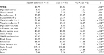
Demographic characteristics of the three groups are presented in Table 1. The three groups were similar as regards as age but not sex, with fewer vaMCI subjects being male. The control group had more education than the stroke patients. Age, sex, and education were controlled for in the inter-group analyses. As expected, the stroke patients had more vascular risk factors, which included history of coronary artery disease, atrial fibrillation, hypertension, smoking, diabetes, and hypercholesterolemia. The vaMCI group had the most vascular risk factors, followed by the NCI group and the control group. Compared with the NCI group, the vaMCI group had lower NART-IQ scores and baseline MMSE scores. Baseline neuropsychological profiles of the three groups are presented in Table 2.
Demographic characteristic of the sample and rates of conversion to dementia after 3 years

Note
Only those with both index and 3-year assessments are included. The three groups are healthy controls, no cognitive impairment (NCI), and vascular mild cognitive impairment (vaMCI) post-stroke. AMI = acute myocardial infarction; NART = National Adult Reading Test; MMSE = Mini-Mental State Examination; ADL = activities of daily living; IADL = instrumental activities of daily living; IQCODE = Informant Questionnaire for Cognitive Decline in the Elderly (Jorm & Jacomb, Reference Jorm and Jacomb1989).
* Control vs. VaMCI.
† Control vs. VaMCI, NCI vs. VaMCI.
‡ Lower conversion rate in the control group and higher conversion rate in the vaMCI group.
§ Control vs. NCI.
^ NCI vs. VaMCI.
a Mean (SD).
b Compared by Mann-Whitney U test.
Comparison of controls, NCI, and vaMCI in neuropsychological test raw scores at index assessment, controlling for age, sex, and education

Note
Post hoc comparisons were adjusted by Bonferroni corrections. NCI = no cognitive impairment; vaMCI = vascular mild cognitive impairment; MMSE = Mini-Mental State Examination; SDMT = Symbol-Digit Modalities Test; COWAT = Controlled Oral Word Association Test.
* Control vs. vaMCI.
† NCI vs. VaMCI.
During the interval between index and follow-up assessments, strokes occurred in three VaMCI, patients, of whom one was diagnosed with dementia at follow-up, and two NCI subjects. After accounting for participants with interval strokes, VaMCI patients were more likely to convert to VaD (n = 11; 24.4%) than NCI patients (n = 5; 8.5%) 3 years after the index stroke/TIA (χ2 = 5.001; df = 1; p = .025), while no participant in the control group was diagnosed with dementia during follow-up. Incident MCI was diagnosed in 31 (36.5%) of controls and 10 (16.9%) of NCI group (χ2 = 5.321; df = 1; p = .021). Twelve (26.7%) VaMCI patients improved and were diagnosed as NCI at follow-up.
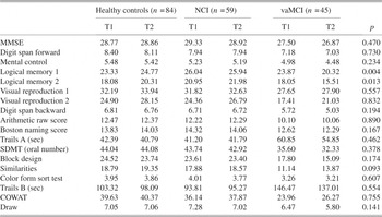
Longitudinal comparisons of cognitive performance among three groups (Table 3) showed significant interactions between group and time in logical memory I (p = .004) and II (p = .012), after controlling for the effects of age, sex, and education, with greater decline in the vaMCI group than NCI and controls. Comparing performance at the two assessments, the vaMCI group showed significant decline in Boston Naming Test (F = 6.827; df = 1; 20; p = .017) and Controlled Oral Word Association test (F = 5.695; df = 1, 20, p = .027). There was a marginal decline in logical memory I (p = .053) and II (p = .072) and visual reproduction II (p = .054).
Comparison of controls, NCI and vaMCI in changes of neuropsychological test raw scores over three years (from index to 3 year assessment), using repeated measures ANCOVA to test interaction of time and group, controlled for age, sex, and education

Note
NCI = no cognitive impairment; vaMCI = vascular mild cognitive impairment; ANCOVA = analysis of covariance; MMSE = Mini-Mental State Examination; SDMT = Symbol-Digit Modalities Test; COWAT = Controlled Oral Word Association Test.
MRI brain measurements at index assessment were compared among the three groups (Table 4). Both NCI and vaMCI had larger volumes of total and deep WMHs in comparison with controls (p < .001). For periventricular WMHs, the significant difference was found between controls and VaMCI only (p < .001). The groups did not differ on brain volumes, ventricular volumes, ventricle-to-brain ratios and hippocampal volumes. The only significant finding was that of smaller amygdala volumes in the vaMCI group.
MRI brain measurements in the study sample at index assessment

Note
ICV = intracranial volume; NCI = no cognitive impairment; vaMCI = vascular mild cognitive impairment; GM = gray matter; WM = white matter; WMH = white matter hyperintensities; VBR = ventricle-to-brain ratio.
* Control vs. NCI, Control vs. VaMCI.
† Control vs. VaMCI, Control vs. NCI.
‡ Control vs. VaMCI.
Comparisons of nonconverters and converters in the vaMCI group did not show significant differences in age, sex, education, proportion of vascular risk factors, or ApoE4 carriers. Nonconverters and converters are comparable in brain volumetric measures. Although converters showed a lower baseline MMSE score than nonconverters, it did not reach statistical significance (p = .087). Converters and nonconverters did not show significant difference in the baseline scores or the slope of decline on any particular cognitive test.
DISCUSSION
We report a longitudinal study of MCI in post-stroke patients. These patients were judged not to have significant cognitive decline prior to their ischemic strokes or TIAs, based on the informant-based questionnaire IQCODE. Furthermore, clinicians decided by consensus that they had sufficient cerebrovascular pathology to account for cognitive impairment. It would, therefore, be reasonable to argue that the MCI in these subjects was vascular in etiology, that is, they had vaMCI.
Our study shows that vaMCI in stroke or TIA patients is progressive, and incident dementia was diagnosed in 24.4% in 3 years, giving a mean value of approximately 8% per year. This was much higher than the rate of 8.5% over 3 years in post-stroke patients who did not meet criteria for MCI. We note that some cognitive decline occurred in all groups. In fact, while no control subject received a dementia diagnosis on follow-up, a significant proportion (36.9%) was diagnosed to have MCI. The rates of transition to MCI were higher in the control group than in the post-stroke NCI group, who showed a categorical transition to MCI or dementia in 25.1%, suggesting that individuals who do not show cognitive impairment following stroke are not particularly prone to cognitive decline in the subsequent years.
The categorical transition during follow-up should be understood in the context of baseline functioning of the different groups. When one examines the performance on individual tests, the decline was not significantly different in the three groups, except on logical memory. Because memory dysfunction was not a necessary requirement for the diagnosis of dementia, this difference is insufficient to account for the differential rates of incident dementia. The difference is mainly due to the fact that the vaMCI group started from a lower baseline function and any decline was more likely to reach the threshold for dementia. At index assessment, the vaMCI performed less well on most cognitive domains except for verbal memory, including working memory, speed on information processing, visual memory, language, visuoconstructive tasks, and frontal-executive function. Those vaMCI subjects who converted to dementia also had greater decline in language function in addition to memory. From this profile, it appears that early memory is relatively spared in early vaMCI and its impairment occurs over time as deficits progress.
The rates of progression and categorical transition of VaMCI have varied in previous reports. In a cohort of patients referred to memory clinic, 40% of vascular cognitive impairment no dementia (VCI-ND) reportedly converted to dementia and 8% improved within 2 years after diagnosis (Hsiung et al., Reference Hsiung, Donald, Grand, Black, Bouchard and Gauthier2006), while 26% improved within 3 years in our study. In the same study, VCI-ND progressed to various type of dementia, including VaD, AD, and Lewy body dementia or Parkinson disease with dementia, suggesting heterogeneity in the diagnosis. Similar results have been found in another longitudinal population-based study, in which 46% of VCI-ND developed dementia while 52% died during 5 years after diagnosis (Wentzel et al., Reference Wentzel, Rockwood, MacKnight, Hachinski, Hogan and Feldman2001). A longitudinal study in post-stroke patients found that 9% non-demented patients developed dementia within 15 months after stroke while 50% improved in cognition (Ballard, Rowan, Stephens, Kalaria, & Kenny, Reference Ballard, Rowan, Stephens, Kalaria and Kenny2003). In another study, based on different criteria of diagnosis, approximately 12–15% patients who were diagnosed vaMCI within 3 months after stroke had developed dementia within 2 years of follow-up, while the rate was only 0–1.4% in cognitively normal post-stroke patients (Serrano et al., Reference Serrano, Domingo, Rodriguez-Garcia, Castro and del Ser2007). Another study reported that 32.7% of those who presented with impaired cognition at baseline assessment after first stroke, compared with 10.6% cognitively normal post-stroke patients, developed dementia at the second-year assessment (Srikanth et al., Reference Srikanth, Quinn, Donnan, Saling and Thrift2006).
The neuropsychological characteristics of vaMCI show many different patterns, owing possibly to a variety of mechanisms involved. When cognitive impairment is directly related to the presence of strategic infarcts affecting a particular structure in the brain, the cognitive pattern is closely related to the corresponding structure. For example, patients with infarcts in angular gyrus were often diagnosed as Alzheimer’s disease (Benson & Cummings, Reference Benson and Cummings1982). The cognitive pattern may also present with accumulated effects of multiple infarcts (Hachinski, Lassen, & Marshall, Reference Hachinski, Lassen and Marshall1974). Post-stroke patients also demonstrate subcortical cognitive impairment, which is caused by various degrees of small vessel obstruction or ischemia, leading to lesions such as lacunar infarcts or white matter hyperintensities. Such ischemic lesions are generally present before the first clinically overt stroke, and the index stroke serves to accentuate previous impairment. It has been reported that MCI due to vascular disease tends to produce greater deficits in the domains of attention, processing speed, and executive function than nonvascular MCI (Nordlund, Rolstad, Klang, Lind, Hansen, & Wallin, Reference Nordlund, Rolstad, Klang, Lind, Hansen and Wallin2007), consistent with the cognitive profile of VaD(Looi & Sachdev, Reference Looi and Sachdev1999). Our findings are not inconsistent with this, with working memory, processing speed, and executive function being more impaired in the early stage of vaMCI, and memory and language showing greater decline in the subsequent follow-up.
Clear determinants of progression did not emerge in our study, except impairment at baseline tended to predict greater decline. Incident dementia was not explained by recurrent strokes or TIAs, the rates of which were low in this sample, arguing that this did not fit the picture of multi-infarct dementia. The decline was slow and appeared to occur in all groups, with the vaMCI group showing greater decline in memory. In our study, converters and nonconverters of VaMCI to VaD did not differ by age, sex, education, burden of vascular risk factors, or structural changes in brain. It is noteworthy, however, that the vaMCI group had more vascular risk factors and more white matter hyperintensities at baseline than the NCI and control groups. Previous studies have suggested some risk factors were associated with cognitive decline and risk of dementia in VaMCI, for example, age (Srikanth et al., Reference Srikanth, Quinn, Donnan, Saling and Thrift2006), white matter hyperintensities (Bombois et al., Reference Bombois, Debette, Bruandet, Delbeuck, Delmaire and Leys2008; Wolf, Ecke, Bettin, Dietrich, & Gertz, Reference Wolf, Ecke, Bettin, Dietrich and Gertz2000), recurrent stroke (Srikanth et al., Reference Srikanth, Quinn, Donnan, Saling and Thrift2006), and low baseline scores of cognitive tests (Ingles, Wentzel, Fisk, & Rockwood, Reference Ingles, Wentzel, Fisk and Rockwood2002; Srikanth et al., Reference Srikanth, Quinn, Donnan, Saling and Thrift2006; Ballard, Reference Ballard, Rowan, Stephens, Kalaria and Kenny2003). Our data suggest that global cognitive and functional impairment at baseline may be of importance in predicting dementia.
The contribution of Alzheimer’s pathology to cognitive impairment and decline in our sample is unknown. Alzheimer’s dementia is common in this age group, and mixed dementia on neuropathology is much more common than clinically diagnosed (Neuropathology Group of the Medical Research Council Cognitive Function and Ageing Study, 2001). A postmortem study of memory clinic patients showed that, while the clinical diagnosis of VaD was common, pure VaD was diagnosed on neuropathology in only one-fifth of these cases (Bowler, Munoz, Merskey, & Hachinski, Reference Bowler, Munoz, Merskey and Hachinski1998). It can be argued that the decline in memory and language function seen in the vaMCI patients in our sample could be due to concomitant AD pathology. Although this cannot be ruled out, we argue that this is unlikely to be the complete explanation. The subjects in the vaMCI group were so diagnosed because they had significant cerebrovascular pathology in the form of white matter hyperintensities and infarcts for a clinical diagnosis by experienced clinicians. They did not show brain atrophy or hippocampal atrophy relative to the control group. Also, subjects when recruited did not have a history of cognitive decline over the previous 5 years. The incidence of dementia of 8% per year, compared with an incidence of approximately 1.8% for this age group in the general population (Jorm & Jolley, Reference Jorm and Jolley1998), is unlikely to be solely due to the progression of AD pathology, and cerebrovascular disease is likely to be the dominant explanation for the cognitive impairment.
Our study has several limitations. First, vaMCI participants in the study are post-stroke or post-TIA in-patients and we did not include vaMCI from other settings. While stroke or TIA can contribute to cognitive impairment, this effect would have been mitigated by the timing of our detailed assessments, some 3–6 months after their cerebrovascular event. In the absence of stroke, vaMCI is more likely to relate to subcortical vascular lesions, and the cognitive impairment will reflect this pathology. It is likely that there is greater heterogeneity of neuropsychological deficits in our sample than would be seen in groups with subcortical ischemic lesions. Second, our sample size was small, and this could account for the lack of significant predictors of decline. Third, the sample had considerable attrition due to severe illness or death, suggesting that more severely affected participants were preferentially lost to follow-up. While this is unavoidable in a longitudinal study, the rates of decline are likely to be under-estimates of the true rates of decline in post-stroke patients.
In conclusion, we report that MCI is common in post-stroke patients and when followed longitudinally, approximately a quarter of these “progress” to dementia over 3 years, a rate which is much higher than that of post-stroke patients who do not show cognitive impairment. Lower cognitive function at baseline appears to be the major risk factor for such conversion.
ACKNOWLEDGMENTS
The authors thank the neurologists at Prince of Wales and St George Hospitals for permission to study their patients; SEALS Laboratories for assistance in blood collection; and Karen Berman, David Gillies, Leslie Howard, Megan Jones, Nicole Kochan, Jeffrey Looi, Eveline Milne, Amy Ross, Penny Sawdy, Jamie Sims, and Alessandro Zagami for their contributions to the study; and Lauren Norton for manuscript preparation. The study was supported by grants from the National Health and Medical Research Council of Australia, the Rebecca Cooper Foundation and Fairfax Family Foundation.