Case presentation
A 31-year-old woman was seen in the clinic with an 18-month history of a gradual reduction in exercise tolerance and worsening breathlessness. The patient described abdominal bloating and dyspnoea while walking slowly along a flat surface, putting her in World Health Organisation functional class III. Initial investigations demonstrated right-axis deviation on electrocardiogram and a dilated right ventricle on echocardiogram.
The patient had a past medical history of a spinal disc herniation, and treated hyperthyroidism. Her medications included a progestogen-only contraceptive pill, omeprazole, aspirin, and occasional co-codamol. There was no significant family history. She was an ex-smoker, having smoked 10 cigarettes/day for 4 years. She did not drink alcohol and denied the use of illicit drug and alternative or herbal medications including anorexigens.
On examination there was a right ventricular heave, and a widely split second heart sound with an accentuated pulmonary component. The chest was clear to auscultation, and there was no peripheral oedema; the jugular venous pulse was at 2 cm above the sternal angle.
The patient was admitted for further investigations:
∙ Blood samples showed a normal full blood count and electrolyte levels. Renal and hepatic function was normal. Virology, autoimmune, coagulation, and haemoglobinopathy screens were negative. Thyroid function was normal.
∙ Electrocardiogram showed sinus rhythm with evidence of right-axis deviation, and T wave inversion in lead V2–V6.
∙ Chest X-ray showed clear lung fields and pleural spaces. The pulmonary arteries were enlarged.
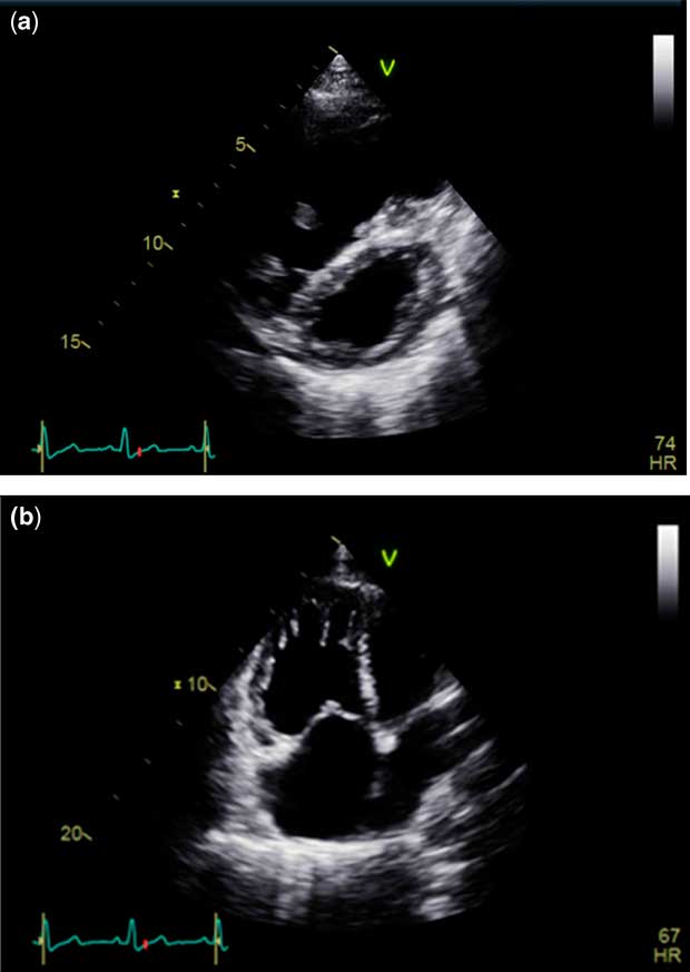
∙ Transthoracic echocardiogram (Fig 1) showed a dilated right ventricle with a diastolic basal diameter of 52 mm (normal range 20–28 mm). The right ventricular systolic function was poor, with a tricuspid annular plane systolic excursion of 14 mm. There was mild tricuspid regurgitation, with a maximum velocity of 3.8 m/second. The left heart structures appeared normal, with normal left ventricular systolic function.
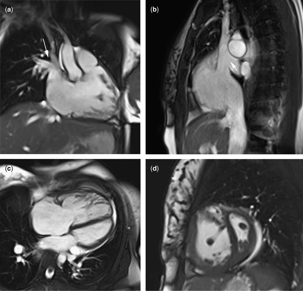
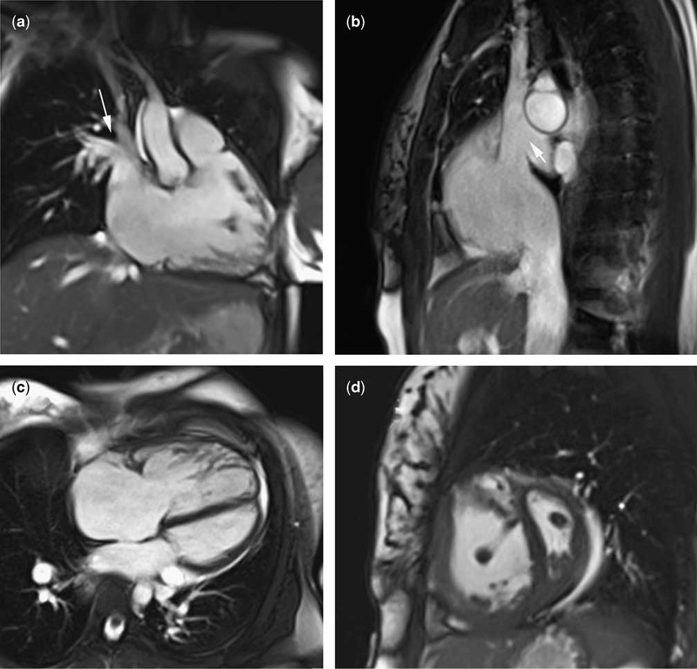
∙ Cardiac MRI (Fig 2) showed situs solitus with atrio-ventricular and ventriculo-arterial concordance. There were bilateral superior caval veins with the left superior caval vein draining to a dilated coronary sinus and the right superior caval vein draining normally to the right atrium. There was a superior sinus venosus atrial septal defect, with partial anomalous pulmonary venous drainage. The right superior and middle pulmonary veins drained to the right-sided superior caval vein, with normal drainage of the right inferior and left-sided pulmonary veins to the left atrium. The right ventricle was dilated and hypertrophied (end-diastolic volume 165 ml/m2), with moderate global impairment of systolic function (ejection fraction 40%). The left ventricle was small and “D”-shaped in short axis, with paradoxical septal movement. The main pulmonary artery was dilated (40 mm), with a pulmonary artery:ascending aorta ratio of >1. Flow analysis estimated a Qp:Qs ratio of 2.3:1.
∙ CT pulmonary angiogram revealed no evidence of pulmonary embolism.
∙ Abdominal ultrasound was normal.
∙ A 6-minute walk test was performed, during which the patient reached 192 m. The test was stopped at 4 minutes and 10 seconds. Oxygen saturations were 97% at the start of the test and 96% at peak exercise. The heart rate increased from 93 to 97 beats per minute. Dyspnoea as measured on the Borg scale was 1 at baseline, rising to 4 immediately post exercise. The blood pressure was 117/84 mmHg at baseline, and 119/85 mmHg immediately post exercise.
∙ Cardiac catheterisation showed a baseline mean pulmonary artery pressure of 54 mmHg, with a mean pulmonary artery wedge pressure of 10 mmHg. These pressures dropped to 53 and 8 mmHg, respectively, with the administration of 100% oxygen and 20 parts per million inhaled nitric oxide. The pulmonary vascular resistance indexed to body surface area was 19.4 Wood units.m2 at baseline, and 14.0 Wood units.m2 with pulmonary vasodilation.
Transthoracic echocardiogram showing a dilated right ventricle from ( a ) parasternal short-axis view and ( b ) apical four-chamber view.

Cardiac MRI. ( a ) Skewed coronal view showing the right ventricle in long-axis, diastolic frame. An anomalous connection is seen between the right-sided superior caval vein and the right upper and middle pulmonary veins (arrow). ( b ) Sagittal view of lateral right atrial chamber, diastolic frame. A large sinus venosus atrial septal defect is seen at the base of the superior caval vein (arrow). ( c ) Four-chamber view diastolic frame. This shows dilated right atrial and right ventricular chambers. At the plane of this image, the interatrial septum appears intact. ( d ) Mid-ventricular short-axis view, early diastolic frame. This demonstrates a dilated, hypertrophied right ventricle, with flattening of the interventricular septum.
The patient was commenced on dual oral pulmonary vasodilator therapy using tadalafil 40 mg once daily and ambrisentan 10 mg once daily. In addition, rivaroxaban 20 mg once daily was commenced for prophylactic anticoagulation. The patient was discharged from hospital, and kept under regular review in the pulmonary hypertension clinic.
Repeat investigations were performed after 3 months:
∙ Cardiac MRI scan showed no significant changes, with a right ventricular end-diastolic volume of 160 ml/m2, and an estimated Qp:Qs ratio of 2.2:1.
∙ Transthoracic echocardiogram demonstrated the superior sinus venosus atrial septal defect with left-to-right shunt. The tricuspid regurgitation velocity was similar to the previous study at 3.6 m/s. The right ventricle remained dilated with impaired systolic function.
∙ Cardiac catheterisation demonstrated a mean pulmonary artery pressure of 42 mmHg, with a mean pulmonary artery wedge pressure of 12 mmHg. The pulmonary vascular resistance indexed to body surface area was 4.2 Wood units.m2. The Qp:Qs ratio derived from the Fick equation was 2:1.
The results of these investigations were discussed in multicentre meetings. The consensus was that these findings indicate a mixed picture of both idiopathic pulmonary arterial hypertension and pulmonary hypertension secondary to CHD. The feeling of the group was that the patient should continue on medical therapy, rather than undergo surgical repair of the atrial septal defect in view of right ventricular dysfunction and surgical risk despite the reduction in pulmonary vascular resistance index.
Currently, her exercise tolerance had improved, and she is able to walk for up to 10–15 minutes at a steady pace. Abdominal bloating persists with ankle oedema. Her dual oral therapy was continued, and furosemide 40 mg once daily was commenced. The patient remains under follow-up of both the pulmonary hypertension and adult CHD teams.
Discussion
Superior sinus venosus atrial septal defects are a rare form of atrial septal defect, characterised by a structural defect in the wall separating the distal superior caval vein and right-sided pulmonary veins.Reference Jost, Connolly and Danielson 1 In addition to the intra-cardiac shunting due to the septal defect, the drainage of the right-sided pulmonary veins is often anomalous to the superior caval vein or right atrium, creating an additional level of left-to-right shunt.Reference Van Praagh, Carrera, Sanders, Mayer and Van Praagh 2 Compared with secundum atrial septal defects, sinus venosus defects are more difficult to diagnose by transthoracic echocardiography,Reference Pascoe, Oh, Warnes, Danielson, Tajik and Seward 3 and are often only diagnosed with advanced imaging techniques.
Owing to later diagnosis, sinus venosus defects are more frequently described as resulting in pulmonary arterial hypertension than other forms of atrial septal defect.Reference Vogel, Berger, Kramer, Alexi-Meshkishvili and Lange 4 Pulmonary arterial hypertension associated with CHD is an increasingly common cause of pulmonary arterial hypertension, and in some registries has a prevalence equal to idiopathic pulmonary arterial hypertension.Reference Dimopoulos, Wort and Gatzoulis 5 Excessive pulmonary blood flow results in endothelial dysfunction and imbalance in cell signalling molecules favouring vasoconstriction and cell proliferation. Sustained haemodynamic stress leads to progressive vascular remodelling and increased pulmonary vascular resistance.Reference Diller and Gatzoulis 6 The degree of pulmonary arterial hypertension in these patients is highly variable, with the most extreme end of the spectrum developing Eisenmenger syndrome,Reference Diller and Gatzoulis 6 in which both pulmonary artery pressure and pulmonary vascular resistance are severely increased with a reversal of shunt direction, and subsequent cyanosis and exercise intolerance.Reference D’Alto and Diller 7 Although sinus venosus atrial septal defects are more likely to result in raised pulmonary vascular resistance compared with secundum atrial septal defects, they are still a relatively rare cause.Reference Vogel, Berger, Kramer, Alexi-Meshkishvili and Lange 4 , Reference Cherian, Uthaman and Durairaj 8 In the seminal series by Wood, Eisenmenger syndrome did not develop until adulthood in the vast majority of atrial septal defect patients, compared with much earlier in those with post-tricuspid level shunts.Reference Wood 9 It is not fully understood why some patients, such as ours, with a fairly small left-to-right shunt develop such a profound degree of pulmonary hypertension.
Surgical repair of the congenital defect in cases of pulmonary arterial hypertension associated with CHD is controversial. In the presence of clearly increased pulmonary vascular resistance (>8 Wood units·m2), the closure of these defects is contra-indicated, as this has been shown to worsen survival when compared with unrepaired patients.Reference Dimopoulos, Wort and Gatzoulis 5 , Reference D’Alto and Diller 7 , Reference Wood 9 Shunt patency allows decompression of the right heart and reduces the likelihood of right ventricular failure. In contrast, in patients with raised pulmonary artery pressure but normal pulmonary vascular resistance (<4 Wood units·m2), surgical closure is usually advocated to prevent the development of increased pulmonary vascular resistance. Difficulties arise in patients such as ours, who have a raised pulmonary artery pressure with borderline pulmonary vascular resistance. Such cases should be assessed on a case-by-case basis by a team skilled in both CHD and pulmonary hypertension.
Although idiopathic pulmonary arterial hypertension is similar to pulmonary arterial hypertension associated with CHD in terms of lung histology, the underlying pathophysiology and treatment strategies are different.Reference Dimopoulos, Wort and Gatzoulis 5 Despite the fact that idiopathic pulmonary arterial hypertension and pulmonary arterial hypertension associated with CHD are classified as distinct disease entities, in some patients, such as ours, there is a degree of overlap. In patients with small defects but severe pulmonary arterial hypertension, as in our case, the degree of pulmonary arterial hypertension is deemed to be out of proportion to the modest increase in pulmonary blood flow.Reference Dimopoulos, Wort and Gatzoulis 5 It is likely that our patient had a greater degree of pulmonary over-circulation before her pulmonary vascular resistance started to rise, although this cannot be proved retrospectively. Although it is difficult to rule out any association between the sinus venosus atrial septal defect and idiopathic pulmonary arterial hypertension in our patient, it was felt extremely unlikely that this defect alone could result in such a profound degree of pulmonary arterial hypertension.
The differentiation between the two forms of pulmonary arterial hypertension is important, as the treatment varies. As outlined above, in some situations in pulmonary arterial hypertension associated with CHD, the correct course of action would be to surgically repair the underlying heart defect, in this case a sinus venosus atrial septal defect. In idiopathic pulmonary arterial hypertension, however, the presence of an atrial level shunt is felt to be associated with improved survival, and some cases a balloon atrial septostomy is performed to maintain cardiac output by allowing right-to-left shunting.Reference Dimopoulos, Wort and Gatzoulis 5 Because of this, surgical repair of such a defect would rarely be advocated.
Conclusion
It is important to recognise when the degree of pulmonary hypertension is out of proportion to the degree of left-to-right shunting, which may indicate the coexistence of idiopathic pulmonary arterial hypertension. This will lead to a more aggressive medical management strategy, and is likely to mean that surgical repair of the congenital heart lesion is not recommended.
Acknowledgements
The authors are grateful to Dr Ekaterina Yarovova and Sandra Ladkin for their help in the preparation of this manuscript.
Financial Support
This research received no specific grant from any funding agency, commercial, or not-for-profit sectors.
Conflicts of Interest
None.