INTRODUCTION
The genus Hydroides Gunnerus, 1768 is the most species rich serpulid genus with 89 valid species (ten Hove & Kupriyanova, Reference Hove and Kupriyanova2009). The Hydroides species from the Western Atlantic and Eastern Pacific (including Hawaii) have been taxonomically revised by Bastida-Zavala & ten Hove (Reference Bastida-Zavala and Hove2002, Reference Bastida-Zavala and Hove2003), but species from the most diverse region (Indo-Pacific) are still understudied. Most of the Hydroides are readily distinguished by opercula structures; otherwise they are very similar morphologically with exception of a few species with more than 7 thoracic chaetigers (i.e. Hydroides bisectus Imajima & ten Hove, Reference Imajima and ten Hove1989 and Hydroides sp. 2 sensu Bastida-Zavala & ten Hove, Reference Bastida-Zavala and Hove2003).
Hydroides perezi is the only Hydroides species previously recorded in checklists of Marshallese polychaetes (Bailey-Brock, Reference Bailey-Brock1999, Reference Bailey-Brock2003), but detailed description of this species in Guam is lacking. We provide descriptions and scanning electron microscopy (SEM) photographs of opercula and chaetae of H. perezi from Pago Bay in addition to two new records H. albiceps and H. bannerorum from Apra Harbor, Guam.
MATERIALS AND METHODS
Specimens were collected by SCUBA divers in December 2010 at several stations within Apra Harbor, Guam. The sediment was preserved in 15% buffered formalin with rose Bengal and later stored in ethanol 70%. Coral chips and rubble pieces were removed from sediments and examined for tubes which could be scraped off and dissolved in an acid bath as described in Brock & Brock (Reference Brock and Brock1977). Hydroides specimens were removed from the debris after dissolution, therefore material treated in the acid bath had calcareous tubes completely dissolved. Preserved specimens were examined with light microscopy and prepared for SEM. Specimens were dehydrated in a graded ethanol series, dried in a SAMDRI-795 critical point dryer using liquid CO2 and gold-coated for about 2 minutes. SEM observations were carried out using the Hitachi S-4800 at the Biological Electron Microscopy Facility (BEMF), University of Hawaii at Manoa. Descriptions are following most recent standards by Bastida-Zavala & ten Hove (Reference Bastida-Zavala and Hove2002, Reference Bastida-Zavala and Hove2003). Voucher material of the three species is deposited at the Bernice Pauahi Bishop Museum, Honolulu, Hawaii, USA (BPBM).
RESULTS
SYSTEMATICS
Family SERPULIDAE Rafinesque, 1815
Genus Hydroides Gunnerus, 1798
Hydroides albiceps (Grube, Reference Grube1870)
(Figures 1 & 2)
Serpula (Eupomatus) albiceps Grube, Reference Grube1870: 520–521.
Hydroides albiceps—Straughan, Reference Straughan1967a: 220, figure 6m; Pillai, Reference Pillai1971: 112–113; Imajima, Reference Imajima1976a: 133–135, figure 8a–v; Imajima, Reference Imajima1976b: 234–235; Imajima, Reference Imajima1978: 53–54; Imajima, Reference Imajima1979: 168; Mak, Reference Mak, Morton and Tseng1982: 601–602; Imajima, Reference Imajima1982: 44; Imajima & ten Hove, Reference Imajima and ten Hove1984: 43–44; Vine & Bailey-Brock, Reference Vine and Bailey-Brock1984: 139, figure 2k; Imajima & ten Hove, Reference Imajima and ten Hove1986: 5; Imajima, Reference Imajima1987: 77; ten Hove, Reference Hove1990: figures 10–12; ten Hove, Reference Hove and Land1994: 107–108; Meng et al., Reference Meng, Hong and Wu1994: 47; Ishaq & Mustaquim, Reference Ishaq and Mustaquim1996: 164–166, figure 2a–g; Fiege & Sun, Reference Fiege and Sun1999: 112–114, figure 2d–f.
Scanning electron microscopy of Hydroides albiceps: (A) operculum in lateral and top view; (B) detail of collar chaeta; (C) thoracic uncini; (D) anterior abdominal uncini; (E) flat trumpet abdominal chaetae; (F) posterior abdominal uncini.

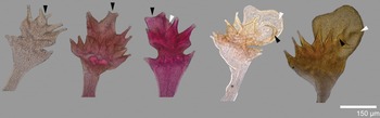
Ontogenetic variation on opercula of Hydroides albiceps. Black arrows show development of two dorsal-most verticil spines and white arrows show development of bulbous part of dorsal spines.

MATERIAL EXAMINED
Pacific Ocean—Apra Harbor, Guam, Mariana Islands, December 2010: Station B3, 13°26′46.08″N 144°39′44.34″E, 14.6 m (3, BPBM R3656); Station B6, 13°27′15.9″N 144°39′35.57″E, 16.5 m (2, BPBM R3657); Station B11, 13°27′12.51″N 144°39′32.7″E, 17.1 m (2, BPBM R3658); Station B14, 13°27′7.8″N 144°39′48.40″E, 18.3 m (5, BPBM R3659); Station B20, 13°26′54.37″N 144°39′40.4″E, 15.1 m (1, BPBM R3660); Station B22, 13°26′57.2″N 144°39′55.9″E, 18 m (1 on stub); Station B25, 13°26′51.5″N 144°39′55.4″E, 16.5 m (4, BPBM R3661).
DESCRIPTION
Tube
Only one fragmented tube observed; white to pale yellow, with faint transversal and 2 longitudinal ridges.
Branchiae
With five radioles left (N = 13, μ = 5 ± 1.73) and five right (N = 14, μ = 4.85 ± 1.56); terminal filament very long in all specimens. Branchial eyes and inter-radiolar membrane absent.
Peduncle
With left (N = 9, 64.3%) or right (N = 5, 35.7%) insertion; with well-defined constriction. Pseudoperculum present in 10 specimens (83.3%), lacking in two (16.6%).
Operculum
Funnel with 14 radii (N = 15, μ = 14.5 ± 4.18) with straight and expanded tips (Figure 1A). Interradial grooves 1/3 of funnel length. Verticil with large dorsal vesicular spine and 8 long, straight and slightly expanded laterally (N = 9, μ = 8.44 ± 0.88); six specimens lacking verticil. Dorsal spine with a bulbous median part and two lateral processes triangular in dorsal view; median bulb with verticil spines inserted at base (Figure 1A).
Collar and thoracic membranes
Collar smooth, tri-lobed with undulating margin. Thoracic membranes well-developed; apron present.
Thorax
With collar chaetiger and 6 uncinigerous chaetigers; collar chaetae 3–5 bayonet with two pointed-elongate teeth and basal processes (Figure 1B), distal blade denticulated; 6–8 limbate capillaries present. Thoracic uncini saw-shaped with 4–5 teeth including a simple large anterior fang (Figure 1C).
Abdomen
With 38 (N = 12, μ = 38 ± 18.49) chaetigers. Anterior and middle abdominal chaetigers with 3–4 flat trumpet chaetae (Figure 1E). Posterior 8–10 chaetigers with long ‘capillary’ chaetae. Anterior uncini saw-shaped with 5–6 teeth including large anterior fang (Figure 1D); posterior uncini rasp-shaped with 4–5 teeth organized in 7–8 transversal rows in addition to single large anterior fang (Figure 1F).
Colour and size
Preserved specimens pale yellow; 1.5–6 mm in length, up to 1.0 mm width of thorax, operculum 0.2–0.5 mm length.
DISTRIBUTION
Red Sea and widely distributed in the Indo-Pacific (ten Hove & Kupriyanova, Reference Hove and Kupriyanova2009). It has been found in Japan (e.g. Imajima, Reference Imajima1976a), South China Sea (Yang & Sun, Reference Yang and Sun1988), Marshall Islands (Imajima & ten Hove, Reference Imajima and ten Hove1986) and newly recorded to Guam in the Mariana Islands.
REMARKS
This species is most similar to Hydroides trivesiculosus Straughan, 1967, Hydroides steinitzi Ben-Eliahu, 1972 and Hydroides bulbosus ten Hove, Reference Hove1990 by the presence of a large bulbous dorsal spine in the verticil. A detailed comparison of these three species can be found in ten Hove (Reference Hove1990: 119, figures 7–22). Fiege & Sun (Reference Fiege and Sun1999) provide a comparative table with H. albiceps, Hydroides malleolaspinus Straughan, 1967, H. trivesiculosus and Hydroides ancorispinus Pillai, Reference Pillai1971.
The material collected in Apra Harbor, Guam presented different developmental stages of opercular structures (Figure 2). Several specimens only presented the funnel, lacking the verticil. Others had a simple verticil (see Figure 1A; ‘priscus’-type sensu ten Hove & Ben-Eliahu, Reference Hove and Ben-Eliahu2005), which made difficult identification as it resembles several other Hydroides species (e.g. Hydroides nodosus Straughan, 1967 from Australia). Details of collar and abdominal chaetae, thoracic and abdominal uncini clarified with SEM helped to ascertain that specimens lacking verticil and with a ‘priscus’-type operculum actually belonged to earlier developmental stages of H. albiceps and not to a different species. For instance, early stages of H. albiceps opercula seem to begin with a single crown followed by a ‘priscus’-type operculum; later on, two of the verticil spines differentiate on the dorsal side and the bulbous part of the dorsal spine start to growth (Figure 2). Well-developed opercula have a large dorsal bulbous part with two lateral triangular processes (modified dorsal-most verticil spines).
Hydroides bannerorum Bailey-Brock, Reference Bailey-Brock1991
(Figure 3)
Hydroides bannerorum Bailey-Brock, Reference Bailey-Brock1991: 199, figure 1a–r.
Hydroides bannerorum—Bastida-Zavala & ten Hove, Reference Bastida-Zavala and Hove2003: 72–73, figure 2 a–g; Bastida-Zavala, Reference Bastida-Zavala2008: 22, figure 6b.
MATERIAL EXAMINED
Pacific Ocean—Apra Harbor, Guam, Mariana Islands, December 2010: Station B33, 13°26′45.05″N 144°40′6.25″E, 18 m (1, BPBM R3662; 1 on stub).
DESCRIPTION
Tube
Not observed in material from Guam.
Branchiae
With four radioles left (N = 2, μ = 4 ± 0) and four radioles right (N = 2, μ = 4 ± 0); terminal filament long in both specimens. Branchial eyes and inter-radiolar membrane absent.
Peduncle
With left (N = 4, 50%) or right (N = 4, 50%) insertion; with ill-defined constriction. Pseudoperculum present in both specimens.
Operculum
Funnel absent. Verticil with 14 (N = 2, μ = 14.5 ± 3.53) dark brown spines, pointed, slightly curved out (Figure 3A). Spines with one small basal internal spinule.
Scanning electron microscopy of Hydroides bannerorum: (A) operculum in lateral view; (B) detail of collar chaeta; (C) thoracic uncini; (D) anterior abdominal uncini; (E) flat trumpet abdominal chaetae; (F) posterior abdominal uncini.

Collar and thoracic membranes
Collar smooth, tri-lobed. Thoracic membranes not well-developed; apron present.
Thorax
With collar chaetiger and 7 uncinigerous chaetigers; collar chaetae 3–4 bayonet with 2–4 pointed-elongate teeth and basal processes (Figure 3B), distal blade denticulate; 4–6 limbate capillaries present. Thoracic uncini rasp-shaped with 7–8 teeth including a simple large anterior fang organized in 2 rows (Figure 3C).
Abdomen
With 15 (N = 1) chaetigers. Anterior and middle abdominal chaetigers with 3–4 flat-trumpet chaetae (Figure 3E). Posterior end chaetigers with long ‘capillary’ chaetae. Anterior uncini saw-shaped with 6–7 teeth including large anterior fang (Figure 3D); posterior uncini rasp-shaped with 4–5 teeth organized in 6–7 transversal rows in addition to large anterior fang (Figure 3F).
Colour and size
Preserved specimens pale yellow, verticil with light to dark brown spines; 3 mm in length, 0.3 mm width of thorax, operculum 0.1–0.2 mm length.
DISTRIBUTION
Oahu, Hawaii (Bailey-Brock, Reference Bailey-Brock1991) and newly recorded to Guam in the Mariana Islands.
REMARKS
This species is unique in the genus Hydroides because it lacks the opercular funnel and by the presence of more than seven thoracic chaetigers. The collar chaeta in H. bannerorum may have up to four teeth and proximal rasps covering completely the base of main teeth, unlike most Hydroides species that normally have only two teeth and proximal rasps.
Hydroides perezi Fauvel, Reference Fauvel1918
(Figure 4)
Eupomatus perezi Fauvel, Reference Fauvel1918: 342, figure 2.
Eupomatus perezi—ten Hove, Reference Hove1970: 58–59, figures 9–16.
Hydroides perezi—Fauvel, Reference Fauvel1953: 457, figure 240a–j; Straughan, Reference Straughan1967a: 221, figure 6o; Straughan, Reference Straughan1967b: 30; Pillai, Reference Pillai1972: 14, figure 3m; Ben-Eliahu, Reference Ben-Eliahu1976: 106, figure 2; Amoureux et al., Reference Amoureux, Rullier and Fishelson1978: 145–146; Vine & Bailey-Brock, Reference Vine and Bailey-Brock1984: 142, figure 2i, j.
MATERIAL EXAMINED
Pacific Ocean—Pago Bay, Guam, Mariana Islands, 8 April 1981, coll. JHB-B (2 on stub; 1, BPBM R3663).
DESCRIPTION
Tube
Opaque, white, with two longitudinal ridges and faint transversal ridges; peristomes absent.
Branchiae
With five radioles left (N = 3, μ = 5 ± 1) and five radioles right (N = 3, μ = 5.33 ± 0.57); terminal filament long in all specimens. Branchial eyes and inter-radiolar membrane absent.
Peduncle
With right in one specimen and two specimens with two opercula fully developed; with well-defined constriction. Pseudoperculum fully developed in two specimens and not observed in one.
Operculum
Funnel with 21 radii (N = 3, μ = 21.33 ± 1.15) with slightly curved out spines with light brown tips (Figure 4A). Interradial grooves 1/3 of funnel length. Verticil with four inflated spines and one large dorsal spine, curved inwards (Figure 4A). One specimen with two opercula had one fully chitinized operculum without dorsal blunt tubercles and one soft operculum with blunt tubercles (tuberculatus-type) on two of the inflated spines.
Scanning electron microscopy of Hydroides perezi: (A) operculum in lateral view; (B) detail of collar chaeta; (C) thoracic uncini; (D) anterior abdominal uncini; (E) flat trumpet abdominal chaetae; (F) posterior abdominal uncini.

Collar and thoracic membranes
Collar smooth, tri-lobed. Thoracic membranes well-developed; apron present.
Thorax
With collar chaetiger and 6 uncinigerous chaetigers; collar chaetae 3–4 bayonet with 2 pointed-elongate teeth and basal processes (Figure 4B), distal blade denticulate; 4–6 limbate capillaries present. Thoracic uncini saw-shaped with 6–7 teeth including a simple large anterior fang (Figure 4C).
Abdomen
With 35 (N = 3, μ = 35.33 ± 4.72) chaetigers. Anterior and middle abdominal chaetigers with 3–4 flat–trumpet chaetae (Figure 4E). Posterior 6–8 chaetigers with long ‘capillary’ chaetae. Anterior abdominal uncini saw-shaped with 6–7 teeth including large anterior fang (Figure 4D); posterior uncini rasp-shaped with 5 teeth organized in 7–8 transversal rows in addition to large anterior fang (Figure 4F).
Colour and size
Preserved specimens pale yellow, funnel and verticil spines yellow to light brown; 2.6–3 mm in length, up to 0.3 mm width of thorax, operculum 0.2–0.3 mm length.
DISTRIBUTION
Widespread in the Indo-Pacific, from Iranian and Persian Gulf to Australia (e.g. Fauvel, Reference Fauvel1953; Straughan, Reference Straughan1967a, Reference Straughanb; Amoureux et al., Reference Amoureux, Rullier and Fishelson1978) and newly recorded to Guam in the Mariana Islands.
REMARKS
This species is very similar to Hydroides tuberculatus Imajima, 1976, Hydroides uniformis Imajima & ten Hove, Reference Imajima and ten Hove1986 and Hydroides dafnii (Amoureux, Rullier & Fishelson, Reference Amoureux, Rullier and Fishelson1978). For instance, dorsal tubercles on verticil spines are absent in H. perezi (but see Discussion) and present in H. tuberculatus. Hydroides uniformis does not have a distinct dorsal spine that is curved inwards as in H. perezi and H. tuberculatus. Hydroides dafnii was described as being a member of Eupomatus (thus lacking lateral spinules on verticil spines) and it was compared to Hydroides gracilis (Bush, 1905) and Hydroides helmatus (Iroso, 1921) although it is most similar to H. perezi and could be synonymous to the latter; however, it is out of the scope of this paper to clarify the relationships among these similar taxa.
DISCUSSION
The genus Hydroides has its most diversification in the Indo-Pacific region with about 52% of the described species (Bastida-Zavala & ten Hove, Reference Bastida-Zavala and Hove2002). Most of these species have not been analysed with SEM imaging to observe the fine structure of the chaetae. The two new records represented herein are well-known species, one by its uniqueness of having full grown opercula with only the verticil and more than 7 thoracic chaetigers (H. bannerorum) and the other by its opercular variability that may lead to taxonomic confusion as not completely developed opercula can show similarities to closely related species (H. albiceps).
Bailey-Brock (Reference Bailey-Brock1991) and Bastida-Zavala & ten Hove (Reference Bastida-Zavala and Hove2003) agree that Hydroides bannerorum is an atypical member of the genus and that it has secondarily lost the funnel instead of not having developed a verticil. Another interesting feature of this species is its collar chaetae with up to four pointed teeth with proximal rasps covering completely the base of main teeth (compare Figures 1B, 3B and 4B). The presence of more than 3 teeth on collar chaetae had led Uchida (Reference Uchida1978) to propose a new genus although ten Hove (Reference Hove and Hutchings1984) has discussed the variability of this character.
One of the most common species in the Indo-Pacific region, Hydroides albiceps has a wide variability in shape and development of the bulbous part of the dorsal spine (ten Hove, Reference Hove1990). We have observed on specimens with different developmental stages that the two dorsal-most spines of the ‘priscus’-type opercula are joined together to form the bulbous part of the operculum but remain as lateral triangular processes (see Figure 2).
Hydroides perezi has been previously recorded to Guam by Bailey-Brock (Reference Bailey-Brock1999, Reference Bailey-Brock2003) and it is described and illustrated herein. The operculum of this species seems to be closely related to the operculum of H. tuberculatus, but differs by the absence of dorsal tubercles on verticil spines of the former. One specimen studied from Guam with soft operculum showed discrete dorsal elevations on some of the verticil spines. However, additional information on the junction of verticil spines is necessary for both species as it has not been observed in Marshallese material and is believed to be absent in H. tuberculatus. The presence of this ‘waistcoat-pocket’ structure in H. perezi is emphasized in Straughan (Reference Straughan1967a, Reference Straughanb) and ten Hove (Reference Hove1970).
ACKNOWLEDGEMENTS
This is contributed paper WRRC–CP–2013–08 of the Water Resources Research Center (WRRC), University of Hawai'i at Mānoa, Honolulu. Collections within Apra Harbor were made by CSA International and in Pago Bay by the author (J.H. B.-B.) and Richard E. Brock. We also thank the efforts of Drs L. Eldredge and C. Birkeland. The staff at the Wormlab in the Department of Zoology at the University of Hawaii at Manoa processed the samples collected from Apra Harbor. This research was funded by a grant from CSA International administered through the WRRC. Jonathon Roberts, UH Manoa Office of Technology Transfer & Economic Development (OTTED), prepared the contract with CSA International. One anonymous referee improved a previous version of this manuscript.