Atrioventricular nodal re-entrant tachycardia accounts for 13–30% of cases of supraventricular tachycardia.Reference Ko, Deal, Strasburger and Benson 1 – Reference Blaufox, Warsy, D’Souza and Kanter 3 Treatment for paediatric supraventricular tachycardia by cardiac ablation is a common procedure. Historically, fluoroscopy was used to guide cardiac ablations. The effects of ionising radiation are dose-related and cumulative. They include dermatitis, genetic effects, and both fatal and non-fatal malignancies. 4 – Reference Koenig, Wolff, Mettler and Wagner 7 Radiation exposure is of particular concern in children, who are more radiosensitive than adults and have a longer lifespan over which radiation doses accumulate. Numerous investigators have explored and evaluated methods to decrease fluoroscopy times during cardiac ablations.
To decrease fluoroscopy times during cardiac ablations, three-dimensional mapping as an alternate way to continuously visualise catheter locations is being used. Several groups have shown that procedures using three-dimensional mapping have shorter fluoroscopy times and have similar safety and efficacy to those using traditional fluoroscopy methods.Reference Cummings, Mahle and Strieper 8 – Reference LaPage, Saul and Reed 14 Many of these studies were prospective trials carried out with an explicit goal of using zero fluoroscopy.
At our institution, we have used three-dimensional mapping (EnSite NavX Mapping System, St. Jude Medical Inc., St. Paul, Minnesota, United States of America) since June, 2007, but without an explicit goal of zero fluoroscopy use. In this study, we sought to describe a single institution’s fluoroscopy and total catheterisation times 5 years after the introduction of three-dimensional mapping without an explicit goal of zero fluoroscopy.
Materials and methods
After obtaining institutional review board approval, the cardiac ablation database at the Children’s Hospital of Pittsburgh was queried for all patients who underwent ablation for atrioventricular nodal re-entrant tachycardia between January, 2004 and December, 2011. Patients ⩽21 years of age were included.
Exclusion criteria were haemodynamically significant congenital heart disease (CHD) and any previous ablation attempt. An institutional shift in treatment for atrioventricular nodal re-entrant tachycardia from radiofrequency ablation to cryoablation in 2006 prompted the division of patients into three separate groups. Group 1 included 18 patients who underwent radiofrequency ablation without three-dimensional mapping from January, 2004 to September, 2006. Group 2 consisted of 13 patients who underwent cryoablation without three-dimensional mapping from October, 2006 to July, 2007. Group 3 included 62 patients who underwent cryoablation with three-dimensional mapping from June, 2007, when three-dimensional mapping was introduced, to November, 2011.
Ablations in our institution are carried out by one of the two electrophysiologists with an agreed-upon approach to cryoablation for atrioventricular nodal re-entrant tachycardia. Our institutional strategy is to use a 6-mm tip cryoablation catheter and to place a 4-minute lesion at the site of effect on the slow pathway followed by a 4-minute freeze–thaw–freeze lesion in the same location. We then place three to five 4-minute insurance lesions surrounding this location for a minimum total of five to seven lesions and 20–28 minutes of ablation.
Data collected included demographics, fluoroscopy time, total catheterisation time, ablation time, acute and chronic procedural success, time from ablation to the recurrence of supraventricular tachycardia or until the patient’s most recent clinic appointment, and any adverse events or complications that occurred during or soon after the procedure. Total catheterisation time was defined as time from insertion of the first catheter to removal of the last sheath. Acute procedural success was defined as absence of an inducible arrhythmia at the completion of the procedure. Chronic success was defined as absence of supraventricular tachycardia at the time of the patient’s most recent follow-up appointment.
Comparisons among the three groups were made with an α level of 0.05. χ2 tests were used for categorical data, and Kruskal–Wallis tests were used for continuous data. Pairwise comparisons between two groups were performed at an α level of 0.0167. χ2 tests were used for categorical data, and Wilcoxon’s rank-sum tests were used for continuous data. A simple linear regression model of fluoroscopy time relative to calendar time – that is, days after January 1, 2004 – was created and the significance of the slope was evaluated. Calculations were made using SAS 9.2.
After data analysis, three outliers with much longer fluoroscopy times were identified within Group 3. Of these patients, one had undergone device closure of a secundum atrial septal defect in the past. The patient was included in the study due to the absence of what was considered to be haemodynamically significant CHD; this history, however, may have made the case more difficult. A second patient’s reported fluoroscopy time of 70 minutes was thought most likely to have been documented erroneously as the procedure was uncomplicated with a procedure time of only 175 minutes and a recorded fluoroscopy dose of 1187 cGy/cm2. The third patient underwent placement of 11 full lesions during what was felt to have been a difficult case.
Results
Patients
A total of 100 patients were initially included in the study. Among them, six patients were excluded for having had a previous ablation attempt and one patient was excluded due to haemodynamically significant CHD. Therefore, 93 patients were included in the analysis.
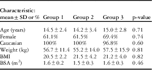
Baseline demographic data are shown in Table 1. There were no significant differences in patient demographics among the groups.
Patient characteristics.

BMI=body mass index; BSA=body surface area
Fluoroscopy and catheterisation times
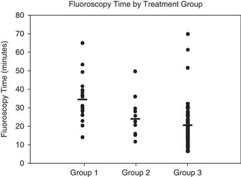
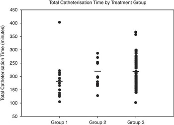
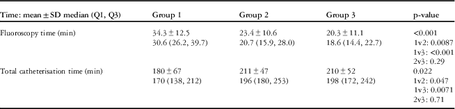
Mean fluoroscopy and total catheterisation times are shown in Table 2. Mean fluoroscopy times in Groups 1, 2, and 3 were 34.3±12.5, 23.4±10.6, and 20.3±11.1 minutes, respectively (Fig 1). Mean fluoroscopy times varied significantly by group when all the groups were compared (p<0.001). Mean total catheterisation times for Groups 1, 2, and 3 were 180±67, 211±47, and 210±52 minutes, respectively, p=0.02 (Fig 2). When Groups 2 and 3 were compared directly, their fluoroscopy times (p=0.29) and total catheterisation times (p=0.72) were not significantly different. The means of the difference between the total catheterisation time and the ablation time in Groups 1, 2, and 3 were 183.7±78.6, 188.2±40.2, and 175.1±43.0 minutes, respectively. These differences were comparable among all groups (p=0.41).
Multiple scatter graph demonstrating decreased fluoroscopy time in patients who underwent cryoablation with 3D mapping (Group 3) compared with Groups 1 and 2.

Multiple scatter graph demonstrating increased catheterisation time in Groups 2 and 3 compared with Group 1.

Fluoroscopy and total catheterisation times.

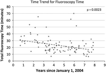
On the basis of an unadjusted linear regression model, there was a significant decrease in fluoroscopy time (Fig 3, estimated slope=−2.00 minutes per year, p=0.0023) across the years.
Unadjusted linear regression model showing a progressive decrease in fluoroscopy time throughout the study period.

Acute and chronic success
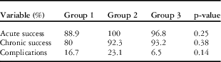
The percentages of patients who achieved acute and chronic success are shown in Table 3. Acute success was achieved in 89, 100, and 97% of patients in Groups 1, 2, and 3, respectively (p=0.25). Chronic success was achieved in 80, 92, and 93% of patients for whom this information could be obtained in Groups 1, 2, and 3, respectively (p=0.38). The mean times from catheterisation to most recent follow-up appointment were 779±584, 711±610, and 398±374 days in Groups 1, 2, and 3, respectively (p=0.08).
Acute and chronic success and complications.

Complication rates
The percentages of patients who experienced complications are shown in Table 3. These were not statistically different among groups (17, 23, and 7% in Groups 1, 2, and 3, respectively, p=0.15). Of the complications in Group 1, two patients had transient right bundle branch block that normalised by the morning after ablation, and one patient had second-degree atrioventricular block that was treated with steroids and normalised by the time of the patient’s follow-up appointment 3 days after the procedure. Of the complications in Group 2, one patient had intermittent second-degree atrioventricular block that began several hours after the procedure and resolved by the most recent follow-up appointment, one patient developed complete atrioventricular block during the procedure that was treated with steroids and lasted for 33 hours, and one patient had both a thrombus of the inferior caval vein and severe circumferential dermatitis at the location of the blood pressure cuff that was treated with steroids. Of the complications in Group 3, three patients developed second-degree atrioventricular block several hours after the procedure that resolved by the time of each patient’s first follow-up appointment. Another patient had a brief episode of non-sustained ventricular tachycardia before discharge, and one patient had atrial fibrillation after isoproterenol administration that resolved with synchronised cardioversion.
Discussion
The purpose of this study was to describe fluoroscopy and total catheterisation times after 5 years of use of three-dimensional mapping at an institution where there has not been an explicit goal of zero fluoroscopy use. In addition, we describe the safety and efficacy of our current techniques of cryoablation with three-dimensional mapping for ablation of atrioventricular nodal re-entrant tachycardia as compared with two previous groups: patients who underwent radiofrequency ablation without three-dimensional mapping and patients who underwent cryoablation without three-dimensional mapping.
Several studies have shown that the use of three-dimensional mapping decreases fluoroscopy times. Most of these studies, however, have been carried out with a goal of zero fluoroscopy use. In 2006, Papagiannis et alReference Papagiannis, Tsoutsinos and Kirvassilis 12 reported one of the earliest studies comparing fluoroscopic catheter navigation techniques with three-dimensional mapping techniques. They concluded that the use of three-dimensional mapping resulted in decreased fluoroscopy and catheterisation times in patients who underwent ablation for atrioventricular nodal re-entrant tachycardia, although the difference was not statistically significant. Miyake et alReference Miyake, Mah and Atallah 13 later showed that fluoroscopy times decreased and total catheterisation times did not change when ablation using non-fluoroscopic imaging was compared with traditional methods. This study included patients with supraventricular tachycardia of any type, and the majority of patients underwent radiofrequency ablation. The study cited historical data of fluoroscopy times between 28.5 and 38.3 minutes. The study group using three-dimensional imaging had a significantly lower median fluoroscopy time than the control group, but the control group’s median fluoroscopy time of 18.3 minutes was also much lower than their cited historical data. This suggests that performing an active prospective study may result in decreased fluoroscopy times in and of itself. Our data show that three-dimensional mapping decreases fluoroscopy times even when this is not an explicit goal of its use.
We sought to describe the decrease in fluoroscopy times, and therefore radiation exposure, with our current techniques of cryoablation and three-dimensional mapping without having an explicit goal of zero fluoroscopy. We chose to evaluate current techniques of cryoablation with three-dimensional mapping in patients undergoing ablation for atrioventricular nodal re-entrant tachycardia, because it is a relatively homogeneous population in that all patients have similar substrates. Although total radiation dose has been found to be of greater clinical importance than fluoroscopy time, the authors were unable to obtain dose data for all patients. Before an equipment upgrade in 2010, radiation doses were entered manually into the electronic record. Many of the doses entered manually after 2010 were found to be incorrect when compared with the doses transferred directly from the equipment, often times because only one plane was accounted for. The manually entered data were therefore considered to be unreliable and fluoroscopy time was used as a proxy.
Among complications we chose to include events such as transient, intermittent second-degree atrioventricular block that some would argue are not true complications. Our complication rates may therefore be overestimated. In 2008, Avari et alReference Avari, Jay and Rhee 15 showed that cryoablation is as safe and effective as radiofrequency ablation, reporting a 97% acute success rate. In their study, 26% of patients experienced temporary second-degree atrioventricular block and one patient (2.6%) developed chronic asymptomatic first-degree atrioventricular block. Total catheterisation times of procedures involving radiofrequency ablations and cryoablations were similar, but median fluoroscopy time was shorter in those patients undergoing cryoablation. In 2010, LaPage et al also reported that cryoablation is a safe and effective therapy for atrioventricular nodal re-entrant tachycardia, with a 100% acute success rate and a 6.5% recurrence rate and with 17% of the patients experiencing procedural complications. These percentages of success rates and complications were similar to those found in our study.
In this study, we show that fluoroscopy times decreased significantly as our techniques transitioned from radiofrequency ablation without three-dimensional mapping to cryoablation without three-dimensional mapping and, finally, to cryoablation with three-dimensional mapping. When Groups 2 and 3 were compared directly, the decrease in fluoroscopy times was not significant (23.4 and 20.3 minutes in Groups 2 and 3, respectively, p=0.29). It is likely that this difference was not significant due to the small number of patients in Group 2 and due to three outlier patients included in Group 3 who had much longer fluoroscopy times than the remainder of the group. Our study is limited by the relatively small number of patients included in Group 2; this likely led to the results being statistically underpowered. There was a short time period between the transition from radiofrequency ablation to cryoablation in 2006 and the introduction of three-dimensional mapping in June, 2007, during which the patients in Group 2 underwent ablation. An unadjusted linear regression model showed a progressive decrease in fluoroscopy time over the entire study period. Although the reduction in fluoroscopy times began with institution of cryoablation, the authors believe that three-dimensional mapping helped in maintaining a progressive decline in fluoroscopy times over the study period. With the continued use of three-dimensional mapping, the authors noted increasing familiarity with and trust in the technology, which allowed for decreasing fluoroscopy use during cases.
Total catheterisation times significantly increased when all groups were compared but were similar when Groups 2 and 3 were compared directly. This increase in total catheterisation time is most likely due to the longer ablation time inherent to cryoablation techniques. To further investigate this, the differences between total catheterisation times and ablation times that were available in Groups 1, 2, and 3 were statistically compared and were found to be similar. This supports the postulation that the difference in total catheterisation times among the groups was due to differences in ablation times and that three-dimensional mapping, by itself, does not significantly increase catheterisation time. The introduction of cryoablation coincided with the introduction of a second electrophysiologist, which may have affected our fluoroscopy times.
For additional centres to adopt this technology, which allows a reduction in radiation exposure and has non-inferior efficacy and safety profiles, it would be helpful to assess the cost differential between the use of standard fluoroscopy and the use of three-dimensional mapping.
Conclusion
The use of three-dimensional mapping helps decrease fluoroscopy times, and therefore radiation exposure, during cardiac ablations for atrioventricular nodal re-entrant tachycardia in young patients even without an explicit goal of zero fluoroscopy use. This study shows an ongoing benefit of reduction in fluoroscopy times and the continuous utility of a three-dimensional mapping system.
Acknowledgements
None.
Financial Support
The project described was supported by the National Institutes of Health through Grant Numbers UL1RR024153 and UL1TR000005.
Conflicts of Interest
Dr Mori Brooks has received research grants from Gilead Sciences Inc. and GenWay Biotech. The remaining authors declare that they have no conflicts of interest.
Ethical Standards
The authors assert that all procedures contributing to this work comply with the ethical standards of the US Department of Health and Human Services and with the Helsinki Declaration of 1975, as revised in 2008, and has been approved by the University of Pittsburgh Institutional Review Board.