Introduction
The high increase in the consumption of processed foods and soft drinks rich in fructose, which has occurred because obtaining fructose is cheap and it has an excellent sweetening power, is paralleled by the development of risk factors associated with metabolic problems.Reference Cirillo, Pellegrino and Conte 1 , Reference Taskinen, Soderlund and Bogl 2
Epidemiological and experimental studies have confirmed the influence of maternal diet on the intrauterine environment and on offspring health.Reference Bringhenti, Schultz and Rachid 3 – Reference Barker 5 The type of sugar (sucrose or fructose) and the timing of maternal exposure (prenatal or suckling periods) are both critical for determining the impact on metabolic health outcomes in the offspring.Reference Toop, Muhlhausler, O’Dea and Gentili 6 In rodents, the father also influences some metabolic alterations in adult offspring.Reference Ng, Lin and Laybutt 7 A combination of both maternal and paternal diet-induced obesity leads to a hypothalamus inflammation and alters leptin signaling, causing hyperphagia and obesity in the adult mice offspring,Reference Ornellas, Souza-Mello, Mandarim-de-Lacerda and Aguila 8 which is relevant to the health problems that countries across the world face today.Reference Ornellas, Carapeto, Mandarim-de-Lacerda and Aguila 9
Maternal programming with a high-fructose diet (HFR) has many effects on offspring, including hypertriglyceridemia, hypertension, hyperinsulinemia and liver fat infiltrates.Reference Rodriguez, Panadero and Rodrigo 10 , Reference Tain, Chan and Hsu 11 However, little is known about the effects on the offspring of a high-fructose diet consumed by the father or both the mother and father. The present study aimed to evaluate the effects of a high-fructose diet consumed by the mother, by the father and by both the mother and father on the metabolism and liver health of adult male offspring, including the β-oxidation, lipogenesis and inflammation pathways.
Materials and methods
Animal and diet
Animal care and procedures were performed following the current guidelines for experimentation with animals (NIH Publication No. 85–23, revised 1996). The experimental protocol was approved by the Ethics Committee of the University of the State of Rio de Janeiro (Protocol number CEUA/021/2016).
C57BL/6 non-consanguineous mice at 4 weeks of age were randomly separated into two groups according to the diet offered, control (C) or HFR (45% of energy). This content of fructose was adapted from previous studies.Reference Sharma, Li and Ecelbarger 12 , Reference Schultz, Neil, Aguila and Mandarim-de-Lacerda 13 Thus, four groups of parents were created: C mother (n=10), HFR mother (n=10), C father (n=10) and HFR father (n=10). Animals were kept in ventilated cages under controlled temperature conditions in a NexGen system (Allentown Inc., PA, USA) at 21±2°C and 12 h/12 h dark/light cycle (light off at noon and light on at midnight) with free access to food and water.
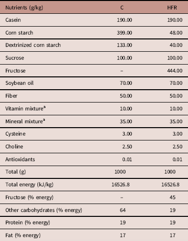
The compositions of the diets are detailed in Table 1. They adhered to the standards of the American Institute of Nutrition for a rodent to support growth during pregnancy, lactation and the postweaning period of life (AIN-93G).Reference Reeves, Nielsen and Fahey 14 The diets were manufactured by PragSolucoes (Jau, SP, Brazil).
Composition and energy content of the diets (AIN 93 G-based diets)

C, control; HFR, high-fructose diet.
a Vitamin and mineral mixtures are in accordance with AIN 93 G.
Twelve-week-old males and females (who spent 8 weeks feeding on experimental diets) were mated (one couple per box), forming four experimental groups with all possibilities of mothers and fathers: C mother and C father, C mother and HFR father, HFR mother and C father, and HFR mother and HFR father. Males were immediately removed from cages after the appearance of the vaginal plug, which was considered the first day of pregnancy. After confirmation of gestation, the fathers were sacrificed. Maternal diet continued until weaning, when the mothers were also sacrificed.
At birth, the pups were separated by genderReference Wolterink-Donselaar, Meerding and Fernandes 15 and weighed. On day 1, the litter was adjusted to contain six pups (three females and three males when possible), and at 21 days old (weaning), only one male was randomly taken from each litter to compose the adult offspring groups. The offspring were fed the C diet after weaning. Four groups were formed: (a) male offspring of a C mother and a C father (C/C offspring, n=10); (b) male offspring of a C mother and a HFR father (C/HFR, n=10); (c) male offspring of a HFR mother and a C father (HFR/C, n=10); and (d) male offspring of a HFR mother and HFR father (HFR/HFR, n=10). The denomination of the offspring groups was based first on a diet given to the mothers and then on a diet given to the fathers.
Body mass (BM), food intake and water intake
The BM of parents and offspring was measured weekly. Food and water consumption were monitored daily (estimated as the difference between the offered and the rest).
Blood pressure (BP)
Systolic BP during the pre-mating period of the parents and at 3 months of age in offspring were measured by tail-cuff plethysmography (Plethysmograph version 2.11; Insight Equip., Ribeirao Preto, SP, Brazil).
Oral glucose tolerance test
In parents 2 days before mating and in offspring 2 days before euthanasia, an oral glucose tolerance test (OGTT) was run after 6 h of fasting (starting at 6 am, during the dark cycle). First, glucose (25% in sterile saline, 0.9% NaCl) at a dose of 1 g/kg was administered by orogastric gavage to glucose overload. Blood was sampled in the caudal vein at 0, 15, 30, 60 and 120 min after glucose administration, and glycemia was measured with a glucometer (Accu-Chek, Roche, Germany). Glucose values at time zero were considered fasting glucose levels. The glucose tolerance analysis was based on the area under the curve from 0 to 120 min (GraphPad Prism v. 7.04 for Windows, La Jolla, CA, USA).
Sacrifice and tissue extraction
As mentioned, parents were sacrificed after weaning of offspring, and offspring were sacrificed at 3 months of age. Animals were fasted for 6 h, heparinized (200 mg/kg), and anesthetized (intraperitoneal sodium pentobarbital, 150 mg/kg). Blood was collected by exsanguination through the section of cervical vessels, and plasma was separated by centrifugation (2000 g, 15 min at room temperature). The liver was removed and weighed, and several liver fragments of all lobes were rapidly cut and frozen at −80°C.
Liver biochemistry
In 50 mg of frozen liver tissue put in an ultrasonic processor (with 1 ml of isopropanol), we measured hepatic triacylglycerol (TAG) level.Reference Marconi, Paolini and Buscaglia 16 The homogenate was centrifuged at 2000 g, and 5 µl of the supernatant was used to measure TAG by the colorimetric enzymatic method using a commercial kit and an automatic spectrophotometer (Bioclin System II, Quibasa Ltda, Belo Horizonte, MG, Brazil).
Plasma analysis
Plasma total cholesterol (TC) and TAG were analyzed in parents. Uric acid, TC and TAG were measured in 3-month-old offspring by the colorimetric enzymatic method using a commercial kit and an automatic spectrophotometer (Bioclin System II). Both parents and offspring had their plasma concentrations of insulin, leptin and adiponectin assessed in duplicate using immuno-enzymatic test kits from Merck KGaA and its affiliates (Darmstadt, Germany) (insulin: #EZRMI-13K ELISA kit (precision: inter-assay: 6.0–17.9%; intra-assay: 0.9–8.4%); leptin: #EZML-82K ELISA kit (precision: inter-assay: 3.0–4.6%; intra-assay: 1.1–1.8%); adiponectin: #ZMADP-60K ELISA kit (precision: inter-assay: 1.4–10.8%; intra-assay: 3.8–8.2%)).
Western blots
Total protein from the liver was extracted using lysis buffer and protease inhibitors. Subsequently, the homogenate was centrifuged for 10 min at 4°C, and the supernatant was collected. The protein concentration was determined with the BCA kit (Thermo Scientific, Rockford, IL, USA). After denaturation, proteins were separated by electrophoresis on a polyacrylamide gel and transferred to a nitrocellulose membrane (Amersham Biosciences, Piscataway, NJ, USA). The membranes were blocked with either 5% skim milk or 5% bovine serum albumin diluted in saline buffer with Tris (TBS-T, 20 mmol/l Tris/HCl, pH 7.4 and 500 mmol/l NaCl) at room temperature for 2 h to prevent nonspecific binding. The membrane was incubated for ~12 h at 4°C with the following primary antibodies: β-actin (43 kDa; Santa Cruz Biotechnology, sc-130300); carbohydrate-responsive element-binding protein (ChREBP) (62 kDa; Santa Cruz Biotechnology, sc-33764); interleukin (IL) 1 β (17 kDa; Santa Cruz, sc-52012); IL6 (26 kDa; Abcam, AB-7737); total c-Jun N-terminal kinases (tJNK) (54 kDa; Santa Cruz Biotechnology, sc-7345); phosphorylated c-Jun N-terminal kinases (pJNK) (49 kDa; Santa Cruz Biotechnology, sc-12882); nuclear factor kappa B (NFκB) (65 kDa; Santa Cruz Biotechnology, sc-109); peroxisome proliferator-activated receptor (PPAR) alpha (55k Da; Santa Cruz Biotechnology, sc-9000); peroxisome proliferator-activated receptor (PPAR) gamma (67 kDa; Santa Cruz Biotechnology, sc-773); suppressor of cytokine signaling 3 (SOCS3) (30 kDa; Santa Cruz Biotechnology, sc-7009); sterol regulatory element-binding Protein (SREBP) 1c (68 kDa; Santa Cruz Biotechnology, sc-367); tumor necrosis factor (TNF) alpha (26 kDa; Santa Cruz Biotechnology, sc-1350). The membrane was incubated with the secondary antibody for 1 h at room temperature. Electrochemiluminescence was used to analyze the protein expression, and the band images were captured with a ChemiDoc XRS (Bio-Rad, Hercules, CA, USA). The band intensity was quantified with ImageJ software (v. 1.50, NIH, ImageJ.nih.gov/ij, USA). The membranes were stripped and reincubated with β-actin (43 kDa; Santa Cruz Biotechnology, sc-81178) to normalize the data.
RNA isolation and real-time PCR
Reverse transcription followed by real-time quantitative polymerase chain reaction (RT-qPCR) was used to measure the expression of mRNA. The total RNA of the liver was extracted with TRIzol (Invitrogen, CA, USA). NanoVue spectroscopy (GE Life Sciences, Buckinghamshire, UK) was used to quantify RNA. Then, 1 µg RNA was treated with DNase I (Invitrogen). Afterward, oligo(dT) primers for mRNA and Superscript III reverse transcriptase (both from Invitrogen) were applied to the synthesis of first-strand cDNA. The endogenous β-actin was used to normalize the expression of the selected genes. After the pre-denaturation and polymerase activation program (4 min at 95°C), 44 cycles of 95°C for 10 s and 60°C for 15 s were followed by a melting curve program (60°C to 95°C with a heating rate of 0.1°C/s). Negative controls consisted of wells in which the cDNA was substituted with deionized water. The relative expression ratio of the mRNA was calculated using the formula
 $$2^{{{\minus}\Delta\Delta _{{Ct}} }} $$
, in which −Δ
Ct
represents the ratio between the number of cycles (Ct) of the target genes with the endogenous control. The primers for RT-qPCR (gene, 5–3', primer) are detailed in Supplementary Material Table S1.
Statistical analysis
After the data were tested for the equality of variance (Browne–Forsythe test) and normality (D’Agostino–Pearson normality test), we expressed the data as the mean and the standard deviation. The differences between the groups were analyzed by one-way ANOVA and the post-hoc test of Holm–Sidak (GraphPad Prism v. 7.04 for Windows; GraphPad Software). A P-value <0.05 was considered statistically significant.
Results
Mother and Father
HFR increases BP without significant changes in BM or glucose tolerance
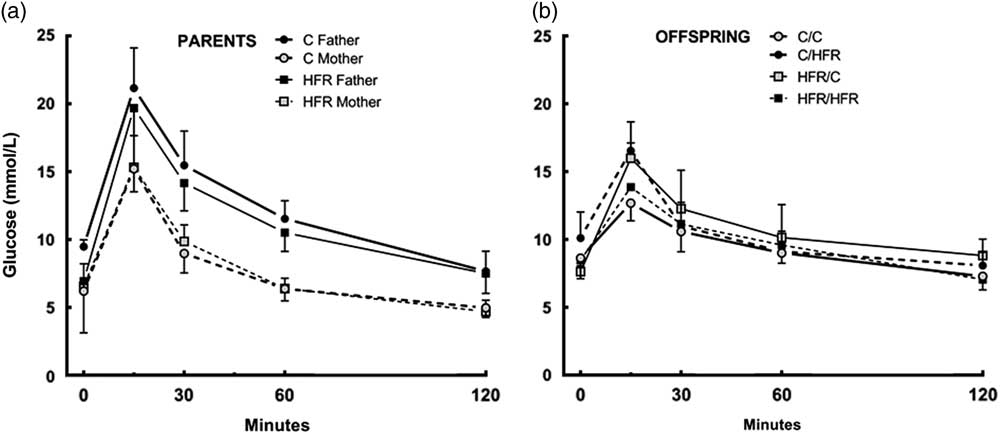
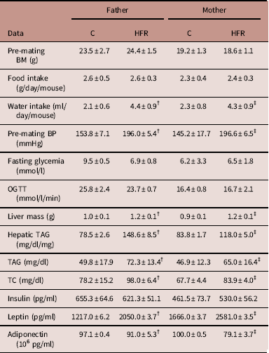
There was no difference in the BM of the parents when the study started or throughout the pre-mating period. The consumption of food and energy was not different between the groups, but HFR parents had higher water intake than those in the C group (+106% in fathers, +86% in mothers, P<0.0001) (Table 2). The BP was higher in the HFR parents (fathers +27%, mothers +35%, P<0.0001) than in the controls, even with no alterations in BM. Fasting blood glucose and OGTT did not show differences between the groups (Table 2, Fig. 1a).
Glucose tolerance is not altered in parents or the offspring of HFR fathers and/or mothers. (a) Oral glucose tolerance test in parents two days before mating. (b) Oral glucose tolerance test in offspring 2 days before sacrifice. Data are the mean±s.d. (one-way ANOVA and post-hoc test of Holm–Sidak). C, control diet; HFR, high-fructose diet.

Parent’s data (mean and s.d., n=5 each group)

BM, body mass; BP, systolic blood pressure; OGTT, oral glucose tolerance teste; TAG, triacylglycerol; TC, total cholesterol.
One-way ANOVA and post-hoc test of Holm–Sidak.
P<0.05 when: †compared with C father, and ‡compared with C mother.
HFR leads to hepatic steatosis, impaired lipid profile and altered leptin and adiponectin secretion
The HFR parents had an enlarged liver (fathers +15%, mothers +18%, P=0.0290) due to a significant increase in hepatic TAG (fathers +89%, mothers +41%, P<0.0001) in comparison to the C parents (Table 2). Plasma insulin was not different between C and HFR parents. However, the HFR parents compared with C parents showed hypercholesterolemia (fathers +25%, mothers +24%, P=0.0158) and hypertriglyceridemia (fathers +45%, mothers +39%, P<0.001). Plasma leptin was higher and plasma adiponectin was lower in the HFR parents than in the C parents (leptin: fathers +68%, mothers +55%, both P<0.0001; adiponectin: fathers −6%, P=0.018, mothers −21%, P<0.0001) (Table 2). Thus, biochemical alterations were observed in parents that received the HFR diet.
Offspring
Offspring of HFR fathers and/or HFR mothers have elevated BP but no gain in BM
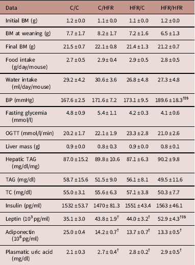
BM, food intake and water intake did not show differences between the groups throughout the experiment (Table 3). However, the BP was increased by HFR. compared with that of the C/C group, BP was 13% higher in the HFR/HFR group (P<0.0001), 10% higher in the C/HFR group (P<0.0001) and 9% higher in the HFR/C group (P<0.0001) (Table 3). There was no significant difference between the groups of offspring in fasting glucose or glucose tolerance (Table 3; Fig. 1b), as was observed in parents.
Offspring’s data (mean and s.d., n=5 each group)

BM, body mass; BP, systolic blood pressure; OGTT, oral glucose tolerance test; TAG, triacylglycerol; TC, total cholesterol.
One-way ANOVA and post-hoc test of Holm–Sidak.
P<0.05 when: †compared with C/C offspring, ‡compared with C/HFR offspring, and §compared with HFR/C offspring.
HFR mother and/or HFR father alters the plasma levels of leptin, adiponectin and uric acid in offspring, even without apparent liver damage
Hepatic mass, liver TAG and plasma insulin, TC and TAG did not show significant differences between groups. Leptin, in comparison with the C/C group, was 25% greater in the C/HFR (P=0.0022), 25% greater in the HFR/C group (P=0.0022) and 50% greater in the HFR/HFR group (P<0.0001). Leptin was also greater in the HFR/HFR group than in both C/HFR (+21%, P=0.0022) and HFR/C (+20%, P=0.0022) groups. Adiponectin, in comparison with the C/C group, was 43% lower in the C/HFR group (P<0.0001), 45% lower in the HFR/C group (P<0.0001) and 47% lower in the HFR/HFR group (P<0.0001). Uric acid, in comparison with the C/C group, was 29% higher in the C/HFR group (P=0.0397), 34% higher in the HFR/C group (P=0.0171) and 38% higher in the HFR/HFR group (P=0.0083) (Table 3). Even without apparent structural damage to the liver, inflammation signaling pathways, lipogenesis and β-oxidation were altered in the offspring of HFR parents.
HFR parents lead to hepatic inflammation in the offspring by activation of NFκB, SOCS3 and JNK
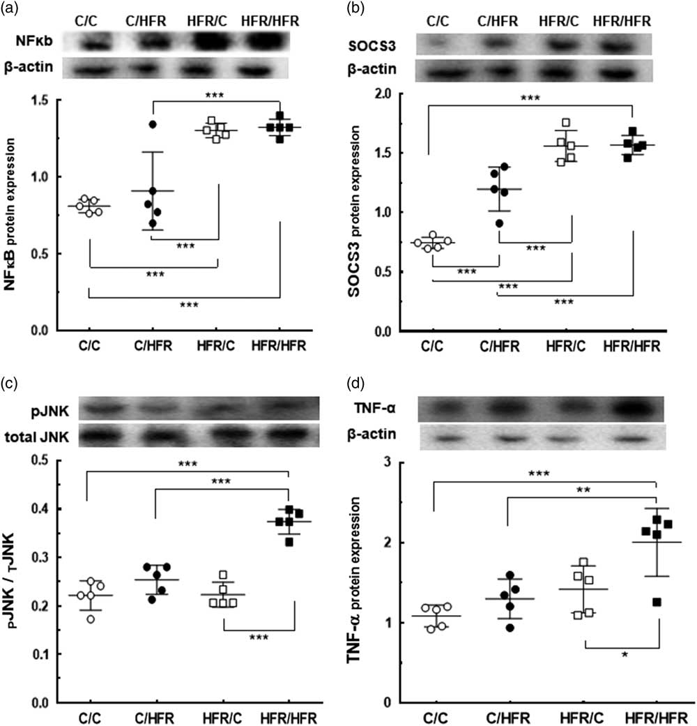
NFκB protein expression was higher in the HFR/C group than both the C/C group (+61%, P=0.0001) and the C/HFR group (+43%, P=0.0008). In the HFR/HFR group, NFκB protein expression was higher than both the C/C (+63%, P=0.0001) and C/HFR (+46%, P=0.0007) groups (Fig. 2a). SOCS3 protein expression, compared with the C/C group, was overexpressed in the C/HFR (+61%, P<0.0001) and HFR/C groups (+109%, P<0.0001). SOCS3 expression in the HFR/HFR group was higher by 111% over the C/C group (P<0.0001) and by 31% over the C/HFR group (P=0.0006) (Fig. 2b). Phosphorylated JNK expression, compared with that of the C/C group, was greater in the HFR/HFR group (+69%, P<0.0001), the C/HFR group (+47%, P<0.0001) and the HFR/C group (+67%, P<0.0001) (Fig. 2c).
Paternal and/or maternal HFR diet activates the offspring hepatic signaling pathways. (a) NFκB protein expression (in arbitrary units, a.u.) and representative immunoblotting with bands. (b) SOCS3 protein expression (in arbitrary units, a.u.) and representative immunoblotting with bands. (c) pJNK protein expression (in arbitrary units, a.u.) and representative immunoblotting with bands. (d) TNF-α protein expression (in arbitrary units, a.u.) and representative immunoblotting with bands. As an endogenous control, β-actin was used to normalize the expression of the selected proteins. tJNK was used to normalize the expression of pJNK. Data are the mean±s.d. **P<0.01, ***P<0.001 (one-way ANOVA and post-hoc test of Holm–Sidak). NFκB, nuclear factor kappa B; C, control diet; HFR, high-fructose diet; SOCS, suppressor of cytokine signaling; pJNK, phosphorylated c-Jun n-terminal kinases; tJNK, total c-Jun n-terminal kinase; TNF, tumor necrosis factor.

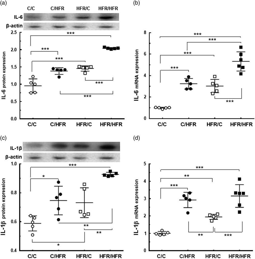
TNF-α expression in the HFR/HFR group was 84% higher than the C/C group (P=0.0009), 54% higher than the C/HFR group (P=0.0080) and 41% higher than the HFR/C group (P=0.0244) (Fig. 2d). IL6 protein expression, compared with that of the C/C group, was higher in the C/HFR group (+45%, P<0.0001) and in the HFR/C group (+53%, P<0.0001). IL6 protein expression was higher in the HFR/HFR group by 114% over C/C (P<0.0001), 48% over C/HFR (P<0.0001) and 40% over HFR/C (P<0.0001) (Fig. 3a). IL6 mRNA analysis showed the same pattern IL6 protein (Fig. 3b). The variation of the IL1-β protein expression between the groups followed the IL6 pattern, but the mRNA expression showed lower expression in the HFR/C group (Fig. 3c and 3d).
Parental HFR diet enhances the offspring hepatic IL6 and IL1-β. (a) IL6 protein expression (in arbitrary units, a.u.) and representative immunoblotting with bands. (b) IL6 mRNA expression. (c) IL1-β protein expression (expressed in arbitrary units, a.u.) and representative immunoblotting with bands. (d) IL1-β mRNA expression. As an endogenous control, β-actin was used to normalize the expression of the selected proteins. Data are the mean±s.d. *P<0.05, **P<0.01, ***P<0.001 (one-way ANOVA and post-hoc test of Holm–Sidak). IL, interleukin; C, control diet; HFR, high-fructose diet.

HFR parents cause increased hepatic lipogenesis in offspring
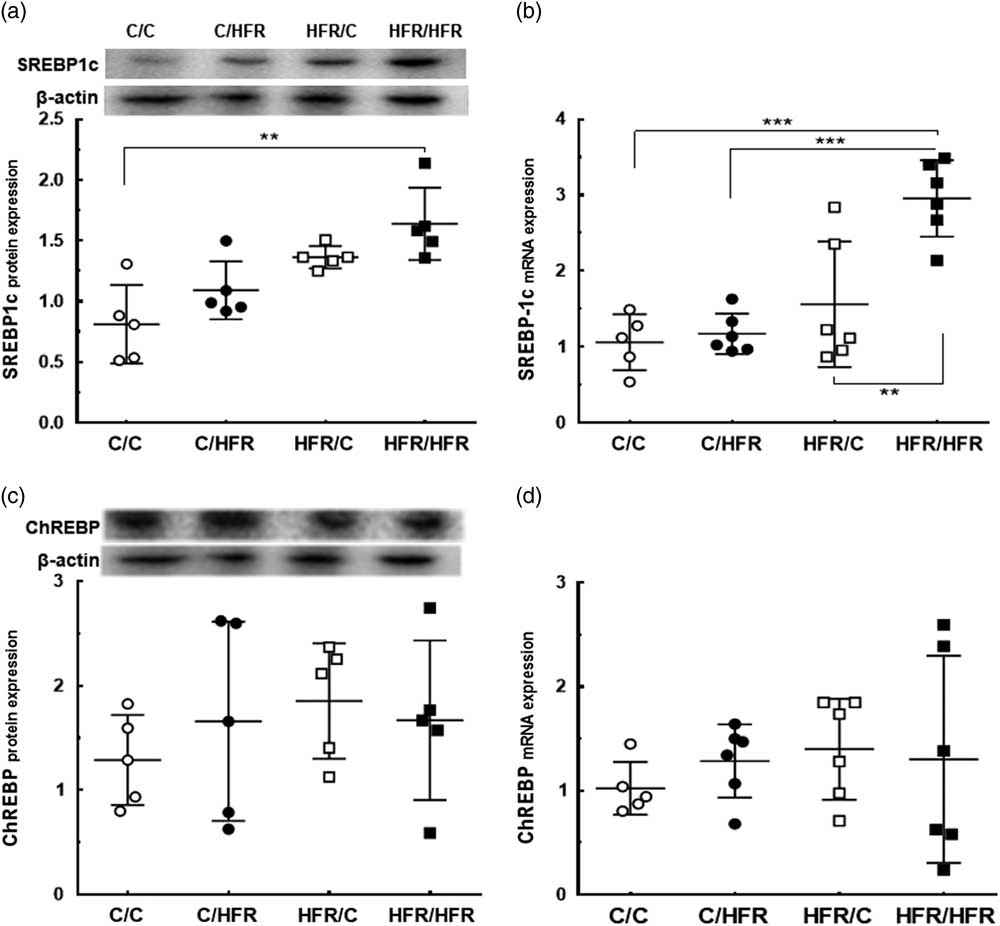
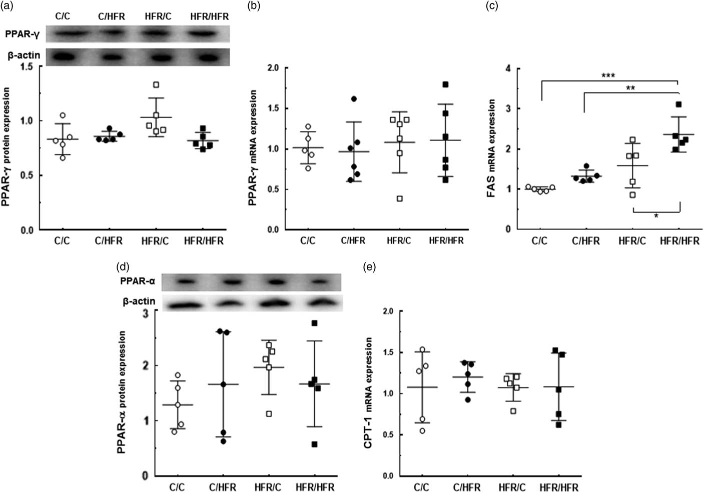
SREBP-1c, compared with that of the C/C group, was overexpressed in the HFR/HFR group (protein expression +102%, P=0.0006, Fig. 4a), and its mRNA expression was higher in the HFR/HFR group (+179%, P<0.0001), the C/HFR group (+152%, P<0.0001), and the HFR/C group (+89%, P=0.0011) (Fig. 4b). ChREBP and PPAR-γ did not show significant differences between groups (Figs. 4c, 4d, 5a, 5b). FAS gene expression was higher in the HFR/HFR group by 136% over the C/C group (P=0.0001), 78% over the C/HFR group (P=0.0017) and 49% over the HFR/C group (P=0.0152) (Fig. 5c).
Parental HFR diet upregulates the offspring hepatic lipogenic marker SREBP-1c. (a) SREBP-1c protein expression (expressed in arbitrary units, a.u.) and representative immunoblotting with bands. (b) SREBP-1c mRNA expression. (c) ChREBP protein expression (expressed in arbitrary units, a.u.) and representative immunoblotting with bands. (d) ChREBP mRNA expression. As an endogenous control, β-actin was used to normalize the expression of the selected proteins. Data are the mean±s.d. **P<0.01, ***P<0.001 (one-way ANOVA and post-hoc test of Holm–Sidak). SREBP, sterol regulatory element-binding protein; C, control diet; HFR, high-fructose diet ChREBP, carbohydrate-responsive element-binding protein.

Parental HFR diet increases the offspring hepatic FAS without alterations in β-oxidation. (a) PPAR-γ protein expression (in arbitrary units, a.u.) and representative immunoblotting with bands. (b) PPAR-γ mRNA expression. (c) FAS mRNA expression. (d) PPAR-α protein expression (expressed in arbitrary units, a.u.) and representative immunoblotting with bands. (e) CPT1 mRNA expression. As an endogenous control, β-actin was used to normalize the expression of the selected proteins. Data are the mean±s.d. **P<0.01, ***P<0.001 (one-way ANOVA and post-hoc test of Holm–Sidak). FAS, fatty acid synthase; PPAR, peroxisome proliferator-activated receptor; CPT, carnitine palmitoyltransferase.

HFR mother and/or HFR father does not compromise the hepatic β-oxidation in offspring
PPAR-α protein expression and CPT1 gene expression did not show significant differences between groups (Fig. 5d and 5e), indicating that the parental HFR diet was not responsible for reprogramming offspring hepatic β-oxidation.
Discussion
This study shows that a HFR father or HFR mother alone might transmit hepatic inflammation to male adult progeny, without alterations in lipogenesis or β-oxidation. A combination of HFR father with HFR mother (parental HFR diet) maximized the effects on progeny, allowing the appearance of adverse effects that did not occur when only the father or mother was fed the HFR. Therefore, the HFR/HFR offspring had more pronounced liver inflammatory markers and some altered pathways of lipogenesis, as indicated by the FAS and SREBP-1c expression.
Considering that both C and HFR diets were isoenergetic, no differences were found in the BM of parents, and therefore, no association was seen in fructose intake or obesity. The gain in BM observed in connection with industrialized sources of fructose, mainly sweetened beverages, may be related to hypercaloric consumption rather than to fructose itself. Furthermore, a high-fructose diet may induce metabolic disorders independent of overweight or obesity.Reference Schultz, Neil, Aguila and Mandarim-de-Lacerda 13 , Reference Schultz, Barbosa-da-Silva, Aguila and Mandarim-de-Lacerda 17 The enlarged liver, increased plasma TC and TAG, and increased hepatic TAG in HFR parents induced nonalcoholic fatty liver disease (NAFLD) in their offspring.
The offspring data demonstrated that maternal, paternal or parental HFR intake can lead to hyperuricemia. Uric acid is synthesized during the degradation of adenosine monophosphate (AMP) in fructose metabolism. The increased level of intracellular uric acid is followed by an acute increase in circulation, probably due to its release from the liver. The uric acid formation is critical to the mechanism of metabolic syndrome. In plasma, uric acid stimulates the NADPH oxidase activity and reactive oxygen species (ROS) production in mature adipocytes. Consequently, inflammatory markers (IL6 and TNF-α) are produced in parallel with the decrease in adiponectin, an important anti-inflammatory agent.Reference Kang and Ha 18 Accordingly, the HFR diet was correlated with increased leptin and decreased adiponectin in parents. These alterations were also seen in their offspring, mostly in the HFR/HFR group.
The HFR diet may contribute to lipid deposition in white adipose tissue (WAT), especially in visceral fat.Reference Stanhope, Schwarz and Keim 19 , Reference Bargut, Santos, Machado, Aguila and Mandarim-de-Lacerda 20 Considering that WAT and hepatic steatosis are etiologically and functionally intertwined,Reference Sondergaard 21 overproduction of TNF-α and IL6 by adipocytes has the potential to affect the liver of the HFR/HFR offspring, compromising the signaling, metabolism and immune process of the descendants.Reference Wree, Kahraman, Gerken and Canbay 22
TNF-α is an inflammatory cytokine that regulates the hepatocyte physiology in a diversity of pathophysiologic conditions,Reference Papa, Bubici, Zazzeroni and Franzoso 23 being both a cytotoxic agent and a protective agent (through NFκB activation). However, the most prominent effect of TNF-α is the induction of NFκB,Reference Wullaert, van Loo, Heyninck and Beyaert 24 whose expression was pronounced in the HFR/C and HFR/HFR offspring, indicating that the mother, more than the father, has a substantial role in the expression of NFκB in offspring. In contrast, TNF-α also induces mitogen-activated protein (MAP) kinases, mainly the JNK family and caspases. Whereas activation of NFκB controls survival, JNK and caspases are effectors of cell death in the TNF-α pathway.Reference Papa, Bubici, Zazzeroni and Franzoso 23 Therefore, phosphorylation of JNK may contribute to cell death. The increased phosphorylated JNK, observed in HFR/HFR offspring, agrees with the expected role of JNK. In turn, overexpression of IL6 stimulates the expression of SOCS3, characterizing the liver inflammation condition.Reference Wullaert, van Loo, Heyninck and Beyaert 24 NFκB and SOCS3 were overexpressed in both HFR/C and HFR/HFR offspring compared with the HFR father group (C/HFR), suggesting the greater participation of the mother in programming offspring inflammatory insults.
According to the ‘two-hit’ theory, hepatic steatosis in NAFLD is primarily caused by lipid accumulation (first hit), due to effects of oxidative stress, ROS, lipid peroxidation and the presence of pro-inflammatory cytokines. The insult progresses to nonalcoholic steatohepatitis (NASH), marked by intense inflammation in the liver (second hit).Reference Paschos and Paletas 25 However, the ‘multiple parallel hits’ hypothesis,Reference Tilg and Moschen 26 suggesting that NASH may reflect a disease in which steatosis is a consequence of inflammation, fits better with our findings.
Some increased markers observed in offspring were derived from parents and were related to the metabolism of the HFR diet. Industrially isolated fructose ingested in food is rapidly absorbed and is directed into the hepatocyte by its GLUT2 receptors.Reference Douard and Ferraris 27 In the liver, fructose is phosphorylated into fructose-1-phosphate (F1P) by fructokinase, which is not inhibited by adenosine diphosphate (ADP) or by citrate and therefore is not regulated by cellular energy status. The fast conversion of fructose in F1P leads to an imbalance between AMP and adenosine triphosphate (ATP), increasing the AMP/ATP ratio.Reference Tappy, Le, Tran and Paquot 28 Consequently, the lack of negative feedback leads to a situation where all fructose ingested is converted to F1P. This mechanism is responsible for almost all deleterious effects on the fructose metabolism. Although the offspring were not fed a HFR diet, some parental adverse phenotype was transmitted to the offspring.
AMP can be converted to uric acid by xanthine dehydrogenase, leading to hyperuricemia, one of the conditions found in metabolic syndrome.Reference Abdelmalek, Suzuki and Guy 29 This state was observed in the offspring of fathers and mothers fed a HFR diet individually or in combination. The phosphorylation of fructose into F1P may also explain the high BP,Reference Takahashi, Shinoda and Furuya 30 as seen in HFR parents and in HFR/HFR offspring (even while receiving a C diet). F1P is cleaved by aldose to generate glyceraldehyde 3-phosphate and dihydroxyacetone phosphate, which are primary sources of methylglyoxal (MG).Reference Liu, Wang, Desai and Wu 31 MG, besides being a highly toxic aldehyde responsible for ROS production, may also change the function of calcium channels, leading to an increase in intracellular calcium that generates vascular resistance.Reference Vasdev and Stuckless 32 Moreover, the intense phosphorylation of fructose into F1P decreases cellular ATP and impairs the synthesis of nitric oxide, promoting ROS formation and elevated BP.Reference DiNicolantonio and Lucan 33 ROS are continuously produced by cells as a part of their metabolic processes.Reference Zhang, Zhang and Chen 34 Hepatocellular oxidative stress caused by excessive ROS production due to inflammation leads to the maturation and secretion of the pro-inflammatory cytokine IL1-βReference Zhang, Zhang and Chen 34 and increased phosphorylation of JNK. Interestingly, IL1-β mRNA expression was higher in C/HFR offspring than in both HFR/C and C/C, emphasizing the importance of the father’s nutrition in epigenetic parameters of fetal programming.
The nutritional stimuli in critical stages of development can influence the expression of various genes by changes in chromatin conformation and the accessibility of transcription factors. Therefore, epigenetics is considered the primary tool for the transmission of paternal phenotypes to offspring.Reference Gallou-Kabani and Junien 35 Also, some epigenetic marks during spermatogenesis may persist throughout embryonic development, inducing irreversible epigenetic changes and phenotypic consequences expressed in the offspring.Reference Soubry 36 Epigenetic methods also modulate the effects of transcriptional regulation through several processes, such as DNA methylation, histone alterations, and transcription of non-coding RNA (e.g. miRNA).Reference Li, Shen and Hua 37 , Reference Zhao, Zhao and Ren 38 However, the lack of observation of epigenetic mechanisms is a limitation of our study.
Herein, we demonstrated an increase in both SREBP-1c and FAS in the HFR/HFR group, with no alterations in ChREBP expression. Both SREBP-1c and ChREBP are transcription factors upregulating lipogenic enzymes such as FAS.Reference Koo, Miyashita, Cho and Nakamura 39 Furthermore, F1P can directly activate SREBP-1c or ChREBP.Reference Denechaud, Dentin, Girard and Postic 40 A parental HFR diet can activate lipogenesis de novo, and consequently, the concentration of malonyl-CoA increases, which could inhibit CPT1 and thus β-oxidation.Reference Nomura and Yamanouchi 41 However, the adult male offspring in the current study did not show differences in hepatic β-oxidation markers. Therefore, it is plausible to conclude that lipid accumulation would be the next step following hepatic inflammation, with preservation of β-oxidation at first. In the long term, the elevated lipogenesis in the offspring may compromise β-oxidation, because the formation of fatty acids leads to a higher intracellular concentration of malonyl-coenzyme A, which may impair β-oxidation.Reference Rasmussen, Holmback and Volpi 42
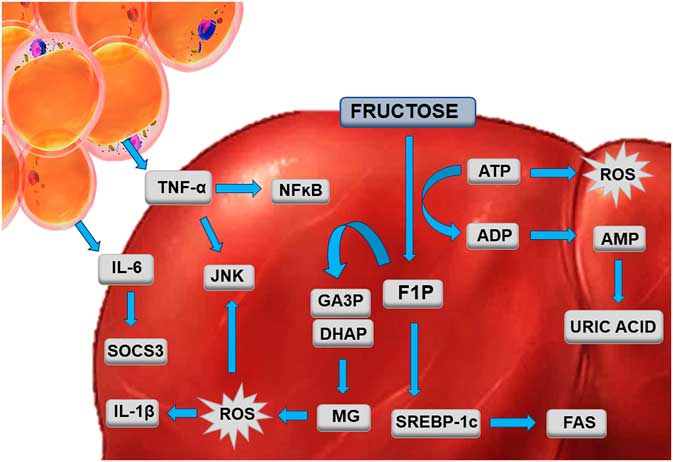
In conclusion, the mechanisms by which a parental HFR diet programs the offspring are proposed in Fig. 6. The father’s participation in the transmission of phenotypes to the descendants is a new and relevant subject for health professionals involved in nutrition together with obstetricians and pediatricians. This study demonstrates that both mother and father, individually or in combination, when fed a fructose-rich diet, affect the metabolism and liver in adult male offspring. Giving the HFR to the mother and father increases the adverse effects on offspring health and has more consequences than the mother or father alone.
The hypothesized mechanism by which a parental high-fructose diet may program male adult offspring. The increased amount of visceral fat contributes to the production of IL6 and TNF-α, which are directed to the liver. In liver, IL6 contributes to overexpression of SOCS3, and TNF-α contributes to overexpression of JNK and NFκB, characterizing hepatic inflammation. Consumption of a high-fructose diet by parents affects some pathways related to the hepatic metabolism of fructose in offspring, even though offspring have only consumed a control diet. Therefore, plasma accumulation of uric acid is found due to excess AMP, which is formed from the dephosphorylation of ATP. The ATP reduction generates ROS, contributing to increases in BP. Moreover, MG is produced by its subunits GA3P and DHAP, further adding to the formation of ROS and hypertension. Hepatic accumulation of ROS is associated with overexpression of JNK and IL1-β, summing to inflammation. Finally, due to fructose metabolism, SREBP-1c, a transcription factor related to de novo lipogenesis, is activated and activates the transcription of FAS. AMP, adenosine monophosphate; ATP, adenosine triphosphate; DHAP, dihydroxyacetone phosphate; FAS, fatty acid synthase; GA3P, glyceraldehyde 3-phosphate; IL, interleukin; JNK, c-Jun N-terminal kinases; MG, methylglyoxal; NFκB, nuclear factor kappa B; ROS, reactive oxygen species; SOCS, suppressor of cytokine signaling; SREBP, sterol regulatory element-binding protein; TNF, tumor necrosis factor.

Acknowledgments
The authors would like to thank Aline Penna de Carvalho and Michele Soares for their technical assistance.
Financial Support
The work was supported by CNPq (Conselho Nacional de Desenvolvimento Cientifico e Tecnologico) grant numbers 302.920/2016-1 to CMAL, and 306077/2013-2 to MBA) and FAPERJ (Fundacao Carlos Chagas Filho do Amparo a Pesquisa do Rio de Janeiro), grant numbers E-26/201.186/2014 to CAML, and E-26/201.335/2014 to MBA).
Conflicts of Interest
None.
Ethical Standards
The authors assert that all procedures contributing to this work comply with the ethical standards of the relevant national guides on the care and use of laboratory animals (NIH Publication No. 85-23, revised 1996) and has been approved by the institutional committee (Ethics Committee of the University of the State of Rio de Janeiro).
Supplementary material
To view supplementary material for this article, please visit https://doi.org/10.1017/S2040174418000235