1. Introduction
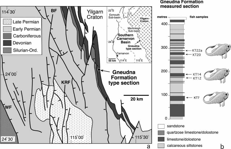
Ptyctodont tooth plates are common fossils in Devonian marine and non-marine assemblages, with most of the known ptyctodontid taxa based solely on them (Denison, Reference Denison1978). Articulated ptyctodonts are less common; however, from the Gogo Formation, NW Western Australia, three genera of articulated, three-dimensionally preserved ptyctodonts, Materpiscis attenboroughi (Long et al. Reference Long, Trinajstic, Young and Senden2008), Campbellodus decipiens Miles & Young, Reference Miles and Young1977 and Austroptyctodus gardineri (Miles & Young, Reference Miles and Young1977), are documented. The species gardineri was first referred to the genus Ctenurella by Miles & Young (Reference Miles and Young1977), then Long (Reference Long1997) placed the species in its own genus Austroptyctodus, following the discovery and preparation of an additional specimen (WAM 70.4.253). A second Western Australian site, in the Gneudna Formation, at Williambury Station, Carnarvon Basin (Fig. 1a), has yielded a diverse early Frasnian fish fauna in which tooth plates are the most abundant components of the ptyctodont material (Long & Trinajstic, Reference Long, Trinajstic, Blieck and Turner2000). However, one bed (KT7), about 90 metres above the base of the succession (Fig. 1b), has abundant, isolated and partially articulated ptyctodontid dermal plates. To date, most of the bones of the head and trunk shields, parts of the axial skeleton and the dermal clasping bones of the males have been retrieved through acid etching of samples. In addition, the occipital and orbital ossifications, preserved in three dimensions, provide precise information of endocranial morphology. We consider that the tooth and dermal plates belong to only one taxon, a new genus and species Kimbryanodus williamburyensis n. gen., n. sp. As complete ptyctodont specimens are rare, this material represents an important addition to our knowledge of this group.
Locality map and stratigraphic column of the Upper Devonian (Frasnian) fish fauna from the Gneudna Formation, Carnarvon Basin, Western Australia. (a) Locality of the type section. (b) Stratigraphic horizons in which ptyctodont remains were found.

An apparent evolutionary trend within the Ptyctodontidae is a reduction in their dermal armour, limiting morphological characters useful for species-level systematics (Carr, Reference Carr1995). In addition, too few complete specimens are known, which has also contributed to our lack of knowledge regarding phylogenetic interrelationships within ptyctodonts. The first phylogenetic hypothesis of relationships within the ptyctodonts, proposed by Johnson & Elliott (Reference Johnson and Elliott1996), was unable to resolve the basal taxon. The interpretation of the nuchal plate (Miles & Young, Reference Miles and Young1977; Long, Reference Long1988) or postpineal plate (Johnson & Elliott, Reference Johnson and Elliott1996) was identified as being paramount in determining ptyctodont relationships (Johnson & Elliott, Reference Johnson and Elliott1996). Depending on each author and their views of this character, arguments both for and against Rhamphodopsis threiplandi Watson, Reference Watson1938 being the least derived of the ptyctodonts were proposed. Long (Reference Long1997) has provided new interpretations for the dermal plate arrangement in the head shield of four ptyctodont genera, two from Australia: Campbellodus (Miles & Young, Reference Miles and Young1977; revised Long, Reference Long1997) and Austroptyctodus Long, Reference Long1997; and two from Germany: Rhynchodus Newberry, Reference Newberry1873 and Ctenurella Ørvig, Reference Ørvig1960. A new phylogenetic analysis and revised classification of the Ptyctodontida are presented here based on the new observations made by Long (Reference Long1997). The outstanding three-dimensional preservation of the Australian taxa is of immense phylogenetic importance in terms of understanding the early evolution of these fishes and their evolutionary relationships.
2. Geological setting and age
The Carnarvon Basin is an elongate arcuate basin, which extends along the W and NW Western Australian coastline between Geraldton and Karratha with a total area of 650 000 km2, 115 000 km2 of which are onshore (Hocking, Moors & Van de Graaff, Reference Hocking, Moors and Van De Graaff1987). It is subdivided into the Northern Carnarvon Basin, which is largely offshore, and the Southern Carnarvon Basin, which is predominantly onshore (Hocking, Reference Hocking1990), and includes the Gascoyne Platform that lies to the west and the Merlinleigh (Fig. 1a), Bidgemia and Byro sub-basins that lie to the east. The Middle to Upper Devonian succession crops out along the eastern margin of the Merlinleigh Sub-basin. Palaeogeographical reconstructions suggest that during Late Devonian times the Carnarvon Basin occupied a low southern palaeolatitude.
The marine Gneudna Formation is exposed for about 100 km along the eastern margin of the Merlinleigh Sub-basin (Fig. 1a) and has been interpreted as a progradational succession. The type section crops out SE of Williambury Homestead at the locality known as Gneudna Paddock (Fig. 1a). The type section is 450 m thick and comprises thinly to thickly bedded limestone and limestone/dolostone alternating with very thick units of siltstones and silty sandstones (Fig. 1b). The siliciclastic units tend to be friable and are either not exposed or only crop out as scree. Only one horizon (KT7, Fig. 1b), a thinly bedded ostracodal wackestone, has yielded articulated ptyctodont remains. The presence of articulated ostracods in the wackestones and the dominance of carbonate mud suggest deposition in low-energy conditions.
The conodont Ancyrodella sp. (R. Nicoll, unpub. internal report, Australian Bureau of Min. Resources, 1979) and the thelodont Australolepis seddoni Turner & Dring, Reference Turner and Dring1981 (Turner, Reference Turner1997; Trinajstic, Reference Trinajstic2001) indicate an early Frasnian age (transitans to punctata conodont Zones) for the Gneudna Formation. Other vertebrate remains known from the Gneudna Formation include: acanthodiforms, Cheiracanthoides spp.; placoderms, Holonema westolli, Groenlandaspis sp., Bothriolepis sp.; actinopterygians, Moythomasia durgaringa and Mimia toombsi; dipnoans, Chirodipterus sp.; the osteolepiform Gogonasus and the onychodontid Onychodus sp. (Long & Trinajstic, Reference Long, Trinajstic, Blieck and Turner2000).
3. Materials and Methods
3.a. Fossil preparation
Twenty-five residues from resistant limestone beds of the Gneudna Formation were extracted through disaggregating the rock in 10% acetic acid. All residues were rich in vertebrate remains and other acid-resistant microfossils. Articulated and associated ptyctodont remains were recovered from a single bed, KT7 (Fig. 1b). Isolated body plates and tooth plates were recovered from beds KT12–14 and KT20–22a (Fig. 1b). Specimens are deposited in the Western Australian Museum. Institutional abbreviations are as follows: WAM – Western Australian Museum, Perth; NHM P – Natural History Museum, London; URP and ARD – Muséum national d'Histoire naturelle, Paris; SUI – Museum of Natural History Geological Repository, University of Iowa.
3.b. Comparative ptyctodont material
The new ptyctodont from the Gneudna Formation was compared with the following supplementary specimens in museum collections:
Austroptyctodus gardineri (Miles & Young, Reference Miles and Young1977; revised Long, Reference Long1997). A single well-preserved and articulated specimen held in the Western Australian Museum WAM 86.9.662. The specimen, due to its delicate nature, is embedded in resin as part and counterpart. The holotype WAM 70.4.253 is incomplete. The remaining specimens are held in the Natural History Museum, London (NHM P50906, NHM P50908, NHM P50910, NHM P57654, NHM P57655). Upper Devonian (Frasnian), Gogo Formation, Western Australia.
Campbellodus decipiens Miles & Young, Reference Miles and Young1977. Three specimens held in the Western Australian Museum, one an almost complete individual (WAM 86.9.672), the holotype, which comprises an incomplete head shield (WAM 70.4.252) and a set of isolated tooth plates (WAM 95.6.112). The Natural History Museum, London holds a single incomplete specimen (NHM P50907). Upper Devonian (Frasnian), Gogo Formation, Western Australia.
Chelyophorus verneuili Agassiz, Reference Agassiz1844. Isolated plates were viewed from the Agassiz collection held in the collections of Muséum national d'Histoire naturelle, Paris (URP.2, URP.4–6, URP.10–14). A second specimen, which is articulated and in which the orbital ossifications are preserved within the head shield, was viewed in the palaeontology collections, St Petersburg, Russia. Disarticulated plates from the Natural History Museum were also examined (NHM P7483). Upper Devonian (Famennian) Dankov–Lebedyan beds, Russia.
Ctenurella gladbachensis Ørvig, Reference Ørvig1960. Two almost complete well-preserved specimens from the collections of the Muséum national d'Histoire naturelle, Paris (ARD232, ARD230a & b). Additional specimens from the Natural History Museum, which include articulated individuals, part and counterpart material (NHM P48255–6, NHM P48238–9), individuals with the skull roof well preserved (NHM P48249) and isolated plates (NHM P53605, NHM P53574). Middle–Upper Devonian (upper Givetian/lower Frasnian), Oberer Plattenkalk, Germany.
Denisonodus plutonensis Johnson & Elliott, Reference Johnson and Elliott1996. The specimen was not viewed; characters were coded from Johnson & Elliott (Reference Johnson and Elliott1996).
Materpiscis attenboroughi Long et al. Reference Long, Trinajstic, Young and Senden2008. Holotype (WAM 07.12.1), a complete articulated specimen including the tail and with a single embryo inside the abdominal region. Upper Devonian (early Frasnian), Gogo Formation, Western Australia.
Ptyctodopsis menzeli Denison, Reference Denison1985. Holotype (SUI 33860), consisting of a partial head and trunk shield, Museum of Natural History Geological Repository, University of Iowa. Middle Devonian (upper Givetian), Cedar Valley Limestone, USA.
Rhamphodopsis threiplandi Watson, Reference Watson1938. Specimens were viewed at the Natural History Museum, London (NHM P48664, NHM P52979, NHM P52954). Scottish Middle Old Red Sandstone, Edderton, Ross-shire, Scotland.
Rhamphodopsis trispinatus Watson, Reference Watson1938. Character states taken from Miles (Reference Miles1967).
Rhynchodus tetrodon Newberry, Reference Newberry1873. Cast of the holotype from the Humboldt University Palaeontology Museum, Berlin (Huf 521a & b) held in the Natural History Museum, London (NHM P48771–2). Upper Devonian (Frasnian), Kellwasserkalk, Germany.
Other comparative taxa. Characters were taken from published descriptions for the brachythoracid arthrodire Antineosteus lehmani Lelièvre, Reference Lelièvre1984 (Lelièvre, Reference Lelièvre1984); the petalichthyids Eurycaraspis incilis Liu Reference Liu, Chang, Liu and Zhang1991 (Liu, Reference Liu, Chang, Liu and Zhang1991) and Lunaspis broilii Gross, Reference Gross1937 (Denison Reference Denison and Schultze1978).
Romundina Ørvig, Reference Ørvig and Lehman1975. A complete, articulated acanthothoracid specimen from the collection of the Muséum national d'Histoire naturelle (MNHN-BR4.5). Early Devonian (Lochkovian), Arctic Canada.
4. Systematic palaeontology
Subclass PLACODERMI M'Coy, Reference M'Coy1848
Order PTYCTODONTIDA Gross, Reference Gross1932
Family ptyctodontidae Woodward, Reference Woodward1891
Genus Kimbryanodus nov
Derivation of name. In recognition of Dr Kim Dennis-Bryan for her pioneering work on placoderms.
Diagnosis. See species.
Kimbryanodus williamburyensis sp. nov.
Figures 2–9
1997 Ptyctodontidae gen. nov. Long
2000 Ptyctodontidae gen. nov. Long & Trinajstic, p. 482, pl. 2, figs 1–16
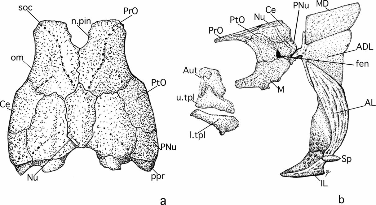
Composite reconstruction of the body armour of Kimbryanodus williamburyensis gen. et sp. nov. (a) Dorsal view of the head shield. (b) Lateral view of the dermal exoskeleton. See Appendix 1 for key to abbreviations.

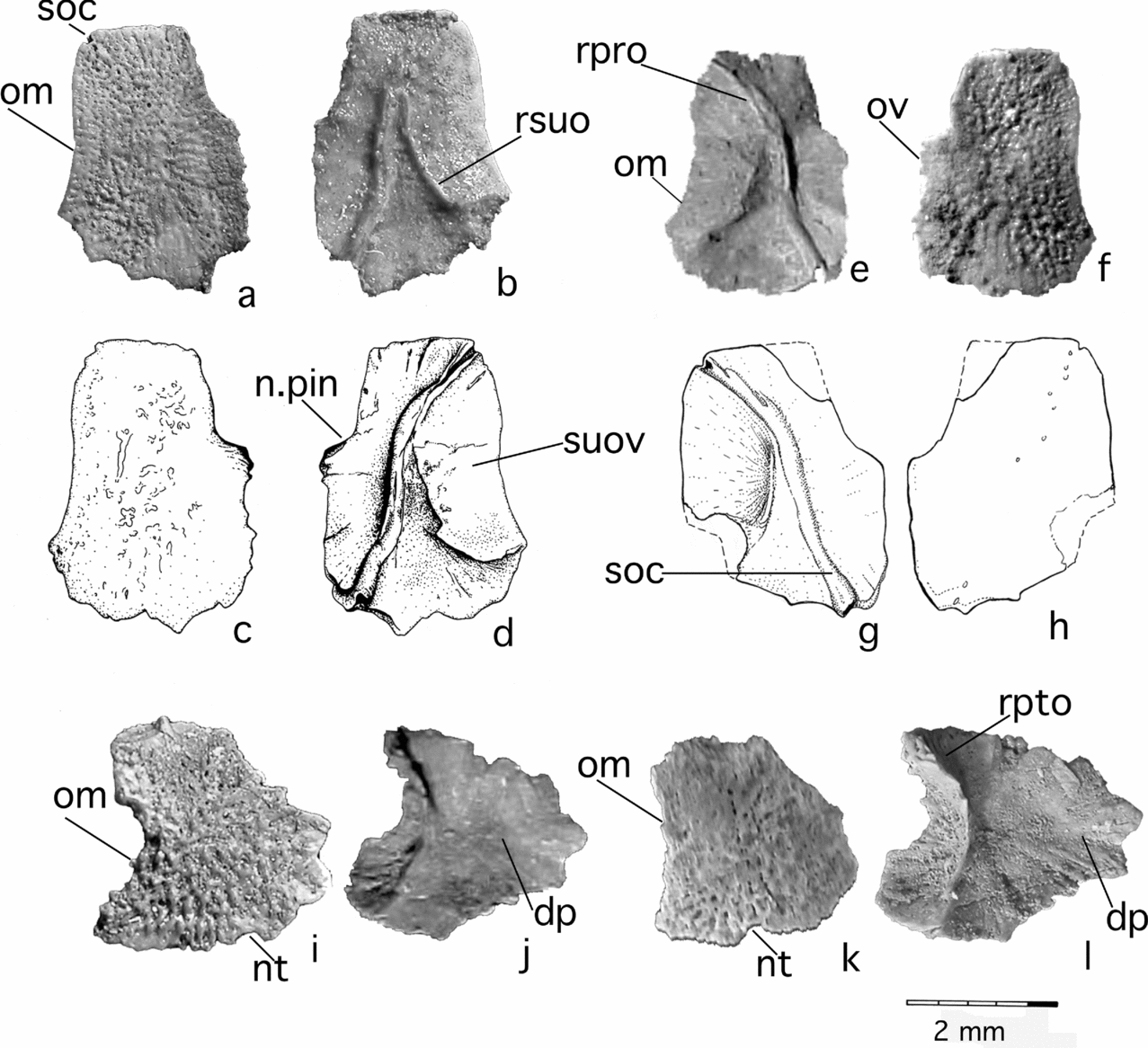
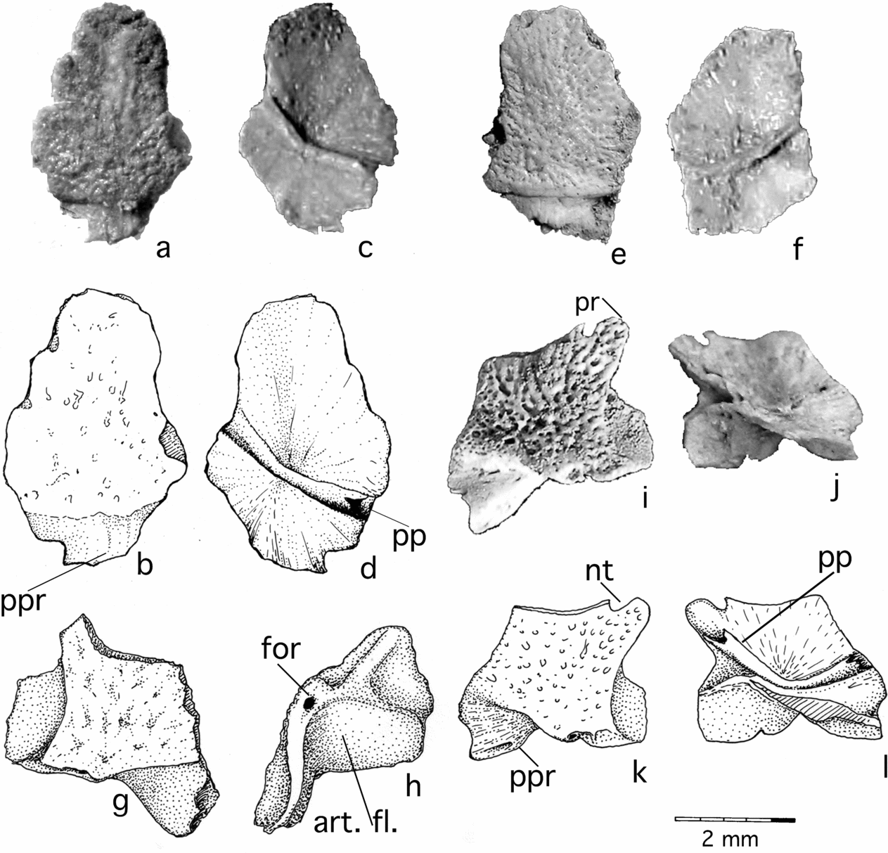
Kimbryanodus williamburyensis gen. et sp. nov. preorbital and orbital plates. Preorbital plates: (a) WAM 96.4.8, dorsal view; (b) visceral view; (c) WAM 96.4.8, line drawing, dorsal view; (d) visceral view; (e) WAM 96.4.7, visceral view; (f) dorsal view; (g) WAM 99.8.5, line drawing, visceral view; (h) dorsal view. Postorbital plates: (i) WAM 99.8.15 dorsal view; (j) visceral view; (k) WAM 99.8.16 dorsal view; (l) visceral view.

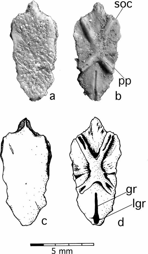
Kimbryanodus williamburyensis gen. et sp. nov. nuchal plate WAM 96.4.9: (a) dorsal view; (b) visceral view; (c) line drawing, dorsal view; (d) line drawing, visceral view.

Kimbryanodus williamburyensis gen. et sp. nov. central and paranuchal plates. Central plates: (a) WAM 99.8.18, dorsal view; (b) line drawing, dorsal view; (c) visceral view; (d) line drawing, visceral view; (e) WAM 99.8.13 dorsal view; (f) visceral view. Paranuchal plates: (g) WAM 99.8.12, line drawing, dorsal view; (h) line drawing, visceral view (i) WAM 96.4.12, dorsal view (j) ventral view; (k) line drawing, dorsal view; (l) line drawing, visceral view.

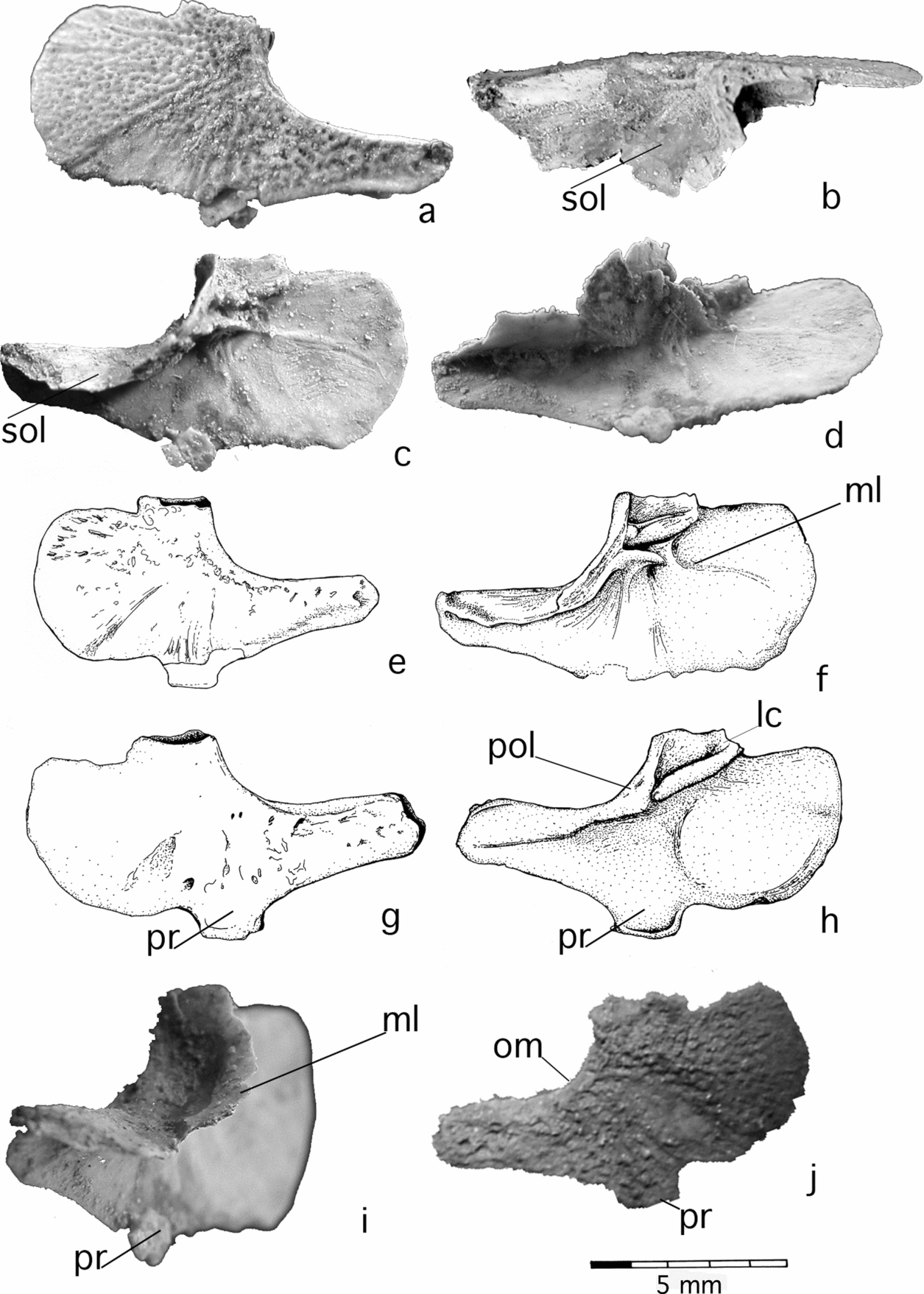
Kimbryanodus williamburyensis gen. et sp. nov. marginal plates: (a) WAM 99.8.1, lateral view; (b) dorsal view; (c) mesial view; (d) ventral view; (e) line drawing, lateral view; (f) line drawing, mesial view. (g) WAM 96.4.19, line drawing, lateral view; (h) line drawing, mesial view; (i) anteromesial view; (j) lateral view.

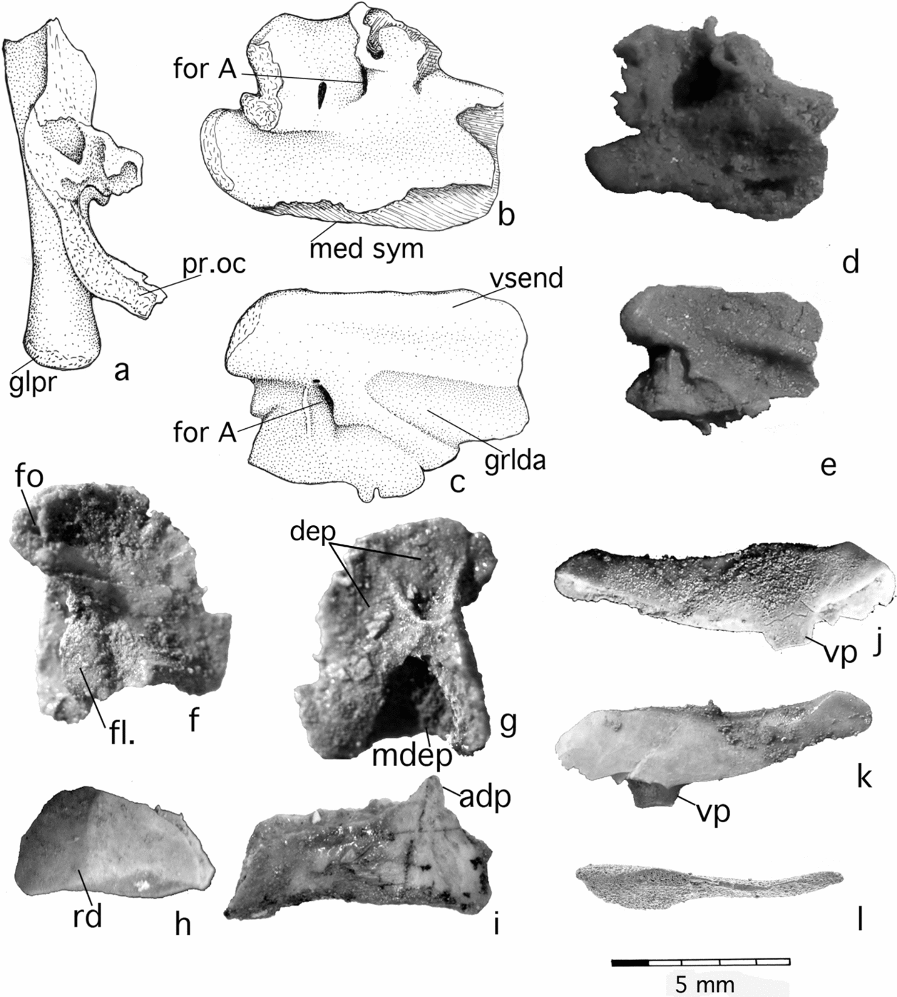
Kimbryanodus williamburyensis gen. et sp. nov. endocranial and jaw elements. Left occipital ossification WAM 96.4.18: (a) line drawing, mesial view; (b) line drawing, dorsal view; (c) line drawing, ventral view; (d) dorsal view; (e) ventral view. Autopalatine WAM 96.4.13: (f) mesial view; (g) lateral view. Upper tooth plates: (h) WAM 96.4.16, lateral view; (i) WAM 96.4.14 lateral view. Lower toothplates: (j) WAM 96.4.15, lateral view; (k) WAM mesial view; (l) WAM 99.8.9, dorsal view.

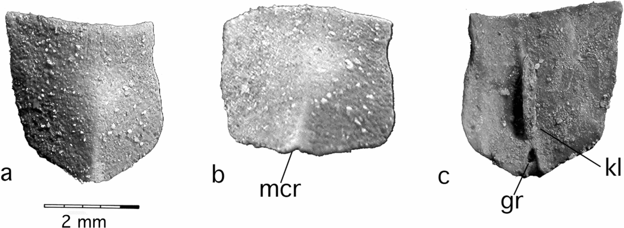
Kimbryanodus williamburyensis gen. et sp. nov. Median dorsal plate WAM 99.8.22: (a) dorsal view; (b) posterior view; (c) visceral view.

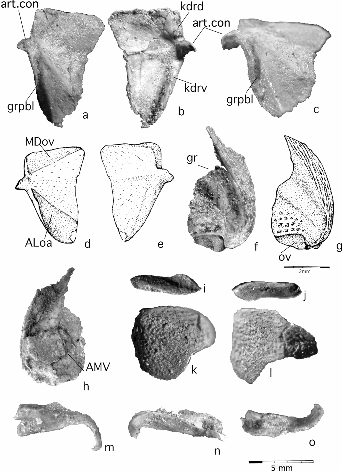
Kimbryanodus williamburyensis gen. et sp. nov. dermal plates of the trunk shield. Anterior dorsolateral plate, WAM 99.8.20: (a) lateral view; (b) mesial view; (c) ventral view; (d) line drawing lateral view; (e) mesial view. Anterior lateral plate, WAM 99.8.12: (f), lateral view; (g) line drawing, lateral view (h) visceral view with anterior median ventral plate adhered to the visceral surface. Spinal plate, WAM 99.8.3: (i) lateral view; (j) visceral view. Interolateral plate, WAM 99.8.7: (k) antero-ventral view; (l) ventral view. Right dermal element from the male pelvic clasper, WAM 96.4.17: (m) ventral view; (n) visceral view; (o) lateral view.

Derivation of name. After Williambury Station where the type section of the Gneudna Formation is located and from which the specimen was found and in recognition for the assistance that the Percy family, who run Williambury Station, have given the University of Western Australia and the Western Australian Museum over many years.
Holotype. A partial specimen (WAM 99.8.1) comprising associated nuchal, central and postorbital plates of the head shield, ethmoid ossification, and lower tooth plates and an incomplete median dorsal plate (the only thoracic material preserved).
Paratypes. One partial articulated specimen (WAM 99.8.20) comprising anterior dorsolateral plate; anterior lateral plate; anterior median ventral plate. Numerous isolated plates: preorbital plates (WAM 96.4.7, 96.4.8, 99.8.5, 99.8.4); postorbital plates (WAM 99.8.15, 99.8.16); central plates (WAM 96.4.10, 99.8.14, 99.8.13, 99.8.18); nuchal plate (WAM 96.4.9); paranuchal plates (WAM 96.4.12, 99.8.12); marginal plates (WAM 96.4.19, 99.8.1); tooth plates (WAM 96.4.14, 96.4.15, 96.4.16, 99.8.9, 99.8.10, 99.8.23); autopalatine (WAM 96.4.13); occipital ossification (WAM 96.4.18); median dorsal plate (WAM 99.8.22); anterior lateral plate (WAM 99.8.12); anterior dorsolateral plate (WAM 99.8.24); interolateral plate (WAM 99.8.7); spinal plate (WAM 99.8.3); pelvic claspers (WAM 99.8.11, 99.8.25, 96.4.17).
Locality and horizon. Frasnian, transitans to punctata conodont Zones; type section of the Gneudna Formation (23°58′10″ S, 115°12′30″ E), Williambury Station, Carnarvon Basin, NW Western Australia (Fig. 1a). Articulated specimen collected from a dolomitic bed (KT bed 7), 90 m above the base of the section within the carbonate facies, isolated plates collected from KT beds 7, 12–14 and 20–22a (Fig. 1b).
Diagnosis. Genus with relatively flat dorsal skull roof table which is broadest across the postorbital–paranuchal junction; anteriorly pointed nuchal plate tapers posteriorly and has a short median ventral groove (gr) and two small lateral ventral grooves (lgr) near posterior margin of the visceral surface; preorbital plate having a pineal embayment one-third its length; short paranuchal plate, being only half the length of the large postorbital plate, marginal plate with extensive lateral postorbital division with reticulate ornamented and a distinct median ventral process and dorsally a straight attachment margin to postorbital plate. Shearing dentition, supragnathal deeper than long with short anterior dorsal process and anterior ridge extending from the biting surface to the dorsal margin; infragnathal narrow, splint-like with cutting edge. Trunk shield with short, broad and relatively flat median dorsal plate with a well-developed narrow ventral keel and a rounded posterior margin; anterior dorsolateral plate with a distinct dorsolateral inflexion; interolateral plate with straight external anterior contact margin with the anterior lateral plate; small spinal plate with no spinal process; short broad anterior median ventral plate with anterior median point.
Remarks. As reconstructed (Fig. 2a, b), Kimbryanodus williamburyensis nov. differs from all other ptyctodonts in having a relatively broad, dorsally-flat head shield; a large postorbital plate and the combination of an anteriorly pointed and posteriorly tapering nuchal plate; central groove (gr) with two small lateral grooves (lgr) along the midline of the posterior portion of the nuchal plate (Fig. 4b, d) and the combination of a large postorbital division of the marginal plate with reticulate ornament and a developed square ventral process (Fig. 6j), and a short, relatively flat paranuchal plate (Fig. 2a); differs from Campbellodus, Chelyophorus, Ctenurella, Denisonodus, Ptyctodopsis, Rhynchodus and Rhamphodopsis in having a marginal plate with an extensive postorbital division; from Austroptyctodus, Chelyophorus, Denisonodus, Materpiscis, Ptyctodopsis, R. threiplandi, R. trispinatus, and Rhynchodus in having a small spinal plate; from Campbellodus, Denisonodus, Ptyctodopsis, R. threiplandi, R. trispinatus, and Rhynchodus in lacking a median dorsal spine; a straight overlap margin between the interolateral and anterior dorsolateral plates and the V-shape of the overlap margin between the anterior dorsolateral plate and anterior lateral plate; from Austroptyctodus, Campbellodus, Chelyophorus and Rhynchodus in having an anteriorly pointed nuchal plate; from Austroptyctodus, Campbellodus and Rhynchodus in having the preorbital plates contacting in the midline and by the large pineal embayment; from Denisonodus and Ptyctodopsis in lacking a median point on the anterior margin of the median dorsal plate, which projects in front of the anterior dorsolateral plates and shearing dentition; from Austroptyctodus and Materpiscis by the absence of linear rows of ornament on the postorbital division of the marginal plate and a rounded posterior margin on the median dorsal plate, from Campbellodus in having a single median dorsal plate and an absence of scales on the tail.
Significant variation in the size and shape of dermal plates may indicate that different ontogenic stages are represented with considerable morphological variation between individuals. Sexual dimorphism is already well documented within the group (Denison, Reference Denison and Schultze1978) and may also account for size difference among adults. Miles (Reference Miles1967) recorded variation between individuals of Rhamphodopsis, and Miles & Young (Reference Miles and Young1977) observed morphological variation between different individuals of Austroptyctodus. Additional, complete articulated specimens are needed to test these hypotheses.
4.a. Description of the head shield
The three-dimensional preservation of Kimbryanodus williamburyensis nov. has enabled the head shield to be fully reconstructed (Fig. 2a, b) with the exception of the pineal plate, which is missing (presumed present, but not yet discovered). A large embayment on the anteromesial margin of the preorbital plate for the pineal plate is present (n.pin, Figs 2a, 3d). Anteriorly, between the orbits, the roof is flat and becomes only slightly arched about the rostrocaudal axis passing through the postorbital and paranuchal plates. The head shield is broader than long, being broadest at the suture between the postorbital and paranuchal plates. In lateral view, the cheek shows a narrow postorbital fenestra (fen, Fig. 2b). Unlike Austroptyctodus, Campbellodus and Rhynchodus (Long, Reference Long1997), the preorbital plates meet mesially in Kimbryanodus williamburyensis nov. Most of the plates bear an ornament of elongated tubercles, joined to form radiating ridges, interspersed with a series of pits and grooves.
The ventral surface of the skull-roof has well-developed thickenings, a supraorbital vault and prominent tubes, which enclose the sensory lines. These features will be described in association with the plates on which they occur.
4.a.1. Preorbital plate
The preorbital plates (PrO; Figs 2a, b, 3a–h) are relatively broad and flat, having a rectangular shape which differs from the flat triangular-shaped PrO plate of Materpiscis (WAM 07.12.1). WAM 96.4.8 (Fig. 3a) is the largest plate recovered (107 mm) and WAM 99.8.4 is the smallest plate recovered (51 mm), indicating some variation in the bone proportions. The presence of developed overlap margins (ov) on the medial edge of the right PrO plate (WAM 96.4.7, Fig. 3f; WAM 99.8.5, Fig. 3h) indicates that unlike Austroptyctodus, Campbellodus and Rhynchodus, there was no median gap between them. They show a weakly concave lateral margin that curves around the top of the orbits and differs from the almost straight lateral margin present in Austroptyctodus (WAM 70.4.253, 86.9.662) and Materpiscis; however, as in Austroptyctodus and Materpiscis, the PrO does not extend around the upper anterior margin of the eye as in Campbellodus (WAM 86.9.672). In dorsal view the path of the supraorbital sensory canal (soc) can be recognized as a series of minute pores running diagonally across the plate (Fig. 3a). In visceral view the canal is visible as a bony tube (Fig. 3b, d, e, g). The visceral surface is dominated by the supraorbital vault (suov, Fig. 3d), which extends to the anterolateral corner of the plate anteriorly (and posteriorly continues onto the postorbital plate), and is delineated by the preorbital (rpro) and supraorbital (rsuo) ridges (Fig. 3b, e), although these are not as prominent as those in Austroptyctodus.
4.a.2. Postorbital plate
The postorbital plate (PtO; Figs 2a, b, 3i–l) has a smooth concave lateral margin for the orbit (WAM 99.8.16; om, Fig. 3k) and irregular interdigitating margins between the paranuchal and central plates. It is large, compared to other ptyctodonts, being almost twice the size of the paranuchal plate (Fig. 2a). A small notch (nt, Fig. 3i, k) in the posterolateral margin of the postorbital plate formed a margin for the postorbital fenestra (fen, Fig. 2b), which is high and narrow like that in Materpiscis (Long et al. Reference Long, Trinajstic, Young and Senden2008, Fig. 1h). Unlike Campbellodus there are no linear thickenings associated with the postorbital fenestra. A marked embayment (dp, Fig. 3j, l) on the visceral surface of the PtO corresponds to the downwards folding of the plate to form the lateral face of the head shield as an external angulation between the dorsal and lateral faces of the skull; however, this is not as strongly arched as in Austroptyctodus, Campbellodus, Ctenurella and Rhynchodus. In visceral view, it shows a prominent ridge (rpto) delineating the supraorbital vault (Fig. 3l).
4.a.3. Nuchal plate
The nuchal plate (Nu, Figs 2a, b; 4a–d) in Kimbryanodus williamburyensis nov. forms the median posterior margin of the head shield (Fig. 2a). In this respect it is similar to Austroptyctodus, Campbellodus, Materpiscis and Rhynchodus. The margins of the nuchal plate are complete and gently concave with a narrow underlap area to meet the surrounding bones with which it connects. WAM 99.8.1 (its delicate condition precluded photography) is approximately half the size of WAM 96.4.9 (Fig. 4a, b). An anterior point projects between the posterior margins of the preorbital plates but does not separate them (Fig. 2a). The sensory lines are not clearly visible in dorsal view, but are prominent X-shaped raised tubes of bone in ventral view (soc, pp, Fig. 4b). Unlike Austroptyctodus, Campbellodus and Denisonodus, where the bony tubes end at the posterior corners of the nuchal plate, in Kimbryanodus williamburyensis nov. and Materpiscis the bony tubes end immediately posterior to the centre of the plate (WAM 96.4.9; Fig. 4b, d); however, Materpiscis has a shorter and broader nuchal plate. Along the midline of the posterior portion of the nuchal plate is a central groove (gr) with two small lateral grooves (lgr) at the posterior margin of the nuchal plate (Fig. 4b, d). This character is unique to the genus, being present on both nuchal plates recovered (WAM 96.4.9, 99.8.1).
4.a.4. Central plate
The central plates (Ce; Figs 2a, b, 5a–f) are approximately the same size as the preorbital plates and in this respect similar to Austroptyctodus and Ctenurella. However, they lack the anterior-medial embayment for the posterior processes present on the preorbital plates of Austroptyctodus and Campbellodus. A small anterior underlap is present for the overlap of the preorbital plate and extensive lateral contact margins for the postorbital and paranuchal plates (WAM 99.8.18, Fig. 5a; WAM 99.8.13, Fig 5b). The margin in contact with the paranuchal plate is weakly convex in dorsal view where the skull roof bends down (Fig. 2b). A smooth process of dermal bone, which continues onto the paranuchal plate, forms the posterior process of the skull roof (ppr; Fig. 5a, b, e), which is similar to Austroptyctodus, Ctenurella and Campbellodus.
4.a.5. Paranuchal plate
The paranuchal plate (PNu; Figs 2a, b, 5g–l) is short (WAM 96.4.12, 40 mm), being approximately half the length of the postorbital plate (WAM 99.8.15, 75 mm), whereas in Austroptyctodus and Materpiscis it is the same length as the postorbital plate and in Ctenurella, Rhynchodus, and Rhamphodopsis it is longer. Compared with other ptyctodonts the paranuchal plate is relatively flat. A deep notch (nt) is present in WAM 96.4.12 (Fig. 5k), corresponding to a notch on the postorbital plate for the postorbital fenestra (fen, Fig. 2b), as in Austroptyctodus and Campbellodus. The anterior process (WAM 96.4.12; pr, Fig. 5i) surrounding the notch is longer and narrower than in either of these taxa, resembling instead the process of Materpiscis. The median part of the posterior pit line canal is visible externally as a series of pores and in visceral view as a well-developed tube of bone (pp; Fig. 5l).
4.a.6. Marginal plate
Two marginal plates (M) have been preserved and the straight dorsal margin of this plate forms a contact margin for the postorbital plate (Figs 2b, 6a, e, g, j). The overall shape of the marginal plate most closely resembles those of Austroptyctodus, Ctenurella and Materpiscis with its deeply convex posterior margin; unlike Austroptyctodus and Materpiscis, the denticulated, ridged ornament on the postocular division is absent. On one marginal plate (WAM 96.4.19) there is a well-developed square-ended process on the ventral border (pr, Fig. 6g–j), but on the other plate the ventral border is incomplete (WAM 99.8.1; Fig. 6a, e). Austroptyctodus (WAM 86.9.662, MNH P57637), Ctenurella (MNHN ARD 229, MNH P 53343, 53344) and Materpiscis also have a process on the ventral border of the marginal plate but it is smaller. The orbital division of the M plate is longer than the postorbital division in Kimbryanodus williamburyensis nov., whereas in Ctenurella and Materpiscis it is shorter and in Austroptyctodus they are equal. As in Austroptyctodus there is a well-developed, thick, inwardly projecting mesial lamina on the smooth visceral surface (ml; Fig. 6b–d, f, h, i), comprising subocular (sol) and postocular (pol) divisions (Fig. 6b, c, h).
4.b. Description of the dentition
4.b.1. Supragnathal plate
The supragnathal (u.tpl, Figs 2b, 7h–1) is approximately one-third high as it is long, has a gently curved biting margin with a ‘beak’-like extension that fits into a corresponding depression on the anterior margin of the infragnathal. A small anterior dorsal process forms the attachment to the autopalatine (WAM 96.4.14; adp, Fig. 7i), which when compared to Campbellodus appears quite low, resembling more the condition seen in Austroptyctodus. A prominent anterior ridge (rd) extends from the biting surface to the dorsal margin, a character unique to Kimbryanodus williamburyensis nov. (WAM 96.4.16; Fig. 7h). The occlusal surface is slightly concave, pitted and transversely grooved.
4.b.2. Infragnathal plate
The infragnathal plate (l.tpl, Figs 2b, 7j–l) is narrow and anteriorly sectorial, similar to the condition seen in Rhynchodus and Ctenurella, whereas the lower toothplates of Austroptyctodus, Campbellodus and Materpiscis are higher and wider. The narrow, posterior division of the biting surface is triturating and the trabecular inner body of the gnathal is revealed in an acid-etched specimen (WAM 99.8.1, Fig. 7l). Although a triturating posterior surface is more common in Ptyctodus, Denison (Reference Denison1985) notes that in Rhynchodus the gnathals may have a triturating surface posteriorly. The lower tooth plates have a well-developed ventral process (vp, WAM 96.4.15, 7j, k), which is also present in Ctenurella (MNHP ARD 230A).
4.c. Description of the endoskeleton of the head
4.c.1. Autopalatine
The single perichondrally ossified autopalatine WAM 96.4.13 (Figs 2b, 7f, g) is sub-rectangular in shape, resembling that of Ctenurella (MNHN ARD 229) rather than the triangular-shaped autopalatine of Austroptyctodus (WAM 70.4.253). A deep embayment directly posterior to the anteroventral corner received the broad triangular dorsal process of the supragnathal (mdep; Fig. 7g) and is positioned more anteriorly in both Austroptyctodus and Materpiscis. A ridge is developed along the anterior margin of the autopalatine from which a lamina of strongly folded bone projects on the anterodorsal corner. This ridge of bone is absent in Austroptyctodus (WAM 70.4.253) and very narrow in Materpiscis. Two further embayments, one median and one anterior (dep; Fig. 7g), are present, and all three embayments are separated by thickened perichondrial bone elevated above the surrounding surface. A strong fold is present on the mesial perichondrial lining which results in the ventral portion of the bone flaring outward (fl, Fig. 7f).
4.c.2. Endocranium
Long (Reference Long1997) gave a detailed description and reconstruction of the endocranium in ptyctodonts based on Chelyophorus, Austroptyctodus, Campbellodus, and the then-undescribed Kimbryanodus williamburyensis nov. The endocranium comprises three paired ossifications, the orbital, occipital and ethmoid, and of these, only the occipital (WAM 96.4.18) ossification is well preserved with the margins intact, although it is not in situ (Fig. 7a–e). This material does not significantly differ from that described by Miles & Young (Reference Miles and Young1977) and Long (Reference Long1997), with the exception that the occipital process of the endocranium (pr.oc) is shorter in Kimbryanodus williamburyensis nov. and does not extend past the glenoid process, whereas in Austroptyctodus (NHM P57664 and NHM P50910) the occipital process of the endocranium flares posteromedially.
4.d. Description of the thoracic shield
4.d.1. Median dorsal plate
Only one complete median dorsal plate is preserved (MD; WAM 99.8.22; Fig. 8a–c); it is broad, and shield-like in shape, with concave anterior and posterior margins. In Austroptyctodus and Materpiscis the posterior margin is acutely pointed. A low median crest (mcr; Fig. 8b) is present on the posterior dermal surface. Unlike Austroptyctodus (WAM 70.4.254, NHM P50908), the median dorsal plate has very little ornament on the external surface of the plate and is weakly arched. The overlap area for the median dorsal plate on the anterior dorsolateral plate (Mdoa; Fig. 9d) suggests that the median dorsal plate was steeply inclined in profile (Fig. 2b). There are no overlap areas to suggest the presence of a median dorsal spine, but a deep posterior groove (gr) on the ventral surface suggests an articulation with a posterior basal plate (Fig. 8c). In this respect it is similar to Austroptyctodus (Long, Reference Long1997) and Chelyophorus (D. Goujet, pers. comm.), Ctenurella and Materpiscis.
4.d.2. Anterior dorsolateral plate
The anterior dorsolateral plate (ADL; Figs 2b, 9a–e) has a distinct, dorsolateral angular inflexion (WAM 99.8.20; Fig. 9c) similar to Chelyophorus and Rhynchodus. It is broader along the dorsal margin than the ventral margin and in this respect is similar to Austroptyctodus and Ctenurella, whereas in Materpiscis the dorsal and ventral margins are of similar dimensions and in Campbellodus the ventral margin is broader. A small, thin ventral process of ornamented bone meets the anterior lateral plate (Fig. 9d) and defines a V-shaped insertion for the anterior lateral plate, which is similar to that described for Austroptyctodus, Ctenurella, Chelyophorus and Materpiscis.
4.d.3. Anterior lateral plate
The narrow dorsal margin of the anterior lateral plate (AL, WAM 99.8.12, Fig. 9f–h) forms an acute apex at the contact with the anterior dorsolateral plate. An unornamented groove (gr, Fig. 9f) separates the exterior of the plate from the thin mesial lamina, which has four rows of stellate tubercles. There are a variable number of stellate tubercular rows in Austroptyctodus, with WAM 70.4.253 having seven rows, whereas NHM P50908 has only four rows. A second AL plate (WAM 99.8.17) does not have the mesial lamina preserved so it is impossible to determine if there is any variation in row number present in Kimbryanodus williamburyensis nov.
4.d.4. Interolateral plate
The interolateral plate (IL; anterior ventrolateral plate of Miles & Young, Reference Miles and Young1977; Johnson & Elliott, Reference Johnson and Elliott1996) is similar to that of Denisonodus and Rhamphodopsis, being strongly arched in a dorsoventral direction and lacking a developed postbranchial lamina (WAM 99.8.7; Fig. 9k, l). The reticulate ornament on the external surface is sparse and confined to the central region of the plate. Three rows of small stellate denticles are present on the anterior face of the plate (Fig. 2b). A larger postbranchial lamina is present in Austroptyctodus with seven rows of denticles present and Campbellodus (WAM 86.9.672) with five rows of denticles present.
4.d.5. Spinal plate
The narrow, elongate spinal plate (Sp; Fig. 9i, j) is slightly wider at the posterior end. The dorsal and ventral margins are thickened into ridges and the central region of the plate has a reticulated ornament (Fig. 9i). There is no distinct projection of a spinal process as seen in Rhamphodopsis. The visceral surface is concave and smooth (Fig. 9j). The spinal plate is smaller compared to all other ptyctodonts with the exception of Campbellodus.
4.d.6. Anterior median ventral plate
The anterior median ventral plate (AMV) is not preserved in anatomic position, being adhered to the visceral surface of the anterior dorsolateral plate (WAM 99.8.12) so that only the exterior face is visible (Fig. 9h). The symmetrical plate is extensively covered by the interolateral plates, except for a narrow waisted ornamented region along the midline of the plate. It differs from Austroptyctodus, Campbellodus and Materpiscis in having an acute point on the anterior medial margin. In the other taxa the anterior margin is straight or concave.
4.d.7. Pelvic claspers of males
Dermal pelvic elements comprising hook-shaped ‘claspers’ have been preserved with four hooked tubercles on the posterior end of each laterally curving clasper (WAM 96.4.17, Fig. m–o). This differs from Austroptyctodus, in which there are three pointed tubercles (Miles & Young, Reference Miles and Young1977). There are three to four additional low tubercles on the posterior surface and the rest of the bone is unornamented. Each element is expanded anteriorly with a deep ventral concavity as in Austroptyctodus (Miles & Young, Reference Miles and Young1977).
5. Ptyctodont phylogeny
5.a. Taxon selection
Of the ptyctodont taxa used in the present study, only Denisonodus plutonensis is not known from articulated material of one or more individuals (Johnson & Elliott, Reference Johnson and Elliott1996). The acanthothoracid Romundina stellina Ørvig, Reference Ørvig and Lehman1975 and the petalichthyids Lunaspis broilii Gross, Reference Gross1937 and Eurcaryaspis incilis Liu, Reference Liu, Chang, Liu and Zhang1991 were selected as outgroups. The phylogenetic position of Romundina, as basal to the ptyctodonts, was argued by Young (Reference Young1986), Goujet (Reference Goujet1984, Reference Goujet and Ahlberg2001) and Goujet & Young (Reference Goujet, Young, Arratia, Wilson and Cloutier2004). To assist in character polarization, the more derived Antineosteus lehmani Lelièvre, Reference Lelièvre1984 was included in the ingroup.
5.b. Tree computing method
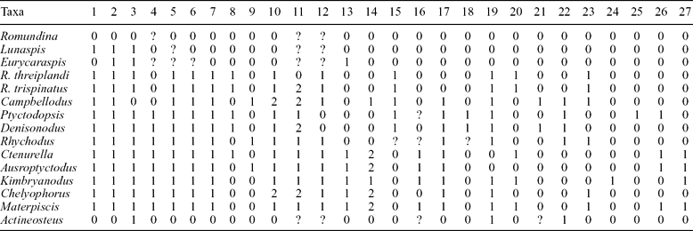
A data matrix of 27 morphological characters coded as in Table 1, unordered and unweighted for 15 taxa, was run through PAUP (Swofford, Reference Swofford1989) using the stepwise heuristic search algorithm and ACCTRAN optimization. One thousand replications with random taxon addition sequences were used. A branch and bound search was also undertaken. Identical results were obtained.
Character data matrix for 15 taxa; for explanation of characters see text Section 5.c

5.c. Character analysis
1. Lateral line canals form an X pattern on visceral surface of the nuchal plate: 0, absent; 1, present.
2. Sensory-line canals expressed on the dermal surface of bones as pores: 0, absent; 1, present. In Chelyophorus pore holes are visible within external grooves.
3. Median dorsal plate: 0, multiple; 1, one plate (modified from character 8 of Johnson & Elliott, Reference Johnson and Elliott1996).
4. Scales: 0, present; 1, absent
5. Only the posterior supragnathals are present: 0, absent; 1, present.
6. Gnathal plates are flat plates with ornament or cusps: 0, present; 1, absent, laterally compressed as a crushing plate.
7. Dermal bone forms a sheath to cartilaginous claspers (males): 0, absent; 1, present.
8. Central plates meet posterior to the nuchal plate: 0, absent (do not meet); 1, present (meet) (character 10 of Johnson & Elliott, Reference Johnson and Elliott1996).
9. Preorbital plates meet mesially: 0, present; 1, absent (character 1 of Johnson & Elliott, Reference Johnson and Elliott1996).
10. Pineal plate is part of anterior margin of skull: 0, absent, rostral plate forms the anterior margin; 1, present, pineal plate forms anterior margin of skull; 2, absent, preorbital plates form the anterior margin of the skull.
11. Upper tooth plates have an anterior dorsal process (state 2 is here defined as when the dorsal process is more than twice the height of the median height of the tooth plate): 0, absent; 1, present short and thick; 2, present high (modified from character 13 of Johnson & Elliott, Reference Johnson and Elliott1996).
12. Tritorial lower tooth plates: 0, present; 1, absent.
13. Median dorsal plate with high spine: 0, present; 1, absent (character 6 of Johnson & Elliott, Reference Johnson and Elliott1996).
14. Spinal plate: 0, present, large; 1, present, small; 2, absent (modified character 5 of Johnson & Elliott, Reference Johnson and Elliott1996).
15. Contact between anterior lateral and anterior dorsolateral plates a simple inverted V-shape: 0 present; 1, absent.
16. Elongated anterior median ventral plate: 0, present; 1, absent.
17. Ginglymoid neck joint: 0, present; 1, absent (character 9 of Forey & Gardiner Reference Forey and Gardiner1986).
18. Median dorsal plate has a straight or concave anterior margin: 0, present; 1, absent, has a median point.
19. Preorbital plates longer than central plates: 0, present; 1, absent.
20. Anterior margin of the nuchal plate is square: 0; is pointed, 1.
21. The point at which the postorbital, central and paranuchal plates meet is anterior to the posterior margin of the nuchal plate: 0; posterior to or level with the posterior margin the nuchal plate, 1.
22. Relative size of the postorbital division of the marginal plate: 0, larger than the orbital division; 1, smaller than the orbital division (marginal plate crescent-shaped).
23. Nuchal plate shape: 0, elongate being more than twice as long as wide; 1, short, relatively broad.
24. Nuchal plate with a median groove: 0, absent; 1, present.
25. Postorbital plate is irregular in outline: 0, postorbital plate is triangular in outline; 1, this character in unique to P. menzeli.
26. Anterior border of the interolateral plate: 0, scalloped; 1, straight.
27. Posterior process present on the marginal plate: 0, absent; 1, present.
6. Phylogenetic reconstruction
6.a. General results
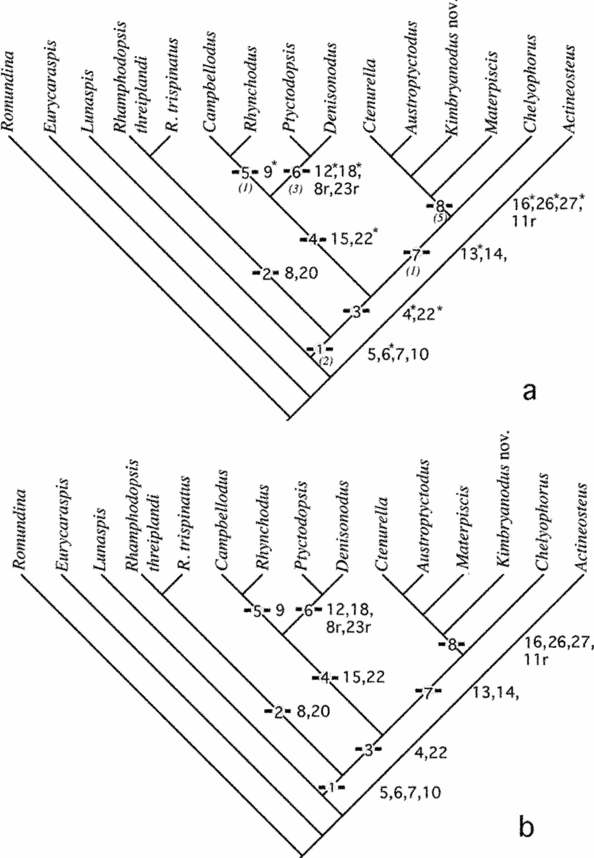
The parsimony analysis produced two equally parsimonious trees (Fig. 10a, b) with a CI = 0.58, RI = 0.73, RC = 0.44 and a tree length of 51 steps. Bremer decay indices showed the trees collapsed after five further steps (Fig. 10a). The hypothesis of relationship shows the ingroup is divided into three polytypic clades; the members of the first clade comprise both species of Rhamphodopsis and the remaining clades comprise taxa that possess a spinal plate and median dorsal spine and those that do not (Fig. 10a, b). The relationship of Kimbryanodus williamburyensis nov. and Materpiscis cannot be resolved with one hypothesis of relationship (Fig. 10a) depicting Kimbryanodus williamburyensis nov. and Materpiscis as successively more remote sister taxa to [Ctenurella + Austroptyctodus], whereas the second hypothesis of relationship (Fig. 10b) switches the positions of Kimbryanodus williamburyensis nov. and Materpiscis. The following sections provide details of the characters, their states and hypothesized state changes that support each node.
Phylogenies showing the hypothesis of relationships among ptyctodontids outlined in the text (a) Hypothesis 1. Character changes are marked on each node; r indicates a reversal; * denotes an unambiguous change, values in italics denote the Bremer support; (b) Hypothesis 2 showing the alternate position for Materpiscis and Kimbryanodus williamburyensis gen. et sp. nov. The tree statistics are: CI = 0.58, RI = 0.73, RC = .44 and a tree length of 51 steps.

6.a.1. Monophyly of the Ptyctodontida
The Ptyctodontida can be defined by the following four synapomorphies (Fig. 10a, b, node 1): possess only one pair of supragnathals, the posterior supragnathals (character 5; 0→1); infragnathal plates without cusps (character 6; 0⇒1); pre-pelvic claspers (males) (character 7; 0⇒1); pineal plate forms part of anterior margin of skull (character 10; 0⇒1);
The upper and lower dentitions of the ptyctodonts are here considered a synapomorphy of the family. All known ptyctodonts possess one pair of supragnathals, considered homologous with the posterior supragnathals because they attach to the autopalatine, whereas in Romundina the single pair of supragnathals is considered homologous with the anterior supragnathals because they attach to the ethmoid ossification. All other placoderms, with the exception of the petalichthyids in which the dentition remains unknown, have both an anterior and posterior pair of supragnathals (Denison, Reference Denison and Schultze1978; Stahl, Reference Stahl, Arratia and Schultze1999; Young, Lelièvre & Goujet, Reference Young, Lelièvre and Goujet2001). The lower dentition of ptyctodontids comprises laterally compressed plates of pleuromic dentine without a growing tritoral surface (Fig. 7l). The dentition therefore differs from the chimearoids, which have numerous hypermineralized tritoral patches growing from a dental lamina and forming a multi-tritored tooth plate. The presence of dental laminae within placoderms remains a contentious issue (Smith & Johanson, Reference Smith and Johanson2003; Burrow, Reference Burrow2003; Young, Reference Young2003).
There has been much discussion as to whether the pelvic claspers of ptyctodontids are homologous with those of the chondrichthyans and holocephalans, particularly the chimaeroids (Miles, Reference Miles1967; Ørvig, Reference Ørvig and Lehman1962), or the result of convergence (Patterson, Reference Patterson1965, Reference Patterson1992; Miles & Young, Reference Miles and Young1977; Denison, Reference Denison1985; Forey & Gardiner, Reference Forey and Gardiner1986; Stahl, Reference Stahl, Arratia and Schultze1999). Goujet & Young (Reference Goujet, Young, Arratia, Wilson and Cloutier2004) cited five characters, which they hypothesized determined the monophyly of the placoderms, although four of the five synapomorphies have been questioned (Zhu, Yu & Janvier, Reference Zhu, Yu and Janvier1999; Zhu & Schultze, Reference Zhu, Schultze and Ahlberg2001; Burrow & Turner, Reference Burrow and Turner1999; Smith & Johanson, Reference Smith and Johanson2003) and their analysis places ptyctodonts within the Placodermi. A connection between the holocephalans and the ptyctodonts is now considered unlikely.
The presence of the pineal plate as the most anterior of the head shield supports the view of Denison (Reference Denison and Schultze1978) that ptyctodonts had an anteriorly unossified cranial roof. It appears there has been a reduction in the number of head shield plates, with the loss of the rostral and post-nasal plates, which are common to other placoderms. A reduction in the number of trunk shield plates compared with other placoderms is also observed.
6.b. Main clades
6.b.1. Clade 1
Rhamphodopsis (Fig.10a, b, node 2) is separated from the other ptyctodonts through the retention of two basal characters: the presence of scales (character 4) which occur on the pelvic fins but are unknown on the tail (Miles Reference Miles1967); a cylindrical condyle on the anterior dorsolateral plate, which forms the trunk articulation of the glenoid joint (character 17; character 9 of Forey & Gardiner, Reference Forey and Gardiner1986). The glenoid condyle in other ptyctodonts is developed as a short shelf, either transversely or longitudinally orientated (Miles & Young, Reference Miles and Young1977; Forey & Gardiner, Reference Forey and Gardiner1986). Two additional characters, the centrals participating in the posterior border of the skull roof (character 8, 0→1) and anterior margin of the nuchal plate square (character 20, 0→1), occur within a number of other taxa.
6.b.2. Clade 2
This clade, comprising Campbellodus, Denisonodus, Rhynchodus and Ptyctodopsis (Fig. 10, node 4), is characterized by the following two synapomorphies: a complex M-shaped articulation of the anterior dorsolateral plate to the anterior lateral plate (character 15, 0→1) and a short postorbital division of the marginal plate (character 22, 0⇒1). This node can be further subdivided into two: the first containing Campbellodus and Rhynchodus (Fig. 10, node 5), the second containing the two North American genera, Ptyctodopsis and Denisonodus (Fig. 10a, b, node 6). Campbellodus and Rhynchodus are united by the presence of preorbital plates with no mesial contact (character 9, 0⇒1, character 1 of Johnson & Elliott, Reference Johnson and Elliott1996) and it has been independently acquired in Austroptyctodus. This character was coded as present in all ptyctodonts in the analysis of Johnson & Elliott (Reference Johnson and Elliott1996); however, Long (Reference Long1997) reinterpreted the arrangement of the bones forming the skull roof in Rhyncodus, Austroptyctodus and Campbellodus, determining that the preorbital plates were separated either side of the mid-line in these taxa.
Two character reversals occur in Campbellodus: the presence of multiple median dorsal plates (character 3, 1⇒0) and the presence of a scale-covered tail (character 4, 1⇒0). New specimens from the Canadian Arctic have revealed that the median dorsal plate in Romundina cf. stellina was also a composite structure comprising two elements and that this may represent the least derived condition (D. Goujet, pers. comm.). The presence of a scale-covered tail is also thought to be the basal condition, with Rhamphodopsis the only other ptyctodont with scales. Austroptyctodus was thought to also have a covering of scales on the tail (Long, Reference Long1997), but these have since been reinterpreted as the anterior lateral plates of three embryos (Long et al. Reference Long, Trinajstic, Young and Senden2008).
The American taxa are united (Fig. 10a, b, node 6) by four characters: the centrals participating in the posterior border of the skull roof (character 8, 0→1); an elongated nuchal plate (character 23, 1→0, also present in Austroptyctodus, Ctenurella and Kimbryanodus williamburyensis nov.); a median point on the anterior margin of the median dorsal plate, which projects in front of the anterior dorsolateral plates (character 18, 0⇒1). Denison (Reference Denison1985) hypothesized that the median notch in the posterior border of the head shield was for the reception of this point; however, this seems unlikely, as a median notch is present in taxa that lack a median point, such as Austroptyctodus, Campbellodus, Kimbryanodus williamburyensis nov. and Rhynchodus; and absent in Denisonodus, in which the median dorsal has a median point.
The presence of crushing, ‘Ptyctodus’-type lower tooth plates (character 12, 1⇒0) also unites these two taxa. Denison (Reference Denison1985) noted that some ptyctodonts have a hybrid tooth type with sectorial blades anteriorly and a tripartite occlusal area on the posterior portion of the biting surface. It is probable that the structure of the tooth plates, both supragnathals and inferognathals, indicated similar feeding patterns rather than relationships among the ptyctodonts.
6.b.3. Clade 3
The remaining clade (Fig. 10a, b, node 7) contains Kimbryanodus williamburyensis nov., which possesses a small spinal plate, and the four aspinothoracid ptyctodontids Austroptyctodus, Chelyophorus, Ctenurella and Materpiscis. Johnson & Elliott (Reference Johnson and Elliott1996) noted differences in the size of the spinal plates between taxa so in this analysis the character was coded as multistate. A small spinal plate has independently occurred in Campbellodus, and spinal plates are large and projecting in Denisonodus, Ptyctodopsis, Rhamphodopsis and Rhynchodus.
In addition, the genera of node 7 (Fig. 10a, b) all possess an anterior dorsal lateral plate with a simple inverted V-shape articulation to the anterior lateral plate (character 15) and they all lack a dorsal spine on the median dorsal plate (character 13, 0⇒1). Austropytctodus, Ctenurella, Kimbryanodus williamburyensis nov. and Materpiscis are further united (node 8, Fig. 10) by having a straight overlap margin between the interolateral and anterior dorsolateral plates (character 26, 0⇒1); a short broad anterior median ventral plate (character 16, 0⇒1); a marginal plate with a ventral process (character 27, 1⇒0) and a short dorsal process on the upper tooth plate, which is considered a reversal (character 11, 2⇒1).
7. Discussion
These phylogenetic conclusions are tentative, as it is probable that there are a large number of taxa not represented, and that these ‘missing’ taxa might influence the relationships obtained. The numerous isolated tooth plates recovered globally, in strata ranging from the Eifelian to the close of the Famennian, support this view. Most ptyctodont genera known are form-genera, based on tooth plates (Miles & Young, Reference Miles and Young1977; Carr, Reference Carr1995), which excludes them from cladistic analysis due to their lack of characters.
Johnson & Elliott (Reference Johnson and Elliott1996) were unable to determine if Rhamphodopsis or Ctenurella represented the most basal clade within the ptyctodontids, as they obtained two equally parsimonious trees (Johnson & Elliott, Reference Johnson and Elliott1996, Fig. 9). Our analysis (Fig. 10a, b) supports the position of Rhamphodopsis as the most basal of the ptyctodonts and therefore agrees with hypothesis two of Johnson & Elliott (Reference Johnson and Elliott1996, Fig. 9.2). Improved knowledge of the patterns of the dermal skull bones in placoderms, based on the discovery of new features described in various placoderms (Long, Reference Long1988, Reference Long1997; Goujet & Young, Reference Goujet and Young1995, Reference Goujet, Young, Arratia, Wilson and Cloutier2004), has resulted in a different interpretation of the presence of a nuchal plate (character 9 of Johnson & Elliott, Reference Johnson and Elliott1996), which is here considered present in all placoderms, including ptyctodonts (Miles & Young, Reference Miles and Young1977; Long, Reference Long1997). In the phylogenetic analysis of Johnson & Elliott (Reference Johnson and Elliott1996), Rhamphodopsis was the only ptyctodont considered to have retained the nuchal plate, and in accordance with Denison (Reference Denison and Schultze1978, Reference Denison1985), they termed the central median bone the postpineal plate. We disagree with this interpretation and consider that the bone they term the nuchal plate is part of the occipital ossification of the braincase. This interpretation is based on the absence of ornamentation on the bone in the Royal Scottish Museum (RSM 1966.37.1; J. Long, pers. obs.), and that this bone could not be seen in other specimens of Rhamphodopsis threilandi (NHM P48664, P52979, P52954; Long, Reference Long1997). In addition, well-preserved in situ braincase material from Austroptyctodus, Chelyophorus and Materpiscis shows the position and orientation of the occipital region of the braincase directly behind the paired central plates (Long, Reference Long1997). The crushed nature of the Edderton specimens (Rhamphodopsis threiplandi, Chicago Natural History Museum PF 1660, 1674–1695) suggests that the occipital ossification was pushed up and has become exposed behind the central plates. It is therefore concluded that the presence of a nuchal plate is retained in all members of the ptyctodonts, and that all placoderms maintain a distinctive pattern of dermal bones in the skull roof (character 2, Goujet & Young, Reference Goujet, Young, Arratia, Wilson and Cloutier2004).
The presence of a long trunk shield, more than one median dorsal plate, scales covering the body behind the shoulder girdle, overlapping dermal plates and a cylindrical condyle on the anterior dorsolateral plate, which forms the trunk articulation of the glenoid joint, are considered primitive characters for placoderms (Miles & Young, Reference Miles and Young1977; Goujet, Reference Goujet1984; Goujet & Young, Reference Goujet, Young, Arratia, Wilson and Cloutier2004). Within the ptyctodonts there is an apparent trend for reduction in the body armour, including the loss of the rostral, postnasal and suborbital plates of the headshield and spinal, anterior and posterior ventral plates and median dorsal plate elements of the trunk shield, and the loss of post-thoracic scales. This suggests that, in respect to the reduction in the number of plates that make up the head and trunk shields, ptyctodonts are more specialized than other placoderms.
Acknowledgements
The new specimens of Gneudna ptyctodonts were collected in 1995 with support from the National Geographic Society of America. We also thank Prof. D. Goujet for access to specimens held in the collections of Muséum national d'Histoire naturelle, Paris, and helpful discussion. Dr G. Young, Dr H. Gill, Dr R. Carr and Dr Z. Johanson are thanked for help and discussion on phylogenetic aspects of this work and Dr Z. Johanson is further thanked for her assistance in accessing specimens in the Natural History Museum. The referees are gratefully acknowledged; their comments and suggestions greatly improved this work. K. Trinajstic would like to acknowledge an ARC grant.
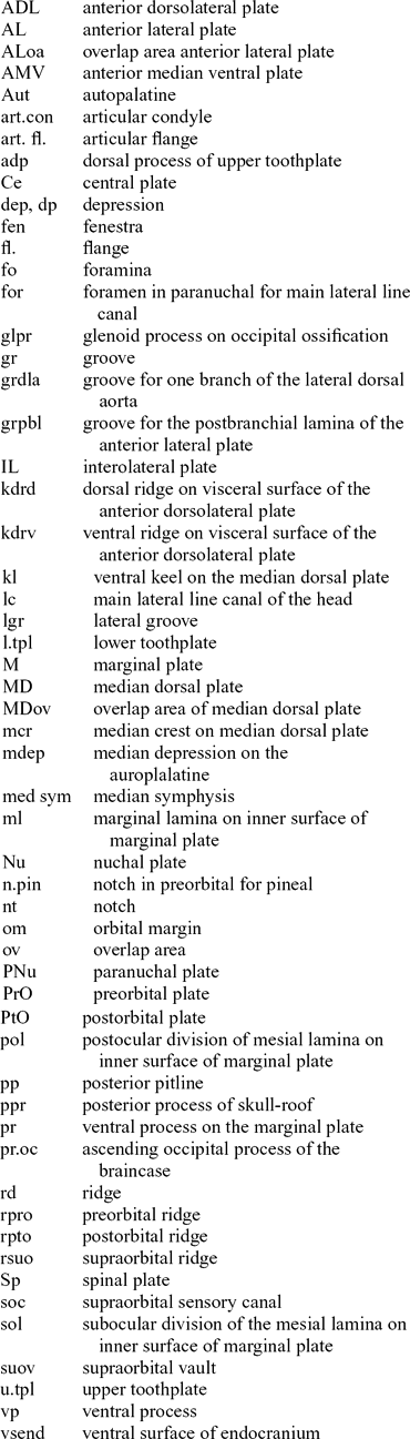
Appendix 1. Abbreviations used in figures