Introduction
Agitation secondary to dementia is a common neuropsychiatric symptom that occurs in nearly 60% of patients with dementia and includes night-time behavioral disturbances and/or behavioral disinhibition (Burgut et al., Reference Burgut, Popeo and Kellner2010). With increasing agitation, patients with dementia increase caretaker burden, risk the safety of themselves and others, and are at risk for premature institutionalization. With the black-box warning of increased mortality associated with antipsychotic use in patients with dementia, alternative therapies for addressing this troubling syndrome have long been debated. Benzodiazepines and mood stabilizers have been used as treatments for agitation in the elderly. Benzodiazepines, while anxiolytic and often sedating, can also disinhibit patients, and increase risk for falls and delirium. Mood stabilizers are often used for agitated patients but take several days to reach therapeutic levels and can be associated with serious side effects requiring close monitoring. Although antipsychotic medications have been associated with elevated mortality when used to treat agitation in dementia, agitation itself has been associated with elevated mortality risk (Peters et al., Reference Peters2015). Therefore, more effective and safer alternatives to current treatments are needed given modest efficacy and considerable tolerability concerns.
Electroconvulsive therapy (ECT) has received negative media coverage due to previous techniques of administration without anesthesia and without muscle relaxants. ECT safety has been carefully studied and improved over decades and is now standard therapy for depression after failure of several medication trials.
While ECT is not indicated for the treatment of agitation in patients with dementia, limited publications have demonstrated its efficacy for this problematic behavioral syndrome in the context of a major neurocognitive disorder. However, there has been no published randomized trial evaluating the safety and efficacy of ECT for treating agitation in dementia. This study will examine the literature supporting evidence for the safety and efficacy of ECT for patients with agitation in the setting of dementia.
Results
Case report
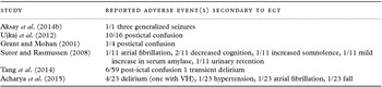
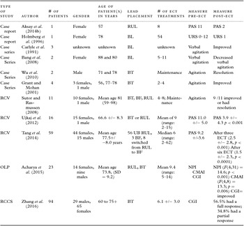
Aksay and colleagues described a case in Germany of a 57-year-old female who had rapidly progressive, severe Alzheimer's disease (Aksay et al., Reference Aksay, Hausner, Frolich and Sartorius2014b) and severe agitation. The patient suffered from significant impairments in language comprehension. The diagnosis of Alzheimer's disease was confirmed by CSF measurement, MRI, and fluorodeoxyglucose positron emission tomography. The patient experienced unspecified transient depressive mood events 26 and 18 years earlier, that were the result of “critical life events.” Those depressive episodes reportedly resolved within a few months without intervention. During this presentation, the patient did experience bouts of crying, anxiety and was suspected of potentially having psychotic symptoms with the agitation. The authors, however, did not state whether the patient's presentation was suspected to be due to an underlying mood disorder. When admitted, she was initially treated with the acetylcholinesterase inhibitor rivastigmine patch (9.5 mg/day) and quetiapine (50 mg/day). The patient failed to respond to multiple medication trials including quetiapine 175 mg/day, risperidone 2.5 mg/day, and melperone 100 m/day. She was also tried on sertraline 200 mg/day and mirtazapine 30 mg/day with no improvement in agitation. The patient's score on the PAS (Pittsburgh Agitation Scale), an evaluation of motor agitation, aberrant vocalizations, resisting care, and aggressiveness, was 11 out of 16 at baseline. No other scales or outcome measures were reported. The authors did not indicate whether there was any history of ECT in this patient. The patient spent nine weeks in the hospital prior to the initiation of ECT. She was given eight ECT treatments, which resulted in significant clinical improvement. While being given ECT, she was treated with rivastigmine 9.5 mg/day, sertraline 200 mg/day, and mirtazapine 30 mg/day. The patient's daughters, who served as her legal guardians, consented to her treatments. The patient was started with twice-weekly, right unilateral electrode placement (RUL) and a pulse width of 0.25 ms using a Thymatron® IV device (Somatics, LLC, Lake Bluff, IL, USA). S-ketamine adjusted to her weight (1.2 mg/kg) was used for anesthesia followed by succinylcholine administration for muscle relaxation. Her seizure threshold was determined to be <50 mC. Her next four sessions were conducted at 150 mC and session six through eight were conducted at 200 mC. No other specific details or parameters for the ECT treatments were provided. It was noted that the patient had a significant decrease in agitation even after two ECT treatments. During the third week of ECT treatment, the patient had a single episode of a generalized seizure. At the end of eight ECT treatments, she had a PAS score of 2. There was no affective, behavioral, or cognitive outcome measure included in this study. The legal guardians refused maintenance ECT (M-ECT). The effect of ECT on the patient's agitation was documented to last 12 weeks. Follow-up every two weeks over a four-month period indicated that the patient encountered two further simultaneous generalized seizures.
Holmberg et al. (Reference Holmberg, Tariot and Challapalli1996) described a 78-year-old Caucasian female from the USA who had vascular dementia, of unknown severity, who experienced a gradual cognitive decline over a five-year period. The authors reported that she had no neuropsychiatric history until the age of 68 years. She developed frontal release signs, an abnormal gait, and posture. The patient also had a CT scan that was consistent with cerebral vascular disease. Besides the mention of a “full dementia evaluation at a tertiary referral center,” no other specific diagnostic studies for dementia were mentioned. There was no report of an underlying mood disorder. The patient scored a 16/30 on the Mini-Mental State Exam. The patient was verbally aggressive, and physically violent toward caregivers and herself, requiring hospitalization and nursing home placement. The patient had seriously harmed a nursing home staff member, causing disability. An unspecified scale was used to monitor the patient's behavior. The assessment included four clinical domains (physical aggression, verbal disruption, abuse of self, and non-compliance with medications and meals) on a four-point scale ranging from none = 0 to severe = 3 for each domain. The patient's scores ranged from 0 to a maximum of 12 per shift. No other rating scales were used. Five months after her admission to the nursing home, the patient's physical aggression worsened, and she began to self-mutilate resulting in skin breakdown. It was determined that patient's life was in jeopardy and ECT was recommended. The authors did not mention the patient having had any previous history of ECT treatments or mood disorder. The patient's husband signed the consent for ECT after the completion of appropriate medical workup. The patient received 11 bilateral, constant current, brief pulse ECT treatments over four weeks. The ECT provided a stimulus at 60–70%. No other ECT details or parameters were provided. The patient's behavior improved after three treatments, as indicated by a decrease in physical aggression and improved nutritional intake. The daily behavior ratings were below 1 before the patient was discharged to the nursing home. After the frequency of ECT was reduced to every other week, the problematic behaviors recurred and she was placed on a M-ECT schedule of every ten days. Over the next two years, the patient underwent a total of 54 ECT treatments, which were well tolerated. The authors did not mention any specific follow-up beyond the two years. The authors noted that attempts were made to decrease the frequency of ECT treatments, but this always resulted in relapse of behavioral symptoms.
Case series
Bang and colleagues (Bang et al., Reference Bang, Price, Prentice and Campbell2008) described two cases of improvement in agitation in patients with dementia with the use of ECT. The first case was an 88-year-old female, in the USA, with a six-year history of “probable” Alzheimer's dementia and Parkinson's disease. The authors did not provide details as to how the diagnosis of dementia was made. The patient did present with global cognitive impairment. Unspecified Parkinsonian symptoms were noted by the authors. There was no report of any previous psychiatric illness, nor any mention of an underlying mood disorder. She had two months of screaming, hollering obscenities, and palilalia (involuntary repetitions of words, phrases, or sentences). The episodes lasted over 45 minute and occurred multiple times a day. CT of the head showed diffuse cortical volume loss with secondary ventricular enlargement. The patient did not respond to quetiapine, risperidone, escitalopram, olanzapine, or divalproex. On admission, she was treated with spironolactone, carbidopa/L-dopa, memantine, lorazepam, ranitidine, and oxycodone. The authors did not mention whether patient had a history of receiving ECT treatments. She underwent a total of 11 BL ECT treatments, with a seizure duration of up to 84 seconds. The authors provided no other ECT details or parameters. The authors described a significant improvement in verbal behavior by the second ECT session. There was no affective, behavioral, or cognitive outcome measurement described. Parkinsonian symptoms remained unchanged from the ECT treatments. No follow-up period was described.
The second patient was an 80-year-old female, in the USA, with a history of multiple medical problems including CHF, unspecified developmental disability and congenital hydrocephalus. The type of dementia and the severity was not described. There was no report of any past psychiatric history, nor was any information given about an underlying mood disorder. The patient attempted to assault staff, had loud speech, and was repetitively yelling “get out.” The authors did not mention whether patient had a history of receiving ECT treatments. The patient received five BL ECT treatments, with seizure durations up to 56 seconds. No other ECT treatment details or parameters were provided. The patient became cooperative and pleasant with staff, with no further signs of verbal agitation or verbal aggression. There was no affective, behavior, or cognitive outcome measurement. No follow-up period was mentioned.
Two cases of dementia-related aggressive behavior were described by Wu and colleagues (Wu et al., Reference Wu, Prentice and Campbell2010). The authors did not use an evaluation scale for agitation. The first patient was a 78-year-old male, in the USA, with a five-year history of Alzheimer's disease, unknown severity. He exhibited global cognitive impairment with jargon aphasia. Computerized Tomography (CT) of the head demonstrated diffuse cortical volume loss with secondary ventricular enlargement. The authors did not provide any other information as to how the patient became diagnosed with Alzheimer's disease. The patient had no known psychiatric history. There was no mention of an underlying mood disorder. He demonstrated agitation and hostility, injuring four nursing staff members. Trials of sertraline, risperidone, olanzapine, intramuscular haloperidol, and valproate were found to be ineffective. The authors did not mention whether patient had a history of receiving ECT treatments. Seven ECT treatments were administered, with bitemporal (BT) electrode placements. Seizures lasted up to 65 seconds. No other ECT details or parameters were provided in the paper. The patient's aggression improved after the fourth ECT treatment. No affective, cognitive, or behavioral outcome measures were provided. He received M-ECT extended to every 28 days and was able to return to the nursing home facility with no behavioral problems within the 3-month follow-up.
Case 2 was a 71-year-old male, in the USA, with no known pre-morbid psychiatric history who presented with a six-year history of frontotemporal dementia, unknown severity. He was noted to have global aphasia, with impaired language comprehension. Head CT showed mild to moderate generalized cortical volume loss with secondary ventricular enlargement. No other information was provided regarding how he was diagnosed with frontotemporal dementia. There was no mention of an underlying mood disorder, but was described as being irritable. He had recently developed physical hostility that included choking, grabbing, and pushing other residents and staff. Trials of sertraline, topical lorazepam, trazodone, and quetiapine had failed. The authors did not mention whether patient had a history of receiving ECT treatments. He received six ECT treatments with BT electrode placement, with seizure durations lasting 58 seconds. No other ECT treatment details or parameters were given. It was noted that the ECT treatments resulted in resolution of aggressive behaviors after the last treatment. There was no affective, cognitive, or behavioral outcome measure described. He was able to return to his nursing home, and was continued on M-ECT every 28 days. No follow-up period was mentioned.
Grant and Mohan (Reference Grant and Mohan2001) described four cases of patients with dementia complicated by agitation and aggression. Each of the four patients was treated with a variety of psychiatric medications including antipsychotics, but failed them. No aggression scale was used in this study; however, all four cases were noted to have a decrease in agitation and aggression by the use of ECT. The patients in this study were treated with brief pulse, MECTA ECT, and BT electrode placement.
Case 1 was a 56-year-old female, in the USA, with unspecified dementia and agitation. Her mother had a history of being diagnosed with Alzheimer's disease. The patient scored a 22/30 on the MMSE during initial evaluation, no other cognitive, behavioral, or affective measurements were used. The patient was suspected of having visual hallucinations. The authors mentioned no underlying mood disorder. The patient did, however, have a psychiatric history of unspecified anxiety. Due to failure of multiple psychiatric medications, ECT was initiated on an emergency basis. The authors did not mention whether patient had a history of receiving ECT treatments. When ECT was started, she was receiving propranolol 20 mg bid, trazodone 275 mg/day, and divalproex sodium 500 mg three times a day. She was found unable to give informed consent, which resulted in her family petitioning the court for guardianship and approval of ECT. She received four bilateral, square-wave, brief-pulse, constant-current ECT treatments with MECTA (MECTA Corp., Tualatin, OR, U.S.A.). The treatments spanned nine days, and she was noted to respond with noticeable improvement after the first ECT treatment session. The first ECT session was at 56.3 J and produced a seizure that lasted 51 seconds by EEG monitoring. The patient's second to fourth ECT treatment produced seizures lasting 37–43 seconds by EEG. The authors provided no other contributory ECT treatment details or parameters. She had no further recurrence of agitated or aggressive episodes five months after treatment. No further follow-up was specified.
Case 2 was a 78-year-old male, in the USA, with agitation, and Alzheimer's dementia that was diagnosed several years prior to hospitalization. The authors did not provide any information as to how the patient was diagnosed with Alzheimer's disease. There was no mention of an underlying mood disorder or history of ECT treatments. The authors did not include any affective, cognitive, or behavioral measures. The patient initially received four bilateral, square-wave, brief-pulse, constant-current device MECTA ECT treatments. The patient's initial treatment was given at 54.6 J, which produced a seizure of 26 seconds on EEG. The patient's next three treatments lasted 33–39 seconds on EEG. There was sustained improvement of aggression that lasted seven months. His aggressive behaviors resurfaced after seven months and he was, therefore, readmitted for ECT. After an additional three ECT treatments, aggressive episodes remitted during the two-month follow-up period. The authors did not mention any details on the type of ECT treatments, or parameters for those subsequent three ECT treatments.
Case 3 was a 77-year-old woman, in the USA, with agitation and Alzheimer's disease, with unspecified severity. The authors did not include information regarding how she was diagnosed with Alzheimer's disease. Head CT, however, showed cerebral atrophy and she had a brother that was diagnosed with Alzheimer's disease at age 64 years. The authors did not report any underlying mood disorder or psychiatric history in this patient. There was no mention of any affective, cognitive, or behavioral outcome measures. She failed multiple psychiatric medications. The family petitioned the court for guardianship and ECT approval. She underwent two bilateral ECT treatments with MECTA. The initial ECT session was at 56.5 J and produced a seizure lasting 52 seconds on EEG, the second treatment was at 57.7 J and produced a seizure of 46 seconds on EEG. The authors did not provide any other details or parameters regarding the ECT treatments. The patient's verbally aggressive behavior diminished and her sleep improved. The patient remained well within the follow-up period of one year, while only being treated with buspirone 15 mg/day and trazodone 150 mg/day.
Case 4 was a 78-year-old female, in the USA, with agitation, unspecified dementia with unknown severity and a history of seizures. There were no details provided by the authors regarding how the patient was diagnosed with dementia. The authors did not include any affective, cognitive, or behavioral outcome measures. She was noted to be anxious by the authors, but there was no other mention of a psychiatric history or an underlying mood disorder. The patient failed multiple psychiatric medications, all of which could not control her agitation. There was no mention of any previous ECT trials. The patient's family requested and was granted permission from the court for the commencement of ECT treatments. The patient received four bilateral, square-wave, brief-pulse, constant-current ECT treatments with MECTA over seven days. ECT was associated with remitted verbal aggression. ECT was started three days after discontinuing phenytoin, and 24 hours after discontinuing lorazepam. The patient was discharged on phenytoin 400 mg/day, trazodone 125 mg/day, citalopram 10 mg/day, and had no further behavioral difficulties for three months. After three months, there was a relapse of symptoms. The patient was re-admitted and given another four ECT treatments; however, the authors did not describe any details or parameters of those treatments. The authors did not mention any follow-up period.
Carlyle et al. (Reference Carlyle, Killick and Ancill1991) submitted a letter to the editor of a journal describing three verbally agitated, screaming patients, in Canada, who had unspecified dementia with unknown severity. No specific selection criteria were mentioned. They all failed multiple trials of psychiatric medications. One patient had a history of major depression, while the other two did not have any reported past psychiatric history. The letter to the editor did not mention whether these patients had an underlying mood disorder at the time of presentation. The genders were not mentioned. The authors did not mention whether any affective, cognitive, or behavioral outcome measures were used. The authors did not state whether these patients had any previous trials of ECT. The authors stated that the patients experienced subjective “dramatic improvement” after receiving bilateral ECT, early in the course of treatments. There were no other details or parameters described by the authors regarding the ECT treatments. There also was no mention of the number of treatments given. The authors did not provide any information regarding a follow-up period.
Retrospective chart review
Sutor and Rasmussen (Reference Sutor and Rasmussen2008) described 11 patients, in the USA, with Alzheimer's disease with unspecified severity, who exhibited agitation. No other selection criteria were mentioned. The data was collected through a retrospective chart review. The diagnosis of Alzheimer's disease was made by a geriatric psychiatrist, neuropsychological testing, and/or neurology consultation, and the DSM IV-TR. Six patients had a history of major depression, three had generalized anxiety disorder, one had bipolar disorder, and three had no previous psychiatric history. The authors did not mention whether there was an underlying mood disorder at the time of the ECT treatments or whether these patients had previous trials of ECT. There were no cognitive, behavioral, or affective outcome measures described. The patients were each treated with ECT for their agitation. Seizures were induced using a Thymatron device. Eight were treated with BT, one with Bifrontal (BF), and two with RUL ECT. Electrode placement was decided by the practitioner responsible for the patient. A dose titration schedule was used to determine seizure threshold with treatments given at 6x the seizure threshold for RUL and 1.5x the threshold for BT and BF. The authors did not provide any other details or parameters relating to the ECT treatments. The patients consisted of ten females and one male, with a mean age of 81.4 and a range of 59–98 years of age. Nine of the patients showed clinical improvement. Seven of the patients were diagnosed with a mood disorder in the past, from which five responded to ECT. The remaining four patients, all of who responded, did not have a prior diagnosis of a mood disorder. Out of the nine responders, six went on to receive M-ECT and were not noted to develop agitation over the next year. Two of the remaining three patients required treatment with “another course of ECT” due to relapse of symptoms. The authors did not provide any details or parameters of that additional course of ECT. There was no other follow-up described by the authors.
A systematic retrospective chart review performed by Ujkaj and colleagues (Ujkaj et al., Reference Ujkaj, Davidoff, Seiner, Ellison, Harper and Forester2012) reported on 16 patients, in the USA, with behavioral and psychological symptoms of dementia (BPSD). Eight patients had Alzheimer's disease, and the remaining eight either had a diagnosis of frontotemporal dementia, vascular dementia, or dementia not otherwise specified. Five patients had severe dementia, four had moderate to severe dementia, four had moderate dementia, and three had mild dementia. Participants had a mean age of 66.6 ± 8.3 years, and were admitted between 2004 and 2007 to a geriatric neuropsychiatry unit. Fifteen of the sixteen patients were women. Subjects treated with ECT for major depressive disorder or for BPSD during a previous admission were excluded. There were two inclusion criteria: a DSM-IV diagnosis of dementia and treatment with ECT for aggression and agitation during admission. Three outcome measures were used pre- and post- ECT. The primary outcome was the PAS. Additional outcome measures were the Clinical Global Impression (CGI) scale and Global Assessment of Functioning (GAF). A MECTA SPECTRUM 5000Q ECT device was used on these patients. Twelve patients received bilateral treatments only. Four patients received RUL, and three of those four were switched to bilateral due to inadequate response. Nine patients received ECT maintenance treatments. Titration to seizure threshold was done on the first treatment. RUL treatment was generally administered at four times the seizure threshold up to six times seizure threshold if necessary. If bilateral lead placement was used it was usually initiated at two to three times the seizure threshold and increased if the response was not sufficient. The treating practitioner made a clinical decision on whether to use bilateral or RUL. A decision to switch from RUL to bilateral was made if a poor response occurred with RUL or if the patient's symptoms worsened. Methohexital was administered for anesthesia followed by succinylcholine for muscle relaxation. Propofol was regularly used to treat postictal agitation and to aid blood pressure control. Patients received from 2 to 15 ECT treatments, with an average of 9. The average number of ECT treatments was 9.3 with a range of 2–15. The PAS total score was reduced from 11.0 +/− 5.0 at baseline to 3.9 +/− 4.3 after ECT ((F = 30.33, df = 1, 15, p < 0.001)). Average CGI score before ECT was 6.0 +/− 0.6 and after ECT was 2.1 +/− 1.6. The GAF score did not have a significant change. The authors did not describe any follow-up, and specifically mentioned that they did not have data on the nine patients who received maintenance treatments.
Tang and colleagues completed a retrospective chart review of 60 patients, in the USA, presenting with agitation associated with dementia that were treated with ECT (Tang et al., Reference Tang, Hermida, Khanh, Laddha and McDonald2014). All diagnoses were made by board-certified geriatric psychiatrists. Patients were selected based the following criteria: (1) on admission, the primary diagnosis on Axis I included dementia (any type); (2) the presenting symptoms on admission included behavioral disturbance or agitation associated with dementia; (3) the PAS Scores were available (four were missing the PAS score but met the first two criteria were also included). All patients had severe dementia. Patients who had major depressive disorder were excluded from the study. Sixty patients (45 women and 15 men, mean age = 77.5 +/− 8.0 years) were included in the analysis. With respect to dementia subtype, 28 were unspecified dementia, 22 were Alzheimer's type, 2 were vascular, 2 were frontotemporal dementia, and 6 were mixed dementia.
Seizure titration method was used at first treatment to establish seizure threshold. All patients were initially treated with RUL, with an ultrabrief pulse width (0.37 ms) at six times the initial seizure threshold (IST). When patients received bilateral treatments, they were given a brief pulse width (1.0 ms) treatments at two times the IST. For anesthesia, methohexital was used. Flumazenil was used in patients who were still taking benzodiazepines. The number of ECT treatments ranged from 2 to 62 with a median of 6. Most patients were discharged after 4–6 treatments from the inpatient unit. Only seven patients began M-ECT, receiving more than 12 treatments on an outpatient basis. Six patients who were treated with RUL had to be switched to BF after 4–9 treatments due to poor response. On average, the change in PAS total from baseline was 7.3 +/− 3.7 with a median of 8. Two cases were re-admitted and one received another acute course of ECT treatment within one year after discharge. No major side effects were reported. Only six patients developed post-ictal agitation and mild confusion. The low rates of confusion after ECT were in part explained by the administration of primarily ultra-brief pulse width.
Retrospective, case-control study
Zhang and colleagues (Zhang et al., Reference Zhang2016) published a retrospective case-control study of 23 patients with dementia, in China, who were treated with ECT. Seventy-one age and sex matched-controls were also included. The two groups were treated for a period of eight years at the National Clinical Research Centre of Mental Disorders in China. Inclusion criteria were dementia, aggression, and/or high risk for suicide. The authors did not specifically mention whether these patients had an underlying mood disorder. There was no affective rating scale used. The severity of the dementia was classified based on MMSE scores. Of the patients who received ECT, 16 had mild dementia, 6 had moderate dementia, and 1 had severe dementia. Eight had Alzheimer disease, eight had vascular dementia, four had mixed Alzheimer disease and vascular dementia, and three had “other types.” The patients were treated with BT lead placement, with a brief pulse wave through Spectrum 5000Q ECT machine; MECTA Corp. The mean number of ECT treatments was 6.1 +/− 3.0. A total of 91.3% responded fully or partially to ECT. The CGI-Efficacy Index was collected as the primary efficacy outcome measure. If the CGI score had marked improvement, it was defined as a “full response,” if there was moderate improvement in the CGI, it was defined as “partial response,” and if there was no change or worsening of the CGI, it was defined as “no response.” It was noted that 56.5% had a full response, and 34.8% had a partial response (as measured by the CGI scale). No post-ECT confusion was reported as an adverse side effect. After controlling for several factors (antidepressants, anti-dementia medication, reason for admission), there was no significant difference in hospital costs or length of hospitalization. The authors concluded that ECT may be a safe and efficacious treatment to treat aggression and suicide associated with patients who have dementia. The authors did not describe any follow-up.
Open label prospective study
Acharya and colleagues evaluated 23 patients, in the USA, with dementia who were admitted to two inpatient facilities (Acharya et al., Reference Acharya2015). The authors did not describe the specific type of dementia and severity. The authors also did not describe whether the patients had an underlying mood disorder. Fourteen of the patients were female, and nine were male. These patients were referred by a psychiatrist who diagnosed dementia, according to the DSM-IV, and determined the need for treatment. Inclusion criteria included a diagnosis of dementia based on the DSM IV, a referral to ECT by the primary psychiatrist, and a baseline Mini-Mental Status Exam (MMSE) of less than or equal to 24. MECTA SPECTRUM 5000Q (MECTA Corporation, Tualatin, OR, USA) and/or Thymatron System devices were used. The primary outcome measure was Cohen–Mansfield Agitation Inventory (CMAI). There was no affective rating scale used. RUL placement was used for 17 patients, two patients were switched from RUL to BT lead placement, and four were treated only with BT lead placement. Brief pulse width of 0.5–1.0 ms at approximately four to six times the seizure threshold was used for RUL, and 1.5 to 2.5 times the seizure threshold for BT. The mean number of treatments was 9.4 with a range of 5–14. Scores on the NPI significantly decreased from baseline to discharge (F(4,31) = 14.64; p < 0.001). By using regression analyses, a significant decline in scores on the Neuropsychiatric Inventory (NPI) subdomains of behavioral dyscontrol (F(4,17) = 11.76; p < 0.001) and agitation (F(4,13) = 15.2; p < 0.001) and CMAI-short form (F(4, 8) =13.3; p = 0.006) was noted from baseline to discharge. As ECT progressed, the need for antipsychotic administration was decreased. On average, there was an average two-point increase in the MMSE score after the completion of ECT. The authors did not describe any follow-up.
Discussion
Although some evidence supports the use of ECT as a promising treatment option for refractory aggression and agitation in patients with severe dementia, interpretation of ECT safety and efficacy data for this population is impacted by methodological limitations of published studies. These study limitations include publication bias, small sample sizes, lack of a control group, short follow-up, inconsistent use of objective rating measures and concomitant psychotropic medications. The available ECT studies often do not clarify the presence of an underlying mood or psychotic disorder prior to ECT treatment intervention, yet alleviation of mood symptoms or psychosis with ECT may result in decreased agitation. The contribution of depression and psychosis in the individual with is at times difficult to establish. When there is a suspicion that mood disorders are a contributory factor all attempts to alleviate these symptoms with behavioral therapy and pharmacotherapy should be attempted prior to embarking on a trial of ECT. Nevertheless, if the medication treatment fails to control symptoms, ECT is a viable option that in fact can treat both conditions and improve quality of life. In order to limit side effects from ECT, the course of treatment should be kept short; however, M-ECT may be required in some patients following acute ECT treatment to reduce the risk of recurrent agitation.
Prior to considering ECT as a treatment option, medical, psychiatric, and environmental causes of agitation and aggression in dementia must be evaluated and treated. Ongoing discussion with the family members should include detailed disclosure of risks versus benefits from the procedure including potential worsening of cognitive function.
BT lead placement has been described as potentially being more effective in treating certain psychiatric disorders when compared to other lead placements, but carries increased risk for cognitive side effects Although the optimal electrode placement for this population of patients with severe aggression has yet to be clarified, some practitioners initiate treatment with RUL electrode placement to minimize the risk of developing cognitive side effects. Transient cognitive impairment, including post-ictal delirium, is the most common side effect of ECT (McClintock et al., Reference McClintock, Staub and Husain2011). Previous data shows that even when patients are given more than 100 ECT treatments over a 25-year period, they may not exhibit cognitive impairment (Devanand et al., Reference Devanand, Verma, Tirumalasetti and Sackeim1991). Most patients participating in these studies have severe cognitive impairment at baseline that may improve after treatment with ECT, based on standard cognitive assessment measures, after reduction of severe agitation and aggression.
Efforts to minimize cognitive side effects include the use of RUL electrode placement and utilizing ultra-brief pulse width. Additionally, the seizure titration method could be helpful in individualizing the treatment parameters according to the subject's seizure threshold. Efforts to enhance seizure duration instead of increasing the charge when the seizure is short include maximizing hyperventilation and modifying anesthetic agents to those with less impact on seizure threshold. Lowering the anesthetic agent and adding a short acting opioid, such as remifentanil (Akcaboy et al., Reference Akcaboy2005; Dinwiddie et al., Reference Dinwiddie, Glick and Goldman2012), or alfentanil (Akcaboy et al., Reference Akcaboy2005), as an anesthesia adjunct could diminish the anticonvulsant effects of anesthetics and enhance seizure quality. Switching the anticonvulsant to ketamine (Janke et al., Reference Janke, Bumb, Aksay, Thiel, Kranaster and Sartorius2015) or etomidate (Singh et al., Reference Singh, Arora, Borle, Varma, Trikha and Goudra2015) could help with seizure quality during ECT. Ensuring hyperventilation (Aksay et al., Reference Aksay, Bumb, Janke, Hoyer, Kranaster and Sartorius2014a) prior to the administration of ECT and using flumazenil (Krystal et al., Reference Krystal, Watts, Weiner, Moore, Steffens and Lindahl1998) for patients receiving benzodiazepines have shown promise in lowering seizure threshold.
Several safety measures should be implemented when treating this population. Delirium is a common side effect of ECT (Reti et al., Reference Reti2014); thus, it is important to monitor for this side effect in a systematic way as a hypoactive delirium could be misinterpreted as reduced agitation. Cognitive function should be monitored throughout the treatment to determine the impact of ECT on various domains of cognition.
A smaller hippocampal volume prior to treatment can potentially be a predictor of a more robust clinical response (Joshi et al., Reference Joshi2016). A recent study evaluating specific neuropsychological symptoms and their relationship to survival time in patients with dementia, determined that aggression/agitation is a robust predictor of mortality and accelerated progression (Peters et al., Reference Peters2015). It is still not clear whether ECT prolongs survival time in patients who exhibit aggression and/or agitation, but it appears to at least improve quality of life of the patient and caregiver based on the available data.
Patients with dementia complicated by severe aggression or agitation often lack capacity to make medical decisions. In patients who lack capacity, involvement of the next of kin or legal guardian is key to ensure appropriate opportunity for ECT treatment.
This review suggests that a double-blinded randomized controlled trial may help identify and establish the efficacy of ECT to improve a patient's quality of life, decrease caretaker burden, and decrease healthcare costs. A future randomized controlled trial will encounter several ethical issues, such as consent, and as a result, may represent barriers for implementing such a study design. A randomized controlled trial must carefully consider the definition of usual care as a comparison group. Well-documented prospective studies and/or additional case series with explicit selection criteria, a wide range of outcome measures, and less selection bias of the study sample that may favor treatment response, is warranted. ECT may be a treatment modality to consider for agitation and aggression in individuals with dementia who are refractory to environmental modifications, behavioral interventions, and pharmacotherapy management.
Study and the adverse events reported

Numerator = number of patients who experienced event; Denominator = total patients in the study; VH = Visual Hallucinations.
Comparing available studies

RCV = Retrospective Chart Review; OLP = Open Label Prospective; RCCS = Retrospective Case-Control Study; RUL = Right Unilateral; BF = Bifrontal; BT = Bitemporal; BL= Bilateral; UB = Ultrabrief; CMAI = Cohen–Mansfield Agitation Inventory; NPI = Neuropsychiatric Inventory; CGI = Clinical Global Impressions Scale; URS = Unspecified Rating Scale.
Conflict of interest
The authors have no relevant affiliations or financial involvement with any organization or entity with a financial interest in or financial conflict with the subject matter or materials discussed in the paper. Brent Forester, MD, MSc: Board of Directors, Secretary/Treasurer: American Association for Geriatric Psychiatry. Research Funding: NIH, Assurex Health, Eli Lilly, Biogen, Rogers Family Foundation Consulting agreements: Eli Lilly, INSYS Therapeutics.
Description of authors’ roles
Dr Glass assisted with writing the paper. Dr Forester assisted with writing the paper. Dr Hermida assisted with writing the paper.