Doty, in 1977,4 had by then described efforts in five neonates at complete repair in a single stage. The interatrial septum was excised, and the right atrium was partitioned, leaving the caval veins on one side of the partition, and the atrial septal defect and tricuspid valve on the other side. A conduit was used to connect the venous portion of right atrium to the pulmonary arteries, and a second conduit was used to connect the proximal pulmonary trunk to the aorta. All five of the patients died very soon after the surgery, undoubtedly because the high pulmonary vascular resistance of the neonate precluded adequate flow of systemic venous blood from the right atrium through the pulmonary arteries.
Norwood had himself performed a palliative procedure for hypoplastic left heart syndrome in 1977 that included enlargement of the atrial septal defect, construction of a modified Glenn anastomosis, ligation of the patent arterial duct, banding of the left pulmonary artery, and side-to-side anastomosis of the aortic arch with the pulmonary trunk. The patient died several hours after surgery. Norwood's first report of successful palliation of hypoplastic left heart syndrome with long-term survival was in 1980 (Fig. 1).5 That procedure included the use of a 10 mm Dacron graft placed from the pulmonary trunk to the descending thoracic aorta. The patent arterial duct was ligated, and a band was placed around the pulmonary trunk distal to the graft. The operation had to be revised two weeks later because of migration of the band, resulting in occlusion of the right pulmonary artery. During the revision, a conduit was placed from the right ventricular outflow tract to the descending thoracic aorta. This resulted in the first reported case of long-term survival after palliation of hypoplastic left heart syndrome. Subsequent to Norwood's early efforts, it became apparent that the essential ingredients of successful initial palliation of hypoplastic left heart syndrome included establishment of a permanent communication between the right ventricle and the aorta, limitation of the flow of blood to the lungs to attenuate pulmonary vascular changes caused by elevated pulmonary blood flow and pressure, and the creation of a satisfactory interatrial communication to ensure unobstructed pulmonary venous return to the tricuspid valve. In 1980, Doty et al.6 described an operation that included placement of a single conduit to connect the pulmonary trunk to the aorta, but with a controlled opening in the conduit to supply the right and left pulmonary arteries. Levitsky et al., also in 1980,7 had renewed the interest in Litwin's operation,3 using the technique to repair a patient with aortic atresia.
Figure 1.
Norwood's original technique as usual in 1980. A conduit was placed between the pulmonary trunk (PA) and the descending thoracic aorta (Ao), and the distal pulmonary trunk was banded to limit pulmonary flow. The arterial duct was ligated. Abbreviations: RV: Right ventricle; ASD: atrial septal defect. From: Norwood WI, Kirklin JK, Sanders SP. Hypoplastic left heart syndrome: experience with palliative surgery. Am J Cardiol 1980; 45: 87–91.
By 1981, at the annual meeting of the American Association for Thoracic Surgery, Norwood was able to describe the evolution of palliative operations, encompassing his experience with 16 patients treated between 1979 and 1981.8 In the first three cases, he had used the procedure initially described in 1980. This included placement of a valved conduit from the right ventricular outflow tract to the thoracic aorta, banding of the pulmonary trunk, creation of a large interatrial communication, and ligation of the arterial duct. In the next three infants, he had placed a valved or non-valved conduit from the right ventricular outflow tract to the distal pulmonary trunk. The ascending aorta and aortic arch were reconstructed by making a longitudinal incision in the ascending aorta and arch, which was anastomosed to the proximal pulmonary trunk and augmented with a baffle of polytetrafluoroethylene. It was the remaining patients who underwent the procedure that has now become widely known as the first stage of the Norwood sequence. It consisted of direct anastomosis of the proximal pulmonary trunk to the ascending aorta and aortic arch. Flow of blood to the lungs was established with a central shunt, utilizing a polytetrafluoroethylene tube graft placed from the newly constructed ascending aorta to the confluence of the pulmonary arteries. As in all other cases, a large interatrial communication was created, and the arterial duct was ligated. At the time of that report in 1981, one patient had undergone a second stage procedure, specifically a modified Fontan procedure, at the age of 14 months. That infant died on the third post-operative day. Three additional survivors of initial palliation were awaiting their second stage procedure at the time of Norwood's report to the American Association for Thoracic Surgery.
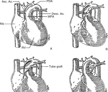
By 1983, Norwood's group had further evolved their approach to palliation, albeit still using the approach now known as first stage palliation (Fig. 2).9 Cardiopulmonary bypass was established by cannulation of the pulmonary trunk for inflow, and the right atrial appendage for venous drainage. The body was cooled to 20°C, and circulatory arrest was established. An anastomosis was performed between the divided proximal pulmonary trunk and the ascending aorta and aortic arch. The diminutive ascending aorta was incised down to a point just several millimeters above the origin of the coronary arteries. A 4 mm shunt of polytetrafluoroethylene was placed between the reconstructed ascending aorta and the right and left pulmonary arteries. The arterial duct was ligated. In describing this early series of patients, Norwood and his colleagues emphasized that only autologous vascular tissue used in the systemic arterial pathway, together with the normally developed pulmonary valve, would maximize both the technical simplicity of the procedure and the potential for growth and development of the reconstruction. They described an infant who made an uneventful recovery from initial palliation and eventually, in 1982, underwent a second reconstructive procedure. The pulmonary and systemic circulations were separated by partitioning the right atrium in such a fashion that the pulmonary venous return was directed through the interatrial communication and into the tricuspid valve. The systemic venous return on the other side of the partition was directed to the lungs through a direct anastomosis between the right atrium and the confluence of the pulmonary arteries. The shunt was removed. Following this modified Fontan procedure, the patient had pleural effusions in the post-operative period that subsequently resolved. The patient was discharged well 21 days after the operation.
Figure 2.
Norwood's modification in 1983 of his original technique. A direct aortopulmonary anastomosis was performed without the use of homograft patch and a central shunt was constructed. Abbreviations: RA: right atrium; Ao: aorta; PDA: patent arterial duct; MPA: main pulmonary artery; RV: right ventricle. From: Norwood WI, Lang P, Hansen DD. Physiologic repair of aortic atresia-hypoplastic left heart syndrome. N Engl J Med 1983; 308: 23–26.
Figure 3.
Norwood's modification in 1988, using a homograft patch to augment the aortic arch, and incorporating this on to the aortopulmonary anastomosis. This modification was introduced to prevent obstruction in the aortic arch as seen earlier in the series. A central shunt was used in this modification. From: Pigott JD, Murphy JD, Barber G, et al. Palliative reconstructive surgery for hypoplastic left heart syndrome. Ann Thorac Surg 1988; 45: 122–128.
Having gained a great deal of experience with treatment of a large number of patients with this anomaly, Norwood and his associates noted some problems from their earlier cases, including the development of obstruction in the aortic arch, and distortion of the pulmonary arteries. Technical modifications were devised to address these issues. A variety of methods were used to construct the shunt, including insertion of a modified Blalock-Taussig graft and use of a central shunt. In an effort to prevent recurrent obstruction in the aortic arch, a variety of types of material were used to augment the neo-aorta beyond the point of amalgamation of the proximal pulmonary trunk with the ascending aorta, through the length of the aortic arch, and down to the descending thoracic aorta beyond the point of insertion of the arterial duct. Cryopreserved pulmonary arterial homografts became the preferred substance for augmentation of the aortic arch, based on their ease of handling, elasticity, and hemostatic properties. Following a rather sizable experience garneted through the late 1980s and early 1990s with central aortopulmonary shunts, use of the modified Blalock-Taussig shunt, or a shunt interposed between the brachiocephalic artery and the proximal right pulmonary artery, eventually emerged as the preferred means of controlling the flow of blood to the lungs. As Norwood and others reported steadily improving rates of survival following first stage palliation, the operative procedure itself became rather standardized.
First stage palliation (Figure 4)
Figure 4.
First stage palliation as it is currently performed. A, The pulmonary trunk (PA) is transected just proximal to the origin of the pulmonary arteries (dotted lines); B, atrial septectomy is achieved through the site of right atrial cannulation. The distal pulmonary trunk is closed with an oval patch of cryopreserved pulmonary arterial homograft. The arterial duct is transected at its entrance into the aorta (Ao). An incision is extended both distally and proximally into the aorta; C, the aortic arch is augmented with a patch of cryopreserved pulmonary arterial homograft; D, proximally, the aorta is anastomosed to the adjacent pulmonary trunk with a few interrupted sutures. The homograft patch is then sutured to the remainder of the circumference of the transected proximal pulmonary trunk; E, a systemic-to-pulmonary arterial shunt is constructed usually by interposing a length of 3.5- or 4-mm polytetrafluoroethylene graft between the brachiocephalic artery and the right pulmonary artery. Abbreviations: LV: left ventricle; RV: right ventricle.
Surface cooling is initiated by maintaining the operating room at approximately 60°F. After the induction of general endotracheal anesthesia, a cooling blanket is used, and bags of crushed ice are placed around the infant's head. Median sternotomy and partial thymectomy are performed. After heparin is administered, the pulmonary trunk is cannulated just above the sinuses of Valsalva. A single venous cannula is introduced through the right atrial appendage. Cardiopulmonary bypass is commenced at flows of 150 to 200 ml/kg/min. With the initiation of bypass, sutures are used as tourniquets to occlude the right and left pulmonary arteries to insure optimal systemic perfusion through the arterial duct. During the period of bypass cooling, the branches of the aortic arch are exposed, and are looped with sutures to be used as tourniquets in preparation for circulatory arrest. Once cooling to the desired temperature has been accomplished, with the nasopharyngeal temperature decreased to 16 to 20°, the branches of the aortic arch are occluded with the tourniquets, and the circulation is temporarily discontinued. Blood is drained via the venous cannula into the bypass reservoir, and cannulas are removed from the field. Atrial septectomy can usually be performed through the site of right atrial cannulation. Rarely, an incision is required in the right atrial appendage, as in instances when the atrial septum is particularly thick, or when there is marked leftward deviation of the superior and posterior attachment of the primary atrial septum. The pulmonary trunk is transected just proximal to the origin of the right pulmonary artery. The distal opening in the confluence of the pulmonary arteries is closed with an oval patch of pulmonary homograft. The arterial duct is exposed by proximal traction on the pulmonary arteries. The duct is isolated, ligated, and transected at its entrance into the thoracic aorta. The resulting opening in the thoracic aorta is extended distally for a centimeter or two into the descending thoracic aorta. Then the opening is extended proximally by carrying an incision along the lesser curvature of the aortic arch into the ascending aorta to a point adjacent to the point of transection of the proximal pulmonary trunk. The friable remnants of the walls of the arterial duct are excised. A gusset of previously thawed cryopreserved pulmonary arterial homograft is used to augment the aortic arch. The augmentation is begun distally in the thoracic aorta, and continued proximally to the aortic arch. The transected proximal pulmonary trunk is anastomosed in side-to-side fashion to the ascending aorta. This aortopulmonary anastomosis is a delicate and crucial technical part of the procedure, which in some instances may be best performed by using multiple interrupted sutures. Then the most proximal portion of the homograft gusset is sutured to the transected proximal pulmonary trunk. With the neo-aortic reconstruction completed, the heart is recannulated and bypass is resumed. The heart generally resumes beating spontaneously, but occasionally requires defibrillation. During the period of perfusion and re-warming, the systemic-to-pulmonary arterial shunt is constructed. As noted, this is generally accomplished by inter-position of a 3.5 or 4 mm polytetrafluoroethylene tube between the proximal brachiocephalic artery and the proximal right pulmonary artery. Alternative methods, including the use of a central shunt from the underside of the augmented aortic arch to the confluence of the pulmonary arteries, may be indicated in some cases. Either the right and left pulmonary arteries, or the shunt itself, are temporarily occluded until the patient has been full re-warmed. At this point, ventilation is commenced, and the shunt is opened to provide flow of blood to the lungs. Separation from cardiopulmonary bypass is generally accomplished with minimal inotropic support. Ventilation is achieved with the fixed inspiratory oxygen gradually being reduced to the lowest level that results in satisfactory oxygenation, ideally with a systemic arterial saturation in the range of 70–80%. The placement of one or more right atrial lines, which are exteriorized through the chest wall, has proved useful in ensuring reliable vascular access for monitoring of cardiac filling pressures, and for infusions. The chest can usually be closed in routine fashion, but it is occasionally useful to delay sternal approximation by closing the wound temporarily with an elliptically shaped patch of polytetrafluoroethylene, which is sutured to the edges of the skin. Several days later, when cardiac function has stabilized, and edema has mobilized, then routine sternal closure can be accomplished.
Intermediate palliation
In the early experience with conversion from the palliated state to the Fontan circulation, it was observed that, even among those infants who were well palliated, there were many who went on to develop a low cardiac output state early after the Fontan procedure. This low output state was characterized by tachycardia and only transient response to administration of volume, with the observation that ventricular filling pressures became progressively higher, with an increase in the volume of fluid in the third space. Echocardiographic evaluation of these patients revealed a very high ratio of ventricular mural thickness to cavity volume. The functionally single ventricle, which had gradually increased its myocardial mass in response to the volume-loaded palliated state, was now contracted, thickened, and poorly compliant. This phenomenon resulted in unacceptably high mortality for the Fontan procedure. We hypothesized that the abrupt reduction of the ventricular volume load after the Fontan procedure resulted in persistence of increased muscle mass in the setting of an acutely diminished volume of the ventricular cavity. This led us to believe that dividing the Fontan operation into two procedures could accomplish earlier reduction of the volume work of the functionally single ventricle, and would allow normalization of the ratio of ventricular mass to volume before completion of the Fontan operation.10 This would then minimize the impact of changes in ventricular geometry on outcome and survival.
Intermediate palliation is accomplished using one of two surgical techniques. A superior cavopulmonary anastomosis, the so-called the bidirectional Glenn anastomosis, is performed by transecting the superior caval vein, creating an end-to-side anastomosis between it and the right pulmonary artery, closure of the cardiac end of the transected vein, interruption of the azygos vein, and elimination of the systemic-to-pulmonary arterial shunt. We prefer to use the hemi-Fontan procedure, an alternative form of superior cavopulmonary anastomosis, which was introduced in 1989 as the first step in a two staged approach to the Fontan operation.11 The principles of the hemi-Fontan operation include connection of the superior caval vein to the pulmonary arteries, elimination of the systemic-to-pulmonary arterial shunt, elimination of other sources of antegrade flow to the pulmonary arteries, augmentation of the central pulmonary arteries to eliminate stenosis or distortion, and maintenance of physical continuity between the right atrium and the superior cavopulmonary amalgamation, facilitating the eventual completion of the Fontan operation (Fig. 5).
Figure 5.
Hemi-Fontan operation. A, The systemic-to-pulmonary arterial shunt is occluded and may be excised. The confluence of the pulmonary arteries is opened anteriorly; B, the right superior caval vein and right pulmonary artery are anastomosed to one another in side-to-side fashion; C, a gusset of cryopreserved pulmonary arterial homograft is used to augment the confluence of the pulmonary arteries, to create a roof over the large anastomosis of the superior caval vein to the pulmonary arteries, and as a dam to close the surgically enlarged junction of the right atrium with the superior caval vein; D, flow from the superior caval vein is into the right and left pulmonary arteries as indicated by the arrows. From: Jacobs M. Hypoplastic left heart syndrome, in Kaiser LR, Kron IL, Spray TL (eds) Mastery of Cardiothoracic Surgery. Philadelphia, Lippincott-Raven, 1998.
Completion of the Fontan operation (Figure 6)
Figure 6.
Completion of the Fontan circulation. A, Before the completion only the flow from the superior caval vein flow enters the pulmonary arteries. Inferior caval venous flow, and pulmonary venous return enter the systemic ventricle; B, the homograft dam, which was previously placed to occlude the junction of the right atrium with the superior caval vein, is widely excised; C, the total caval pulmonary connection is accomplished by a lateral atrial tunnel technique. The lateral atrial tunnel is composed medially of a gusset of polytetrafluoroethylene, and laterally of native atrial wall. From: Jacobs ML. Hypoplastic left heart syndrome, in Kaiser LR, Kron IL, Spray TL (eds) Mastery of Cardiothoracic Surgery. Philadelphia, Lippincott-Raven, 1998.
The Fontan circulation is usually completed 12 to 18 months after the hemi-Fontan operation. After repeated median sternotomy, minimal dissection is required to make preparations for cardiopulmonary bypass. The aorta and right atrium are cannulated, and hypothermic cardiopulmonary bypass is established. During the phase of cooling, enough of the ascending aorta is mobilized to afford access for aortic cross-clamping. The free wall of the right atrium is dissected from the adjacent pericardium and pleura. Bypass cooling is continued until the nasopharyngeal temperature has reached the desired level, usually 16 to 20°C. When cooling has been accomplished, the aorta is cross-clamped and cardioplegic solution is infused. Cannulas are removed. An incision is made in the free wall of the right atrium, parallel and slightly anterior to the terminal groove. The incision extends inferiorly to the level of the Eustachian valve of the inferior caval vein, and superiorly to within a few millimeters of the junction of the right atrium with the superior caval vein. The dam of homograft tissue that was previously placed to separate the right atrium from the confluence of the superior caval vein and pulmonary arteries is excised. The cavopulmonary connection is established as a lateral tunnel using a gusset of polytetrafluoroethylene. In our practice, this is generally trimmed from a 10 mm tube graft that has been split longitudinally. The gusset is sewn inferiorly around the opening of the inferior caval vein into the right atrium, and then along the inferior atrial wall just posterior to the edge of the atrial septal defect. Superiorly, the gusset is sewn to the edges of the opening into the superior caval vein and pulmonary arteries. The lateral tunnel is then completed by incorporating the anterior edge of the gusset in the closure of the atriotomy incision. We choose to fenestrate the lateral tunnel. This appears to result in better preservation of ventricular filling, thus improving cardiac output in the early post-operative period. It also appears that this strategy is associated with less morbidity from effusions.
A more recent modification to complete the Fontan circulation is to use an extra cardiac conduit, a procedure which may be accomplished with or without cardiopulmonary bypass. The extracardiac option involves the inter-position of a prosthetic conduit or aortic homograft between the divided inferior caval vein and the pulmonary arteries. While there is no consensus as to which technique of intermediate palliation to produce the superior cavopulmonary anastomosis, or which technique to complete the Fontan circulation, is optimal for all patients, there is recent data from computational flow dynamics to suggest that the sequence of a hemi-Fontan procedure followed by placement of a lateral atrial tunnel to complete the circulation produces a total cavopulmonary connection characterized by better conservation of energy than that which results from the bidirectional Glenn anastomosis combined with an extracardiac conduit.12