Corticosteroids are widely used for various auto-immunoinflammatory conditions. They have several cardiovascular adverse effects, and the exact mechanism causing this is unknown. Tachy and bradyarrhythmias occur in 1–82% of patients taking steroids. Reference Miura, Ohki and Yoshiba1 These include atrial flutter/fibrillations, ventricular tachycardia, sinus tachycardia, sinus bradycardia, myocardial infarction, and sudden death. Corticosteroid-induced bradycardia is less common than corticosteroid-induced tachycardia. These are less frequent in children. Steroids are also known to reduce serum potassium levels via their mineralocorticoid action and predispose to arrhythmias. Many previous reports describe high-dose intravenous, intramuscular or oral steroid-induced bradycardia, or hypokalaemia in different patients. Here we report a child with warm idiopathic autoimmune haemolytic anaemia who developed treatment refractory hypokalaemia and sinus bradyarrhythmias following low-dose intravenous methylprednisolone therapy.
Case
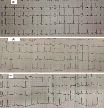
An eight-year-old girl with 7 months past history of repeated episodes of pallor which was treated with monthly blood transfusion at a local hospital, gradually had her condition worsened with frequent need of transfusion every weekly since last 1 month, and was referred to our hospital for further work-up and management. Her high-performance liquid chromatography report was normal. On admission, severe pallor, icterus, moderate hepatomegaly, tachycardia [128 beats/minute] (Fig 1a), and tachypnoea [24 cycles/minute] were present. Emergency packed cell transfusion (PRBC) was done after sending all necessary investigations. Despite receiving two units of blood, pallor persisted with no improvement in haemoglobin concentration and icterus worsened with an increase in indirect fraction of bilirubin. Complete blood count showed normal leucocyte and platelet count with 3 g% haemoglobin. Peripheral blood smear showed normocytic, normochromic red blood cells with acanthocytes, micro-spherocytes, and reticulocytosis. Malarial parasite was absent. Liver function test showed total bilirubin 11.6 mg/dL with an indirect fraction of 10.6 mg/dL normal enzyme levels.
( a ) Electrocardiogram on admission showing sinus tachycardia with heart rate of 128 beats/minute. ( b ) Electrocardiogram showing sinus bradyarrhythmia with average heart rate of 60 beats/minute along with a premature atrial complex during intravenous methylprednisolone therapy. ( c ) Normal sinus rhythm with heart rate of 100 beats/minute at the time of discharge.

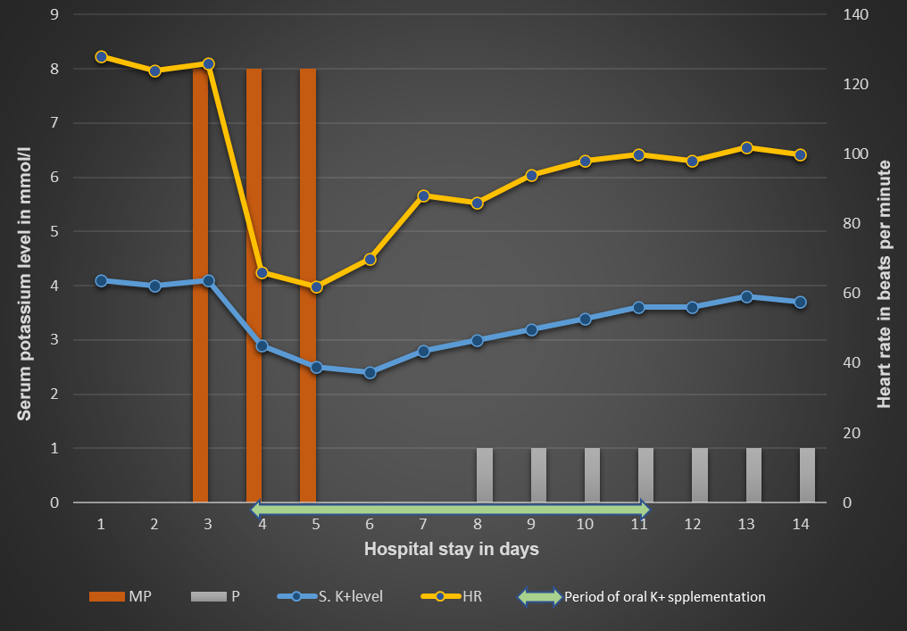
Direct antiglobulin test was positive for IgG and negative for C3. Other tests like basic electrolyte panel, arterial blood gas analysis, urine analysis, renal function test, iron profile studies, thyroid profile, autoimmune profile, and glucose-6-phosphate dehydrogenase levels were normal. Serological markers for hepatotropic viruses, human immunodeficiency virus, and Epstein Barr Virus were negative. Bone marrow study showed normoblastic erythroid hyperplasia with marked dyserythropoiesis. Diagnosis of warm idiopathic autoimmune haemolytic anaemia was made, and treatment was started with methylprednisolone therapy at 8 mg/kg/day. After 24 hours of steroid therapy, child developed bradycardia with pulse rate 50–66 beats/minute (about 60% less than baseline heart rate). Average blood pressure recorded was 88/60 mmHg. She was otherwise asymptomatic. Cardiac markers were normal. Electrocardiogram showed sinus bradyarrhythmia with average heart rate of 60 beats/minute with a premature atrial complex (Fig 1b). A structurally normal heart with optimal ventricular function was proved with echocardiography. Serum potassium level had dropped from 4.1 to 2.9 mmol/L. Since hypokalaemia is known to induce a variety of arrhythmias, immediately oral potassium chloride supplementation was started at 2 mmol/kg/day and was increased upto maximum of 5 mmol/kg/day with regular monitoring of potassium levels. Despite that there was no improvement of hypokalaemia and bradycardia, so steroid therapy was stopped after 3 days. Bradycardia resolved spontaneously over 2 days with normalisation of serum potassium levels. Low-dose oral prednisolone at 1 mg/kg/day was started after stabilisation of heart rate on day 8 (Fig 2) and gradual improvement of haemoglobin, liver function parameters with establishment of normal sinus rhythm of heart rate 100 beats/minute at the time of discharge was documented (Fig 1c).
Variation of heart rate and serum potassium level in relation to steroid therapy (MP = intravenous methylprednisolone at 8 mg/kg/day; P = prednisolone at 1 mg/kg/day; HR = heart rate).

Discussion
Dose, route, rate of infusion, type of steroids, and patient-related factors like cardiac or renal disease are the important risk factors to develop arrhythmias. Methylprednisolone, prednisolone, dexamethasone, and hydrocortisone are the main culprits blamed in the literature. The onset can occur as early as 1 minute and may be delayed up to 6 days. Reference Stroeder, Evans and Mansell2 This late onset can make it difficult to identify corticosteroids as the cause. Dose-dependent effect is seen and they have observed resolution of bradycardia with dose reduction. Reference Al Shibli, Al Attrach and Hamdan3 Rapid rate of infusion of pulse steroid within short time frame of 30 minutes and oral pulse steroids are blamed to the increase in the risk of bradycardia. Reference Al Shibli, Al Attrach and Hamdan3 This is of significance to remember to monitor outdoor patients receiving oral steroids for bradycardia. The exact aetiology underpinning corticosteroid-induced bradycardia is not described properly; however, several plausible mechanisms have been proposed. The most speculated is steroids exerting bradycardic effect through suppressing the cytokine production and function of sympathetic nervous system. Reference Nagakura, Morikawa, Sakakibara and Miura4 Another proposed mechanism is steroids inducing changes in sodium and water physiology resulting in plasma volume expansion and baroreceptor activation leading to bradycardia. Reference Akikusa, Feldman and Gross5 The asymptomatic presentation is more common than symptomatic. Symptomatic patients usually experience dizziness and chest pain. Reference Ohshima, Kawahata, Kanda and Yamamoto6 These arrhythmias are usually transient, and most of them resolve without any medical intervention to administration of anti-arrhythmic drugs or temporary cardiac pacing, Reference Soni, Kumar and Debnath7 either by continuing same dose or reducing dose or stopping steroids by as early as 4 hours to as late as 11 days. Reference Taylor and Gaco8 Abnormal serum electrolytes were also observed in patients who developed bradycardia after receiving corticosteroids. Reference Miura, Ohki and Yoshiba1 Glucocorticoids induce hypokalaemia by transcellular shifting of potassium into the cells by stimulating Na+ K+ ATPase pump or by causing kaliuresis. Reference Perazella, Rastegar, Reilly and Perazella9 Our patient also experienced hypokalaemia after starting steroids. Despite potassium supplementation, imbalance did not improve until steroids was stopped, implying corticosteroids were responsible for both bradycardia and hypokalaemia (Fig 2). Hypokalaemia also independently triggers arrhythmias in addition to those induced by steroids.
We hope this case serves as a reminder to mention that though pulse steroids are known to increase the risk of adverse effects, but even low-dose steroids should be kept in mind as a potential cause to blame for bradyarrhythmias.
Acknowledgements
Dr Sunil Kumar Hemram, MD, Assistant professor, Department of Pediatrics, MMCH, Midnapore and Dr Ashesh Halder, MD, DM (cardiology), Department of Cardiology, IPGMER and SSKM Hospital, Kolkata, for helping to diagnose and manage the patient.
Financial support
This research received no specific grant from any funding agency commercial or not-for-profit sectors.
Conflicts of interest
None.
Ethical standards
The authors assert that all procedures contributing to this work comply with the ethical standards of the relevant national guidelines on human experimentation and with the Helsinki Declaration of 1975, as revised in 2008.