Introduction
Milk protein genes are abundantly and specifically expressed in the mammary gland of lactating mammals. Lactation is essential for the development of newborns. Since the domestication of a few ruminants, milk has become an important food for humans. Milk proteins represent 30% of the proteins consumed by humans in developed countries. On the other hand, milk protein genes are among the most intensively expressed genes and they were soon candidates to study the mechanisms of gene expression. For these reasons, biology of lactation has been extensively studied for the last 50 years. The main factors controlling mammary gland development and milk protein gene expression are well known. These factors are oestrogens, prolactin, progesterone, glucocorticoids, insulin-like growth factor-1, placental lactogen and extracellular matrix (Martinet et al., Reference Martinet, Houdebine and Head1999; Sternlicht, Reference Sternlicht2006). The action of these factors can be studied in vivo, in cultured mammary explants, in cultured primary mammary cells, in mammary cell lines and in transgenic animals.
One of the major biological questions concerning multicellular organisms is that of knowing how well-defined genes are specifically expressed in the different specialised tissues and not in others. It is admitted that in the totipotent and pluripotent cells of early embryos, all the genes are potentially active. Organogenesis and cell differentiation are believed to result from the progressive selection of the genes, which are to remain sensitive to specific activators in adult cells. It was postulated decades ago that different genes are irreversibly silenced in cells not pre-programmed to express them. This implies that specialised genes must remain available during development until late differentiation and must be sensitive to specific inducers. This could be achieved at the chromatin level by the sustained binding of proteins to the genes throughout development to prevent their silencing. Genes themselves may also be specifically modified to become silent. Cytosine methylation in DNA is known to play this role. In no case has the cascade of events controlling gene silencing and activation during development been completely deciphered. The albumin gene can be considered as a model as it remains unmethylated in the endoderm as well as in foetal and in adult hepatocytes. The early binding of two transcription factors, HNF3 and GATA-4, to albumin gene enhancer is believed to maintain the chromatin locally in an open configuration until inducers are present and become capable of initiating expression of the gene (Cirillo et al., Reference Cirillo, Lin, Cuesta, Friedman, Jarnik and Zaret2002; Lomvardas and Thanos, Reference Lomvardas and Thanos2002). Recent data indicate that GATA-3 and FoxA1 transcription factors play a major role in mammary cell differentiation and potentially on the early maintenance of milk gene in an open configuration (Tong and Hotamisligh, Reference Tong and Hotamisligh2007).
Milk protein genes are a good model to study the mechanisms of early differentiation. Mammary gland development occurs essentially during pregnancy, and weaning is followed by an almost complete involution of the organ. Until puberty in female, the mammary gland remains in a state equivalent to that of a foetal organ. After puberty and until pregnancy and lactation, mammary gland growth remains very limited. The equivalent of a foetal development thus occurs in adults at each reproduction cycle. This facilitates the task of experimenters studying foetal development.
For years, the study of gene expression in higher organisms remained impossible for technical reasons. The methods of genetic engineering defined since almost three decades make the study of the differentiation mechanisms easier. The present review summarises the different research steps between the first identification of milk protein mRNAs and the study of milk protein gene control at the chromatin level during development.
Biotechnological applications resulting from the study of milk protein genes are becoming a reality. One possible application is to produce recombinant pharmaceutical proteins in the milk of transgenic animals. Another possibility may be to modify milk composition by transgenesis to optimise this food for humans and for domestic animals. The state of the art in this field is also depicted in this review.
Study of polysome profile
The polysome profile in sucrose gradient reflects the number of ribosomes bound to mRNAs. The systematic determination of the polysome profile in the mammary gland provided a good view of the presence of milk protein mRNA in different physiological situations. The profile of the polysomes from the mammary gland of lactating animals indicated that the dominating mRNAs had a size corresponding to milk protein mRNA. During pregnancy, the polysome profile did not show the presence of milk protein mRNAs. A progressive shift of polysome profile to that observed in lactating animals was induced by prolactin injection into pregnant rabbits and this induction was inhibited by the simultaneous injection of progesterone (Gaye et al., Reference Gaye, Houdebine, Pétrissant and Denamur1973; Assairi et al., Reference Assairi, Delouis, Gaye, Houdebine, Bousquet and Denamur1974).
Identification of casein mRNAs by in vitro translation
Milk from different species contains variable content of proteins. Caseins are present in the milk of all species but at a highly variable concentration. The various caseins are αS1-, αS2-, β- and κ-caseins. The other major proteins are α-lactalbumin involved in lactose synthesis, β-lactoglobulin present mainly in ruminants and whey acidic protein (WAP) present in marsupials, some rodents, lagomorphs, camelids, pigs and likely in some other species. Interestingly, the WAP gene has been recently found in a mutated and inactive form in humans and ruminants (Hajjoubi et al., Reference Hajjoubi, Rival-Gervier, Hayes, Floriot, Eggen, Piumi, Chardon, Houdebine and Thépot2006).
Rabbit reticulocyte lysate proved to be able to translate isolated mRNAs. This tool was used to identify some of the milk protein mRNAs. A fraction from sucrose gradient enriched in mRNA and later a total mRNA fraction obtained by affinity chromatography on polyU-Sepharose were used for this purpose. The mRNAs for ewe α-, β-, and κ-caseins and β-lactoglobulin were identified by increasing the number of identified mRNAs in vertebrates from three to seven (Gaye and Houdebine, Reference Gaye and Houdebine1975). This tool was used to show that milk protein mRNAs are present in polysomes bound to endoplasmic reticulum but not on free polysomes (Houdebine and Gaye, Reference Houdebine and Gaye1975a). It also revealed that half of the casein mRNAs are devoid of polyA (Houdebine et al., Reference Houdebine, Gaye and Favre1974; Houdebine, Reference Houdebine1976).
Centrifugation of cytosol from the mammary gland of lactating rabbits in sucrose gradient revealed that 20% of casein mRNAs were in the form of free mRNPs (ribonucleo-particles). This suggests that during lactation in the rabbit, the translation machinery of mammary cells is saturated. The expression at a high level of foreign proteins resulted in a decrease of natural milk proteins and the total content of protein remained constant. On the contrary, in sheep, the expression of foreign genes did not alter endogenous protein secretion and the total content of proteins in milk was increased.
Purification of milk protein gene mRNAs by polysome immunoprecipitation
In order to characterise more precisely and more easily the variations of milk protein gene mRNA concentration, a purification of some of the mRNAs was attempted. The method originally defined to isolate egg white mRNAs was adapted to casein mRNAs. This method relies on the specific immunoprecipitation of polysomes using antibodies recognising the nascent caseins. The casein mRNAs were isolated from the immunoprecipitated material using affinity chromatography with polyU-Sepharose. This laborious technique made the isolation of ewe αS1- and β-casein mRNAs possible, with a purity estimated to at least 80% (Houdebine and Gaye, Reference Houdebine and Gaye1976). The same technique was implemented to purify the rabbit casein mRNAs. In this case, the casein mRNAs were purified all together since antibodies against each casein were not available. The mRNA fraction obtained in this way was considered as a valuable material to synthesise cDNA probes specific for casein mRNAs. Indeed, repeated observations showed that the synthesis of the four caseins is simultaneously induced.
Measurement of casein mRNA concentration using cDNA probes and liquid-phase hybridisation
The isolated mRNAs were used to synthesise radioactive cDNAs using reverse transcriptase or Escherichia coli Klenow DNA polymerase I. These probes were incubated in capillary tubes in the presence of total RNA fractions obtained from mammary gland at different physiological stages. This laborious method made possible the evaluation of hormone and extracellular matrix effects in vivo and in vitro (Houdebine and Gaye, Reference Houdebine and Gaye1975b; Houdebine, Reference Houdebine1976; Shuster et al., Reference Shuster, Houdebine and Gaye1976; Devinoy et al., Reference Devinoy, Houdebine and Delouis1978; Houdebine et al., Reference Houdebine, Delouis and Devinoy1978a, Reference Houdebine, Devinoy and Delouis1978b and Reference Houdebine, Devinoy and Delouis1978c). A small but detectable amount of casein mRNA was found in the mammary gland of virgin mature rabbit (Houdebine, Reference Houdebine1979).
The measurement of casein mRNA concentration and casein synthesis in mammary explants or cells showed that, as opposed to previous publications, spermidine was probably not the glucocorticoid mediator. The integrity of microtubules appeared necessary for the induction of casein genes. Addition of phorbol esters to a culture medium of mammary explants did not favour or prevent casein gene induction, suggesting that kinases C do not play a major role in the transduction of the prolactin message on milk protein genes (Martel et al., Reference Martel, Houdebine, Teyssot and Djiane1983). A more recent study indicated that protein kinase Cμ is required to obtain activation of the αS1-casein gene in rabbit primary mammary cells by prolactin and extracellular matrix (Praskova et al., Reference Praskova, Jolivet, Houdebine and Mitev2005).
The same techniques revealed for the first time that fish prolactin was capable of inducing milk synthesis in rabbit mammary explants. This showed that the lactogenic properties of prolactin appeared sooner than generally thought before the emergence of mammary gland during evolution (Houdebine et al., Reference Houdebine, Farmer and Prunet1981).
Milk protein gene cloning
Gene cloning techniques provided the possibility of determining the sequence of several rabbit milk protein cDNAs and genes (Devinoy et al., Reference Devinoy, Hubert, Schaerer, Houdebine and Kraehenbuhl1988a and Reference Devinoy, Schaerer, Jolivet, Fontaine, Kraehenbuhl and Houdebine1988b; Schaerer et al., Reference Schaerer, Devinoy, Kraehenbuhl and Houdebine1988; Thépot et al., Reference Thépot, Devinoy, Fontaine, Hubert and Houdebine1990a and Reference Thépot, Devinoy, Fontaine and Houdebine1990b; Jolivet et al., Reference Jolivet, Devinoy, Fontaine and Houdebine1992; Baranyi et al., Reference Baranyi, Aszodi, Devinoy, Fontaine, Houdebine and Bosze1996; Ghareeb et al., Reference Ghareeb, Thépot, Puissant, Cajero-Juarez and Houdebine1998). This allowed the use of these sequences as probes to evaluate the expression level of the corresponding genes.
In the rabbit, all the genes started to be expressed simultaneously during the second part of pregnancy (Puissant et al., Reference Puissant, Bayat-Sarmadi, Devinoy and Houdebine1994), suggesting that they are all under the control of common inducers. A more recent study indicated that the rabbit WAP gene was induced later than casein genes during pregnancy (N. Daniel-Carlier, unpublished data). In vitro induction of the genes revealed that casein genes are modestly sensitive to glucocorticoids, that the WAP gene is highly sensitive to glucocorticoids but only in the presence of prolactin, and that all the genes are totally dependent on extracellular matrix to be induced. Transferrin gene was induced essentially by extracellular matrix (Puissant et al., Reference Puissant, Bayat-Sarmadi, Devinoy and Houdebine1994). The common signal that induces all the milk protein genes during the second half of pregnancy might be the partial drop of progesterone at this period in rabbits. The different genes are not equally under the same direct action of progesterone. One possibility is that progesterone controls the composition and thus the activity of the extracellular matrix. The early decline of progesterone in the rabbit might induce a modification of the extracellular matrix, which occurs only by the end of pregnancy in other species. This could induce all the milk protein genes with the contribution of the circulating lactogenic hormones.
Study of transcription factors
Works carried out in rats identified STAT5 as a major transcription factor activated by a tyrosine kinase, JAK2, which is reversibly activated when prolactin is bound to its receptor (see review by Doppler, Reference Doppler1994). A similar mechanism was found in rabbits. STAT5 binding sites were found in the promoter of αS1-casein and WAP genes. A region of the αS1-casein promoter known as the milk box forming an enhanceosome originally found in the rat and bovine β-casein promoter was found in the rabbit promoter. The STAT5 binding site was present in the rabbit promoter and in a distal region containing the milk box. These data were obtained using in vitro mutations, gene transfection into primary rabbit mammary cells (Devinoy et al., Reference Devinoy, Malienou-N’Gassa, Thépot, Puissant and Houdebine1991) and gel retardation (Pierre et al., Reference Pierre, Jolivet, Devinoy, Théron, Malienou-N’Gassa, Puissant and Houdebine1992 and Reference Pierre, Jolivet, Devinoy and Houdebine1994; Jolivet et al., Reference Jolivet, L’Hotte, Pierre, Tourkine and Houdebine1996).
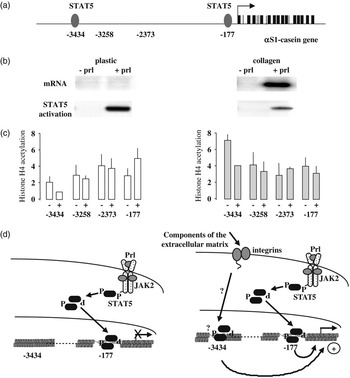
A study carried out in mice indicated that the activation of STAT5 by prolactin in cultured mammary cells occurred only in the presence of the extracellular matrix. It was concluded that the matrix inhibits a tyrosine phosphatase that deactivates STAT5 in the absence of matrix (Edwards et al., Reference Edwards, Wilford, Liu, Hennighausen, Djiane and Streuli1998). The same was not found in the rabbit mammary cells. STAT5 was activated in a similar manner by prolactin in the presence or the absence of the matrix. Yet, casein genes were activated only in the presence of the matrix (Tourkine et al., Reference Tourkine, Schindler, Larose and Houdebine1995; Jolivet et al., Reference Jolivet, Pantano and Houdebine2005). It is not known if the discrepancy between these data is due to the difference of species or to experimental conditions. Interestingly, it was found using the ChIP technique (chromatin immunoprecipitation) that the extracellular matrix induces an opening of the chromatin in the αS1-casein upstream regulatory region as judged by the level of acetylated histone H4 (Figure 1; Jolivet et al., Reference Jolivet, Pantano and Houdebine2005). Prolactin and extracellular matrix might thus deliver independent but complementary messages to the mammary cells to induce milk protein genes. This result is compatible with the idea that chromatin configuration is locally prepared by some inducers allowing other factors to trigger the expression of specific genes.
Transcriptional activation of rabbit αS1-casein gene by extracellular matrix through chromatin structure modifications of a distal regulatory element. (a) Structure of rabbit αS1-casein gene and upstream regulatory regions. The black arrow and boxes represent the transcription start point and the 19 exons of the gene. Four upstream regions are numbered. The −177 and −3434 regions characterised by STAT5 binding sites (round symbols) are both mandatory for high-level stimulation of the rabbit αS1-casein gene transcription by prolactin and extracellular matrix. The two other regions (−3258 and −2373) are dispensable. (b) Prolactin and extracellular matrix dependent activation of rabbit αS1-casein gene transcription. Primary mammary cells isolated from rabbit mammary gland were cultured on thick floating collagen I gel or directly on plastic, then treated or not with ovine prolactin. Upper panel: detection of αS1-casein-mRNA (Northern blots). αS1-casein mRNA was detected only in cells cultured on collagen in the presence of prolactin. Lower panel: STAT5 activation (gel retardation assay). Prolactin induced STAT5 activation was detected in rabbit mammary cell extracts in both culture conditions. (c) Chromatin structure of rabbit αS1-casein gene upstream regions. Chromatin immunoprecipitation by anti-acetylated histone H4 antibody showed relatively high acetylation levels in −177, −2373 and −3258 regions in both culture conditions. In contrast, the −3434 region was characterised by a moderate histone H4 acetylation level in cells cultured on plastic. Thus, an open chromatin structure is present along the distal region in cells cultured on collagen, whereas in cells cultured on plastic, the distal region is embedded in a less open structure. (d) Schematic representation of the possible prolactin and extracellular matrix pathways in rabbit mammary cells: in both culture conditions, prolactin activates the cytoplasmic STAT5 transcription factor through a cascade of protein phosphorylations, including JAK2 tyrosine kinase. In both conditions, the prolactin activated STAT5 might interact with the proximal STAT5 binding site (−177) that is located in an open chromatin structure. In the presence of an appropriate extracellular matrix, a still unknown integrin pathway is activated leading to the opening of chromatin in the distal −3434 region. Finally, the prolactin activated STAT5 interacts with both the distal and the proximal binding sites thus allowing induction of αS1-casein gene transcription. From Jolivet et al. (Reference Jolivet, Pantano and Houdebine2005).

Use of transgenesis
Transgenesis is one of the best ways to study gene function and regulation. Our group participated in 1986 in the generation of transgenic fish (Chourrout et al., Reference Chourrout, Guyomard and Houdebine1986) and during the following years of transgenic mice and rabbits. Transgenic rabbits were generated to create models to study human diseases and to prepare recombinant pharmaceutical proteins in milk proteins (Bösze and Houdebine, Reference Bösze and Houdebine2006).
Our first transgenic rabbits expressed the human α1-antitrypsin gene in their blood at the concentration of 1 mg/ml. The models that are regularly developed refer mainly to diseases due to disorders of lipid metabolism. We also obtained rabbits expressing the green fluorescent gene in all cell types, allowing a direct observation of cells in vivo (Al-Gubory and Houdebine, Reference Al-Gubory and Houdebine2006).
Transgenic rabbits are also generated to produce pharmaceutical proteins at an experimental scale (Bösze and Houdebine, Reference Bösze and Houdebine2006) and expectedly soon at an industrial scale (Koles et al., Reference Koles, van Berkel, Pieper, Nuijens, Mannesse, Vliegenthart and Kamerling2004). The proteins prepared in rabbit milk are as follows: human EPO (erythropoietin) (Massoud et al., Reference Massoud, Attal, Thépot, Pointu, Stinnakre, Théron, Lopez and Houdebine1996), human extracellular superoxide dismutase (EC SOD) (Strömqvist et al., Reference Strömqvist, Houdebine, Andersson, Edlund, Johanson, Viglietta, Puissant and Hannson1996), equine LH/CG (Galet et al., Reference Galet, Le Bourhis, Chopineau, Le Griec, Perrin, Magallon, Attal, Viglietta, Houdebine and Guillou2001), human inhibitor C1 (Koles et al., Reference Koles, van Berkel, Pieper, Nuijens, Mannesse, Vliegenthart and Kamerling2004), rotavirus VP2 and VP6 proteins to be used as vaccine (Soler et al., Reference Soler, Le Saux, Guinut, Passet, Cohen, Merle, Charpilienne, Fourgeux, Sorel, Piriou, Schwartz-Cornil, Cohen and Houdebine2005) and alkaline phosphatase (Bodrogi et al., Reference Bodrogi, Brands, Raaben, Seinen, Baranyi, Fiechter and Bosze2006). Several proteins were also produced in mouse milk at a high level: human α1-antitrysin (Bischoff et al., Reference Bischoff, Degryse, Perraud, Dalemans, Ali-Hadji, Thépot, Devinoy, Houdebine and Pavirani1992), human growth hormone (Devinoy et al., Reference Devinoy, Thépot, Stinnakre, Fontaine, Grabowski, Puissant, Pavirani and Houdebine1994) and bovine growth hormone (Thépot et al., Reference Thépot, Devinoy, Fontaine, Stinnakre, Massoud, Kann and Houdebine1995).
In all cases, the 6.3 kb rabbit WAP promoter region was used to drive the expression of the foreign genes or cDNAs. Several conclusions can be drawn from these examples. As observed with other promoters, transgenes are better expressed when they contain native genomic DNA fragments rather than cDNAs. The 6.3 kb rabbit WAP promoter is sensitive to position effect, similar to most promoters. The rabbit promoter contains a potent enhancer 5.5 kb upstream of the transcription start point (Devinoy et al., Reference Devinoy, Malienou-N’Gassa, Thépot, Puissant and Houdebine1991; Millot et al., Reference Millot, Fontaine, Thépot and Devinoy2001). This explains why it is more potent than the 2.6 kb mouse WAP promoter also used to prepare recombinant proteins in milk. The efficiency of the rabbit WAP promoter is highly dependent on the associated gene in transgenic animals. This strongly suggests that the associated genes may contain enhancers, which contribute on a case-by-case basis to the expression of the transgenes.
Another important observation was that human EPO was found at a low level in the blood of transgenic rabbits irrespective of their sex and particularly during lactation. Proteins of the lactoserum, but not caseins associated in large micelles, can leak from the mammary lumen to blood at a low rate (Grabowski et al., Reference Grabowski, Le Bars, Chene, Attal, Malienou-Ngassa, Puissant and Houdebine1991). The same appears true for recombinant proteins. The health of the transgenic rabbits and mice expressing the EPO genes was thus altered by the presence of the hormone in blood. This reveals one of the limits of transgenic animals to produce active proteins. A better control of the transgenes may be achieved using exogenous inducers such as tetracycline in addition to WAP promoter.
Another observation was that the foreign proteins secreted in rabbit milk were not fully glycosylated. The EC SOD was not fully sialylated but highly active, whereas the porcine LH/CG was active in vitro but not in vivo due to its incomplete glycosylation and short half-life. Such observations have been conducted more recently for other proteins produced in rabbit milk (Koles et al., Reference Koles, van Berkel, Pieper, Nuijens, Mannesse, Vliegenthart and Kamerling2004; Soler et al., Reference Soler, Thépot, Rival-Gervier, Jolivet and Houdebine2006; Houdebine, 2007a).
Design of efficient vectors for the expression of transgenes
The experiments depicted above suggest that the 6.3 kb rabbit WAP gene promoter does not contain all the elements required for successful transgene expression in all or most cases. A longer WAP promoter region did not allow a better expression of the associated genes including rabbit WAP gene in transgenic mice (Bischoff et al., Reference Bischoff, Degryse, Perraud, Dalemans, Ali-Hadji, Thépot, Devinoy, Houdebine and Pavirani1992; Devinoy et al., Reference Devinoy, Montoliu, Baranyi, Thépot, Hiripi, Fontaine, Bodrogi and Bosze2005). This suggests that essential regulators of the WAP gene are located in remote regions of the genome. Several strategies have been followed to improve this situation (Houdebine et al., Reference Houdebine, Attal and Vilotte2002; Houdebine, Reference Houdebine2007b).
The design of vectors for the expression of foreign genes cannot rely only on rational reasoning as all the signals present in genes, in transcribed regions as in promoters, are not known. The construction of a vector suppresses some of these unknown signals and adds other ones. On the other hand, the evaluation of vector efficiency cannot be achieved in cell systems. Indeed, one of the major events controlling gene and transgene expression takes place during early embryo development (Houdebine, 2007a and Reference Houdebine2007b). This implies that new vectors must be tested in transgenic animals and lactating transgenic females for the genes to be expressed in milk. This means that each evaluation is time consuming, allowing the test of only a few number of combinations per year.
Several enhancers and particularly the MMTV (mouse mammary tumour virus) enhancer region were added to vectors. The distal regulatory element from the rabbit αS1-casein was shown to strongly enhance the expression of associated genes in mammary cells. The addition of six copies of this element before the rabbit WAP promoter did not improve transgene expression (Pantano et al., Reference Pantano, Jolivet, Prince, Menck-Le Bourhis, Maeder, Viglietta, Rival and Houdebine2002; Pantano et al., Reference Pantano, Rival-Gervier, Menck-Le Bourhis, Maeder, Viglietta, Houdebine and Jolivet2003). Another enhancer that proved useful was the HTLV-1 IRES (internal ribosome entry site). This element which allows the translation of the second cistron of bicistronic mRNAs is also a strong enhancer. This improved transgene expression (Attal et al., 1999). Several introns and transcription terminators showed variable efficiency (Houdebine, 2007a and Reference Houdebine2007b).
The most important element added to the vectors was the 5′HS4 region from the chicken β-globin locus. This element added upstream of promoters greatly increases the expression level of transgenes. This phenomenon was observed with a promoter active in all cell types (Taboit-Dameron et al., Reference Taboit-Dameron, Malassagne, Viglietta, Puissant, Leroux-Coyau, Chereau, Attal, Weill and Houdebine1999) as with the WAP promoter (Rival-Gervier et al., Reference Rival-Gervier, Maeder, Viglietta, Prince and Houdebine2003). The 5′HS4 element can be used efficiently in most cases. However, a number of other insulators, which may be more or less efficient, remain to be found (Giraldo et al., Reference Giraldo, Rival-Gervier, Houdebine and Montoliu2003).
Regulatory elements favouring gene and transgene expression have generally been identified using long genomic-DNA regions surrounding the gene of interest (Long and Miano, Reference Long and Miano2007). This approach was applied to the WAP gene. Several BAC vectors (bacterial artificial chromosome) containing pig WAP gene were isolated and characterised. One of them containing 150 kb of genomic DNA was used to generate transgenic mice. These animals expressed the pig WAP gene with a high frequency and at a much higher rate than plasmid vectors containing only 20 to 30 kb (Rival et al., Reference Rival, Attal, Delville-Giraud, Yerle, Laffont, Rogel-Gaillard and Houdebine2001; Rival-Gervier et al., Reference Rival-Gervier, Viglietta, Maeder, Attal and Houdebine2002; Saïdi et al., Reference Saidi, Rival-Gervier, Daniel-Carlier, Thépot, Morgenthaler, Viglietta, Prince, Passet, Houdebine and Jolivet2007). Interestingly, a gene construct containing the 6.3 kb rabbit WAP promoter and the firefly luciferase reporter gene was much better expressed in transgenic mice when it was inserted into the pig BAC vector (E. Soler et al., unpublished data). Rabbit gene banks in BAC vectors are now available. The work carried out with pig WAP BAC can be extended to the rabbit gene. The rabbit genomic DNA region might be even more efficient than that of the pig as rabbit milk contains about seven times more WAP than pig milk.
Conclusion and perspectives
The evolution of the techniques during the last 30 years made it possible the study of the mechanisms that control gene expression. Although the most impressive event for milk protein genes is their intense activation by lactogenic hormones during the hours following parturition, major mechanisms are required during embryo and foetal development to prepare these genes to keep their sensitivity towards specific inducers. These events represent the mechanisms of cell differentiation at the chromatin level. The available information strongly suggests that these mechanisms imply not only DNA and transcription factors but also the chromatin structure. The known mechanisms controlling gene regulation indicate that the regulatory elements are spread over a wide range of genomic DNA. The study of long DNA fragments is thus required.
Several recent techniques facilitate the study of chromatin structure. A traditional technique consists of identifying the chromatin sites hypersensitive to DNAse1. These sites correspond to regions more or less devoid of nucleosomes to which transcription factors can bind. The technique known as ChIP (chromatin immunoprecipitation) is able to reveal the presence of any factor locally bound to chromatin as soon as antibodies against these factors are available. This method allows the determination of the biochemical modifications of histones, which maintain chromatin in an open and active or a closed and inactive configuration. Recent improvement of the method showed that it can be implemented with a very small number of cells and thus potentially to early embryos (O’Neill et al., Reference O’Neill, VerMilyea and Turner2006). The technique known as 3C (chromatin conformation capture) makes it possible to identify the remote elements of chromatin which are associated forming a hub at the promoter level through loop formations (de Laat and Grosveld, Reference de Laat and Grosveld2003).
The BAC vectors containing long genomic-DNA fragments surrounding the WAP gene depicted above are an appropriate tool to study chromatin conformation in relation to WAP gene expression. Indeed, the profile of acetylated histones H4 at the WAP locus is essentially the same in the mammary gland of lactating pigs and in the mammary gland of lactating mice harbouring a genomic DNA region containing pig WAP gene (Saïdi et al., Reference Saidi, Rival-Gervier, Daniel-Carlier, Thépot, Morgenthaler, Viglietta, Prince, Passet, Houdebine and Jolivet2007). Moreover, deletions of BAC fragments reduce the expression of the WAP gene in transgenic mice. Interestingly, as judged by the ChIP technique, the chromatin opening of the WAP locus corresponds to the level of WAP gene expression in various tissues and in different physiological situations. A highly conserved region has been found at about 140 kb upstream of the WAP gene in several species. Deletion of this region reduced WAP gene expression and local chromatin opening. This supports the idea that this sequence might operate in the embryo and later to prepare this chromatin region to remain functional in differentiated mammary cells. It becomes potentially feasible to study the WAP locus in early embryos, in embryonic stem (ES) cells, in ES cells differentiated in vitro, in the recently discovered mammary stem cells (Shackleton et al., Reference Shackleton, Vaillnat, Simpson, Stingl, Smyth, Asselin-Labat, Wu, Lindeman and Visvader2006), in cultured primary mammary cells and in mammary glands.
Interestingly, the study of the WAP locus provides researchers and companies with improved tools to produce recombinant pharmaceutical proteins in milk. The preparation of recombinant proteins, particularly in milk, is an important challenge of biotechnology (Houdebine, Reference Houdebine2002, Reference Houdebine2003a and 2007a). The recent approval of the first protein, human antithrombin III, prepared in goat milk further emphasises this fact (Schmidt, Reference Schmidt2006).
The techniques aiming at improving the efficiency of transgene expression developed for recombinant protein production may be extended to vectors implemented to generate relevant models for the study of gene function (Houdebine, Reference Houdebine2003b) and human diseases (Houdebine, Reference Houdebine2005). Similar tools may also be used to improve animal production, including the modification of milk composition for human and animal consumption (Bleck et al., Reference Bleck, White, Miller and Wheeler1998; Hiripi et al., Reference Hiripi, Baranyi, Szabó, Tóth, Fontaine, Devinoy and Bösze2000; Brophy et al., Reference Brophy, Smolenski, Wheeler, Wells, L’Huillier and Laible2003; Houdebine, Reference Houdebine2004; Soler et al., Reference Soler, Thépot, Rival-Gervier, Jolivet and Houdebine2006; Baranyi et al., Reference Baranyi, Hiripi, Szabó, Catunda, Harsänyi, Komäromy and Bősze2007).
Acknowledgements
This paper was published as a main paper of the Sixth Annual INRA-symposium ‘Mammary Gland and Milk’.