Introduction
Rheumatic heart disease (RHD) is the commonest aetiology of mitral regurgitation (MR) in children in low- and middle-income regions of the world. The dominant focus on rheumatic aetiology often overshadows other underlying pathologies, posing major potential challenges in management, as described in this case report of a young Ugandan female.
Case report
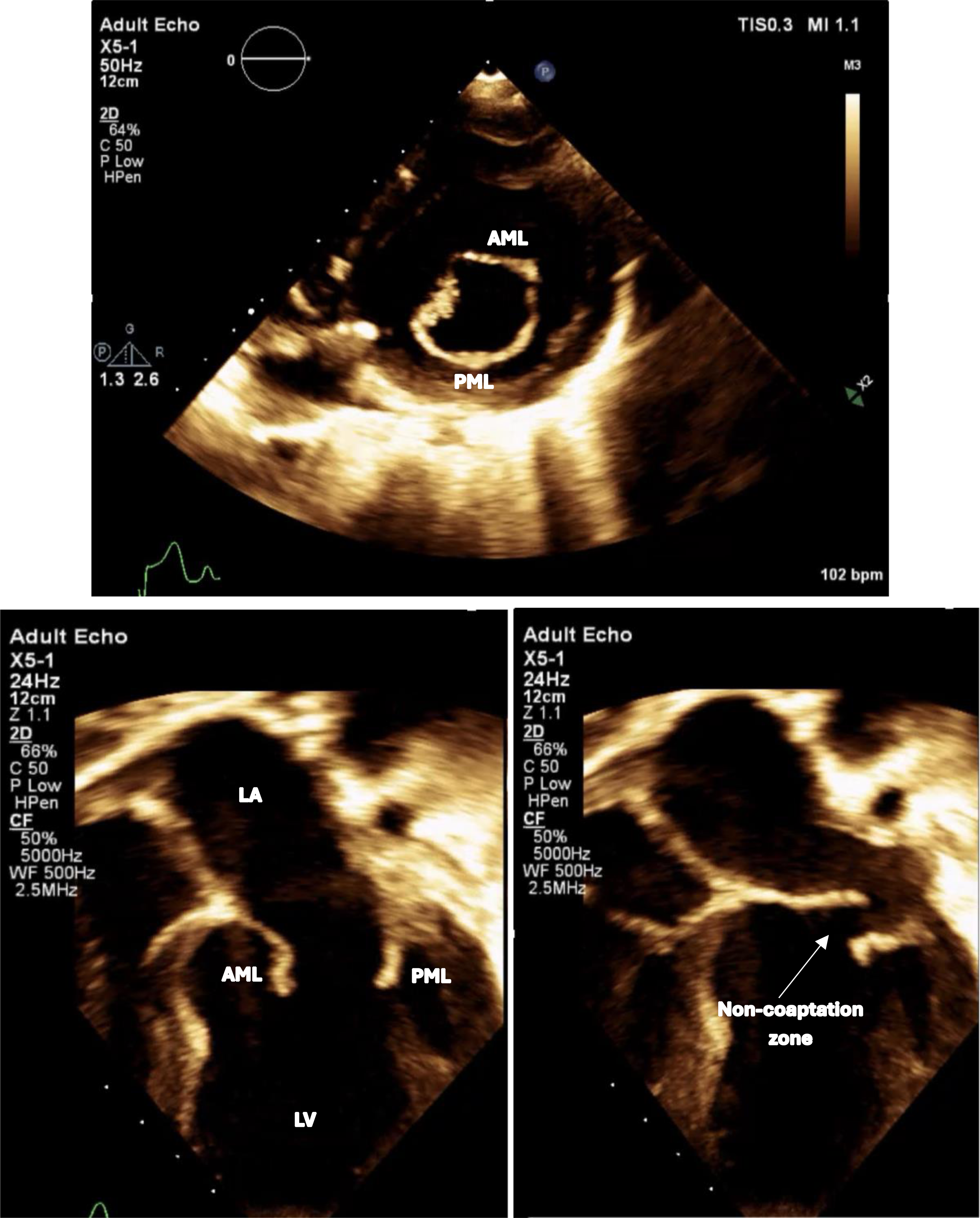
An eight-year-old girl, hailing from a rural village in Uganda, was referred to our institution for corrective surgery in October 2023 after receiving a diagnosis of severe rheumatic MR. Prior to presentation, she had recurrent hospitalisations in her home country for febrile illnesses and was reported to be non-compliant with penicillin prophylaxis. Upon arrival, she appeared chronically malnourished, measuring 107 cm in height and weighing 21 kg, and, despite treatment with oral heart failure medications, she exhibited hepatomegaly with a liver edge 4 cm below the right costal margin as well as palpable splenomegaly. Echocardiography showed ruptured chordae with flail and prolapse of the anterior mitral leaflet (AML) and restricted mobility of the posterior mitral leaflet (PML), causing severe MR through a large area of non-coaptation (Figure 1). Her left ventricle was dilated but had preserved systolic function. Laboratory evaluation showed elevated antistreptolysin O titre (ASO) (200 IU/ml) and Erythocte Sedimentation Rate (ESR) (109 mm/hr), but C-Reative Protein (CRP) (0.43 mg/dl) was normal at admission. Her dengue immunoglobulin M (IgM) was positive, but she remained clinically asymptomatic. Complete blood count and renal chemistry showed normocytic normochromic anaemia (8.9 mg/dl) and renal dysfunction with elevated urea (89 mg/dl) and creatinine (1.25 mg/dl) disproportionate to her cardiac status.
Figure showing flail prolapsing thickened AML with ruptured chordae and fixed restricted PML with large zone of non-coaptation (AML = Anterior mitral leaflet; PML = Posterior mitral leaflet; LA = Left atrium; LV = Left ventricle).

Despite titration of her fluid status and optimisation of heart failure agents in consultation with nephrology, renal parameters continued to be elevated. A renal ultrasonogram was therefore obtained, showing bilateral echogenic kidneys with maintained cortico-medullary differentiation, which, with continued elevation in phosphorus, uric acid, and acidosis, suggested chronic, rather than acute, renal pathology. Her hepatosplenomegaly, anaemia, and raised ESR prompted haematologic workup which revealed no significant findings, including a negative Direct Coombs Test, normal iron stores, and unremarkable peripheral smear, chromatography, lactate dehydrogenase, and reticulocyte count.
On day 12 of her admission, she developed fever spikes with newly elevated CRP (31 mg/dl), stably elevated ESR (111 mm/hr), and ASO (200 IU/ml). Cultures of her urine, oropharynx, and blood were obtained, eventually returning sterile, and she was empirically administered antibiotics. In view of her ongoing fever, persistently elevated ESR, and increasing CRP trend (67 mg/dl), steroids were initiated on suspicion of rheumatic fever recrudescence, to which her fever responded. Surgical intervention on her mitral valve was deferred, and she was discharged back to Uganda on oral heart failure medications and penicillin prophylaxis.
She re-presented six months later in April 2024 for surgery. Upon admission, she continued to have fever spikes, elevated renal and inflammatory parameters (Figure 2), tested positive once more for dengue and now also for falciparum malaria, and was initiated on Artemisinin-combination therapy. With this constellation of findings, rheumatology was consulted, and she was evaluated for underlying primary immune dysregulation. Anti-nuclear antibodies testing with indirect immunofluorescence was 4+ positive for anti-nucleosome, anti-ds DNA, and anti-mitochondrial M2 antibodies, indicating possible early onset Systemic Lupus Erythematosus (SLE). Complement testing, including C3/C4 and CH50, was normal, but antiphospholipid antibody syndrome testing showed elevated anti-cardiolipin IgM levels. A CT scan of her brain showed bilateral choroid plexus calcifications, a non-specific finding that could indicate neuroinflammation. In view of funding constraints, whole-exome sequencing and renal biopsy were deferred. Oral steroids and hydroxychloroquine were initiated, and she demonstrated a gradual decline in ESR and improvement in renal function (Figure 2).
Line graph depicting trend of urea, creatinine and ESR values over time.

Despite these additions, she continued to have severe MR through the large zone of non-coaptation, and consensus among the cardiac team was that her mitral valve pathology was not suitable for surgery, given her ongoing inflammation. She was instead retained on pharmacotherapy and prescribed medical observation to watch for the evolution of SLE symptoms. She was re-assessed in Uganda in June 2024, when she presented with congestive cardiac failure. At that time, her inflammatory parameters and ASO titres (800 IU/ml) remained significantly elevated. A subsequent multi-disciplinary meeting concluded that she most likely had RHD with probable underlying SLE. A follow-up plan was established to continue medical management of her heart failure, obtain further immunological investigations, and consider initiating anti-inflammatory therapy to optimise her systemic inflammation in anticipation of forthcoming surgery for mitral valve repair or replacement with renal replacement therapy backup available if needed.
Discussion
This case highlights the challenges in the identification and management of RHD in the setting of other coexistent but imprecisely defined inflammatory pathologies. In Uganda, like other LMIC settings, RHD remains a highly prevalent problem. Recent estimates place the school-age prevalence of RHD as high as 4%. Reference Weinberg, Beaton, Aliku, Lwabi and Sable1 However, while this patient’s elevated ASO titres established a RHD component to her condition, her marked renal dysfunction and persistently elevated inflammatory markers suggested some type of additional inflammatory pathology, the identification and control of which have important implications for her surgical readiness and long-term prognosis.
One possible explanation for her relapsing hyperinflammatory episodes that we considered was acute rheumatic fever (ARF). ARF is an intermediary, immune-mediated systemic condition that arises following non-treated superficial infections with Group A streptococcus and precedes the development of RHD in susceptible individuals. Reference Kumar, Antunes and Beaton2 In this case, the patient had a long history of reported non-compliance with secondary antibiotic prophylaxis, which can prevent ARF recurrence. Reference Lahiri and Sanyahumbi3 Unfortunately, using echocardiography to differentiate the acute-on-chronic carditis seen in relapsing ARF from the chronic changes seen in RHD can be difficult, and this patient had no other systemic signs, other than fever, that might satisfy the modified Jones Criteria to provide her with a diagnosis of ARF. Reference Gewitz, Baltimore and Tani4,Reference Rwebembera, Marangou and Mwita5 An alternate possibility we considered, in light of her improved inflammatory markers and fever curve following steroid initiation, was the presence of some immune dysregulated condition such as SLE. Her acute-on-chronic, often relapsing disease course was consistent with the natural history of SLE, and her limited laboratory and imaging evaluation, obtained in consultation with rheumatology experts, strongly pointed to this diagnosis. The quick improvements to her clinical status and some laboratory measures after initiation of steroid therapy only further reinforced this line of thinking.
The presence of suspected SLE in this patient introduced substantial uncertainty into the nature of her cardiac presentation. There is a significant overlap in echocardiographic features and natural history between SLE and RHD, and it may be impossible to distinguish the two based on imaging alone. Like RHD, SLE results in fibrosis, thickening, and calcification of valves and chordae tendinae and is strongly associated with pericarditis and pericardial effusion. Reference Chang, Xiao, Mercer-Rosa, Knight and Weiss6 Like RHD, SLE most commonly affects the left-sided valves, including the mitral valve. Based on previous data, out of the 22% of all SLE cases with cardiac valve involvement, 24% will show resolution of valvular dysfunction, 40% will remain stable, and 12% will worsen. Reference Galve, Candell-Riera, Pigrau, Permanyer-Miralda, Garcia-Del-Castillo and Soler-Soler7,Reference Roldan, Shively and Crawford8 Like RHD, cardiac manifestations in patients with childhood-onset SLE most frequently occur early after diagnosis and are more severe than those seen in adult-onset SLE. Most occur within the first year, and there is a strong association between complications and African-American race or nephritis. Reference Chang, Xiao, Mercer-Rosa, Knight and Weiss6,9 Incidentally, however, there are mixed data for the benefit of steroids in SLE-related carditis. While it would be expected that steroids would improve her inflammatory state in SLE, they have in some cases been shown to improve valvular inflammation and function and in other cases to worsen fibrosis, scarring, and thickening of valves and valve apparatuses, resulting in worsening valve dysfunction. Reference Moder, Miller and Tazelaar10 Ultimately, anywhere from 1–8% of patients with SLE-related cardiac involvement will require surgical intervention. Reference Foroughi, Hekmat, Ghorbani, Ghaderi, Majidi and Beheshti11 When valve replacement is done, mechanical protheses are favoured to bioprosthetic valves due to accelerated degeneration, which been reported with bioprosthetic valves due to early degeneration, which seems to be accelerated in the pro-inflammatory SLE state. However, with the need for lifelong anticoagulation, mechanical replacement also presents a significant challenge in LMIC settings where timely and available INR monitoring is not highly accessible. Reference Fluture, Chaudhari and Frishman12
While our team ultimately concluded that her valvular lesions were due to RHD and could be suitable for a straightforward repair, the presence of underlying SLE also introduced uncertainty into her surgical candidacy and long-term prognosis. Generally, due to increased risk of poor outcomes, surgical interventions in ARF, except for emergency situations, are deferred until the acute inflammation has resolved. Reference Ralph, Noonan, Wade and Currie13 Retrospective analyses have shown that patients with SLE are also at elevated risk for suboptimal perioperative outcomes, a risk that improves with adequate control of systemic inflammation. Reference Smilowitz, Katz, Buyon, Clancy and Berger14 Unfortunately,few isolated case reports are available in the literature describing the co-existence of RHD with SLE, and none in paediatric populations. What little data is available suggests that patients have increased risk of cardiovascular problems beyond that of patients with only RHD or only SLE. Reference Diaz, Lim and Liu15,Reference Dhungana, Pandey, Aryal, Kayastha, Pandey and Thakur16 There is need to better understand the immunological overlap between these two diseases to better help delineate their combined prognosis and to better inform evaluations for surgical candidacy.
Given this uncertainty, her case was discussed in a recent multi-specialty international care conference hosted through the Cardiology Across Continents virtual paediatric cardiology web series. In that conference, control of her inflammatory state was prioritised to make her a safe and suitable surgical candidate, with the eventual plan to proceed with mitral valve repair if possible, and mitral valve replacement if required. Referrals were organised to paediatric rheumatologists available in southwest Kenya, and long-term follow-up was arranged locally through the Uganda Heart Institute. Once her SLE-related inflammation has been adequately controlled, she will be transported back to Amrita Heart Institute in Kerala, India, for repeat evaluation prior to expected heart surgery.
Conclusion
Atypical courses in RHD should always raise a suspicion of coexisting additional pathologies. Valvular involvement in SLE can closely resemble RHD and serve as a source of diagnostic uncertainty. A high level of clinical suspicion is essential for diagnosing childhood-onset SLE, as its management and outcomes differ significantly from those of RHD. Coexistence of SLE with RHD poses additional challenges in medical as well as surgical management.
Supplementary material
The supplementary material for this article can be found at https://doi.org/10.1017/S1047951125101248.
Financial support
This research received no specific grant from any funding agency, commercial, or not-for-profit sectors.
Competing interests
None.