Case report
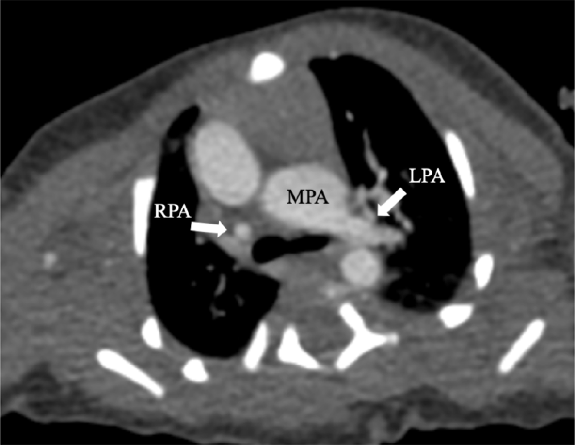
We present a case of neonatal dextro-transposition of the great arteries associated with an anomalous origin of the right pulmonary artery. A full-term female neonate was born through an uneventful vaginal delivery, with a birth weight of 2,650 g and Apgar scores of 9. Physical examination revealed cyanosis of the extremities, hepatomegaly, and a systolic murmur audible in the 2nd-3rd intercostal spaces at the left sternal border. Pulse oximetry showed an oxygen saturation of 85.4%. The chest X-ray examination showed cardiomegaly and prominent increased pulmonary vascularity. Postnatal echocardiography confirmed the diagnosis of dextro-transposition of the great arteries, with an atrial septal defect and patent ductus arteriosus. However, the right pulmonary artery was not identified on echocardiography, likely due to suboptimal patient cooperation during the examination. In view of this, CT angiography was undertaken, which showed atresia of the right pulmonary artery originating from the pulmonary trunk (Figure 1). An anomalous vessel arose from the innominate artery of a left-sided aortic arch, coursing along the trachea to the right pulmonary hilum (Figures 2 and 3).
Discontinuity of the right pulmonary artery and continuity of the left pulmonary artery with the main pulmonary artery in the evaluation by cardiac CT angiography.

( a ) Coronal image demonstrated a left-sided aortic arch with an anomalous right pulmonary artery. ( b ) Sagittal image illustrated the aorta anterior to the pulmonary artery and the anomalous origin of the right pulmonary artery (arrows) from the right innominate artery.

On the image obtained by the 3D reconstruction of CT angiography, the stenosis segment of the abnormal right pulmonary artery.

Prostaglandin therapy was initiated upon hospitalisation due to significant desaturation, which effectively improved the patient’s oxygenation from 85.4 to 91%. To better delineate the complex anatomy, the cinematic volume rendering of cardiac CT angiography was performed. A dynamic three-phase rotational display provided detailed visualisation of the cardiovascular anomalies and the origin of the pulmonary artery (Supplementary Movie S1). The first phase illustrated ventriculoarterial discordance and the anomalous right pulmonary artery origin. The second phase demonstrated bilateral pulmonary vascular markings. The third phase provided an integrated view of cardiac malformation and pulmonary perfusion. Detailed preoperative imaging vividly demonstrated that the surgical repair would carry excessively high risk in this case. Therefore, a conservative and non-invasive management strategy was adopted.
Discussion
Transposition of the great arteries is one of the most common cyanotic congenital heart defects in neonates, with an estimated incidence of 1 in 3,500–5,000 live births. Reference Martins and Castela1 It is frequently associated with additional cardiovascular anomalies, including ventricular septal defects and left ventricular outflow tract obstruction. Reference Samánek, Slavík, Zborilová, Hrobonová, Vorísková and Skovránek2 Without treatment, mortality reaches 30% within the first week and 90% by one year of age. Reference Wernovsky3
In about half of the cases, transposition of the great arteries is an isolated finding. When associated with additional cardiovascular anomalies, it is classified as complex transposition. Anomalous origin of the right pulmonary artery from the innominate artery is exceedingly rare in the context of dextro-transposition of the great arteries. Reference Agati, Sousa and Calvaruso4 This may result from the abnormal neural crest cell migration during embryonic development, disrupting aortopulmonary septum formation. These developmental defects can lead to outflow tract abnormalities and anomalous pulmonary artery branching. Reference Restivo, di Gioia, Marino and Putotto5
The arterial switch operation is the definitive treatment for dextro-transposition of the great arteries, with early postoperative mortality ranging from 1 to 3%. Reference Fricke and Konstantinov6 However, complex anatomical variants present significant surgical challenges and may affect long-term outcomes. In this case, the cinematic rendering technique, along with the cartoon (Figure 4), vividly depicted the cardiovascular structures, providing valuable preoperative information.
(a) and (b). The anterior and posterior view of the volume-rendered images showed the abnormal connection of the great vessels to the ventricles, the right pulmonary artery abnormality (arrows) originating from the right innominate artery and the reduced texture of the right lung compared to the left. (c) The accompanying cartoon illustrated the haemodynamic consequences of the cardiac anomalies. LA = left atrium; LV = left ventricle; RA = right atrium; RV = right ventricle; Ao = aorta; Pa = pulmonary artery; SVC = superior vena cava; IVC = inferior vena cava; INA = innominate artery; LCCA = left common carotid artery; LSA = left subclavian artery; PDA = patent ductus arteriosus.

Supplementary material
The supplementary material for this article can be found at https://doi.org/10.1017/S1047951125109384.
Financial support
This work was supported by the National Natural Science Foundation of China (82471970) and the Sichuan Science and Technology Program (2024YFFK0258).
Competing interests
The authors have no potential conflicts of interest to disclose.
Ethical standards
Permission to publish this report and the included images was granted by the patient’s parents.