Introduction
Tracheal stenosis is a narrowing of the trachea leading to breathing difficulties. Common acquired pre-disposing factors include external neck trauma, prolonged intubation and previous tracheal surgery (e.g. tracheostomy). Congenital tracheal stenosis is rare, and patients usually present at an early stage of life with stridor and/or shortness of breath. Congenital stenosis may be due to extrinsic compression of the airway, for example by cardiovascular malformations.1 More commonly, it is due to innate narrowing of the tracheal lumen causing airway obstruction.Reference Hoffer, Tom, Wetmore, Handler and Potsic2
Congenital tracheal stenosis has different forms. In most cases, the stenosis comprises a funnel-shaped tracheal segment characterised by complete, circular cartilaginous rings.Reference Koopman, Bogers, Witsenburg, Lequin, Tibboel and Hoeve3 The stenotic segment may be short or long, and may affect more than 50 per cent of the trachea.Reference Elliot, Roebuck, Noctor, McLaren, Hartley and Mok4
Surgery is the treatment of choice for moderate to severe tracheal stenosis. Different surgical techniques have evolved over the years. The type and length of stenosis determines the type of surgical procedure.Reference Dodge-Khatami, Tsang, Roebuck and Elliot5
Coblation technology (ArthroCare ENT, Austin, Texas, USA) and its applications are well known in otolaryngology. Coblation has been used in surgical procedures of the upper airways, such as palatal and laryngeal surgery. A few published studies have reported the use of Coblation in the trachea. However, the current report is the first to document the use of an ultra-fine Coblation wand (the PROcise MLW Plasma Wand, ArthroCare ENT) in the trachea.
We report the case of an unusual, delayed presentation of congenital tracheal stenosis in a 13-year-old boy. We describe the multidisciplinary assessment of his condition, and also the novel use of Coblation technology during endoscopic resection of the tracheal stenosis.
Case report
A 13-year-old boy presented to the respiratory clinic with increasing shortness of breath on exertion over a period of 1 year. He complained of occasional stridor on exertion. No voice changes had been noted by the patient or his family. He was otherwise healthy and active, with no other medical issues.
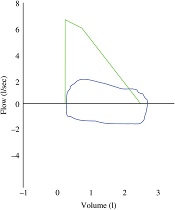
The respiratory physicians organised a pulmonary function test, which revealed severe intra-thoracic airway obstruction (Figure 1).
Pulmonary function test result indicating severe intra-luminal airway obstruction.

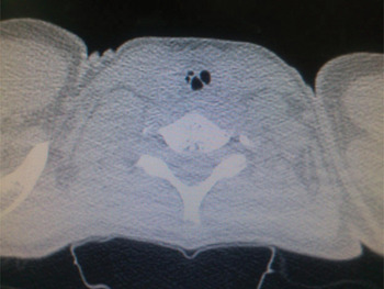
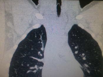
A high resolution computed tomography scan of the patient's neck and thorax did not reveal any external compression of the trachea nor any pulmonary problems. However, there was a focal short segment abnormality in the upper trachea causing stenosis of the tracheal lumen (Figures 2 and 3). These findings suggested congenital tracheal stenosis or post-inflammatory tracheal stenosis.
Axial, high resolution computed tomography neck scan showing tracheal anomaly and lumen stenosis.

Coronal computed tomography scan of the upper chest, showing diverticula causing tracheal stenosis.

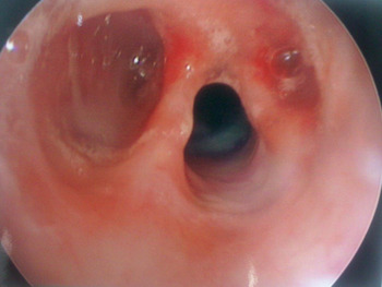
The patient was referred to our department and underwent a combined flexible and rigid laryngotracheobronchoscopy under general anaesthetic. Bronchoscopy revealed an unusual upper tracheal anomaly likely to be congenital stenosis with bilateral diverticula overlying two tracheal rings (Figure 4). There was approximately 50 per cent stenosis of the tracheal lumen. There were no abnormalities of the left or right main bronchi. A biopsy was taken from one of the diverticula to exclude any malignancy or granulomatous cause. Histological examination showed chronic inflammatory changes only.
Endoscopic view of trachea showing bilateral diverticula and tracheal stenosis.

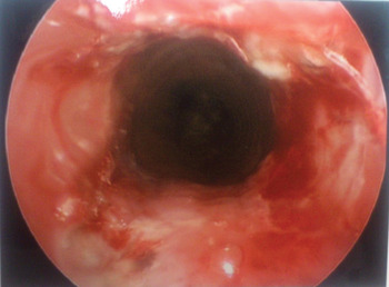
The patient subsequently underwent endoscopic Coblation of tracheal stenosis. The procedure was performed as a standard microlaryngoscopy and bronchoscopy using a Benjamin rigid laryngoscope on suspension and a rigid, 4.0 mm, 0° telescope. An extended length, ultra-fine Coblation wand (the PROcise MLW Plasma Wand) was inserted down to the level of the stenotic site, under direct vision. The default Coblation settings were used, i.e. 7 for ablate and 3 for coagulate, with low-flow saline. The tip of the Coblation wand was faced laterally, and Coblation was performed in a medial to lateral and superior to inferior fashion. Coblation was performed under direct vision to ensure that the stenotic segment was removed as completely as possible while at the same time preserving mucosa over the tracheal cartilages. The left diverticulum was treated first, followed by the right one. Any bleeding was controlled with the Coblation wand (using the coagulation setting) plus topical adrenaline (1:10 000). Triamcinalone (80 mg/mL) was injected submucosally following Coblation (Figure 5).
Endoscopic view of trachea immediately after Coblation resection of the stenotic segments.

Post-operatively, the patient was observed overnight in the intensive care unit. After an uneventful night, he was transferred to the regular ward with instructions for normal mobilisation. He was discharged subsequently with no issues.
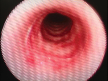
The patient was seen in the out-patient clinic 12 weeks after the procedure. Flexible tracheoscopy was performed in the clinic, revealing a patent trachea (Figure 6). At the time of writing, he was asymptomatic on exertion and had returned to his normal activities. He was being followed up at six-monthly intervals.
Endoscopic view of the patent and healed trachea, 12 weeks after surgery.

Discussion
Tracheal stenosis can be congenital or acquired. Acquired tracheal stenosis is most commonly caused by external neck trauma, intubation and laryngotracheal surgery. Other causes include chronic inflammatory disease, neoplasms and collagen vascular diseases (e.g. Wegener's granulomatosis).Reference Khalid, Goldenberg, Flint, Haughey, Lund, Niparko, Richardson and Robbins6
Congenital tracheal stenosis is rare but can potentially be life-threatening. Three types of congenital tracheal stenosis have been described: short segment stenosis, funnel-like stenosis and long-segment stenosis.Reference Cantrell and Guild7, Reference Blumer, Bauman, Kearns and Smith8
Tracheal stenosis may be isolated or associated with other anomalies such as a pulmonary artery sling. Symptoms vary depending on the severity of the stenosis. The severity of the stenosis usually determines the age of presentation. A long segment stenosis with markedly limited tracheal lumen may present during infancy, while a short segment obstruction may present later in childhood.
Surgical options depend on the severity and type of stenosis.
Coblation technology has been used extensively in ENT surgery, during sinus and laryngeal surgery and within such procedures as adenotonsillectomy and uvulopalatoplasty. Coblation technology uses radiofrequency energy to excite the electrolytes in a conductive medium such as saline solution, creating a precisely energised, ‘focused’ plasma. This plasma's energised particles have sufficient energy to break molecular bonds within tissue, causing tissue to dissolve at a low temperature (approximately 40 to 70oC). This enables volumetric removal of target tissue with minimal damage to surrounding tissues.Reference Woloszko, Stadler and Brown9, Reference Stadler, Woloszko, Brown and Smith10
• Congenital tracheal stenosis is rare but potentially life-threatening
• It may be isolated or associated with other anomalies
• Stenosis severity determines the type of surgical treatment
• In the presented case, tracheal stenosis was treated with Coblation, with good results
• This technique avoids the need for more invasive, open surgery
In the current case, the use of an ultra-fine Coblation wand enabled precise resection of the tracheal diverticula causing the stenosis. We found the PROcise MLW Plasma Wand suitable for our paediatric patient's smaller anatomy, while its extended length enabled satisfactory resection of the tracheal stenosis. Its ultra-fine tip diameter of 2.8 mm enabled precise, pin-point Coblation. It had a shaft working length of 19 cm and bend angles of 50° 3 cm from the handle and 16° at the distal tip. This is in contrast to the PROcise LW Wand, with a 3.8 mm tip diameter, shaft working length of 16.5 cm, and bend angles of 45° 3 cm from the handle and 15° at the distal tip; this bigger wand is more suited to laryngeal procedures in an adult patient. Due to the characteristics of Coblation technology, there was minimal surrounding tissue damage, which minimised the likelihood of post-operative re-stenosis. Coblation treatment also resulted in minimal mucosal loss and therefore faster re-epithelialisation of the resected sites. A post-operative endoscopy 12 weeks after surgery revealed a fully epithelialised, patent trachea.
We believe the current patient represents the first documented case of the use of an extended length, ultra-fine Coblation wand (in this case, the PROcise MLW Plasma Wand) for the treatment of moderate congenital tracheal stenosis. The use of Coblation enabled us to avoid more invasive open surgical treatment in an active, 13-year-old child. Our patient's hospital stay was shorter and his recovery time much quicker than would be expected following conventional surgical techniques.
Conclusion
Patients with congenital stenosis should be managed using a multidisciplinary approach. Each case must be assessed and discussed individually in order to determine the surgical technique most suitable for the patient's anomaly.Reference Elliot, Roebuck, Noctor, McLaren, Hartley and Mok4, Reference Andres, Cotton, Bailey, Myer and Vester11 Coblation technology causes minimal damage to the mucosa and surrounding tissues. The extended length, ultra-fine PROcise MLW Plasma Wand is suitable for tracheal procedures in paediatric patients. Coblation technology should be considered for the surgical treatment of short to moderate segment tracheal stenosis, failing which more invasive, open procedures may be considered.