Introduction
Head and neck cancers commonly metastasise via the cervical lymph nodes to distant sites. Haematogenous spread occurs less frequently, accounting for only 10 per cent of all distant metastases. The incidence of distant metastasis in patients with head and neck squamous cell carcinoma (SCC) is relatively low. The risk of distant metastasis is influenced by age, primary tumour site, loco-regional extension, grading and the achievement of loco-regional control with treatment.Reference Garavello, Ciardo, Spreafico and Gaini1, Reference Spector2
The most common site for SCC metastasis from the head and neck is the lungs, followed by the liver and skin.Reference Spector, Sessions, Haughey, Chas, Simpson and El Mofty3 Reported unusual sites of laryngeal cancer metastasis have included the forearm,Reference Korkmaz, Dursan, Saylam, Akmansu and Eryilmaz4 scapular muscle,Reference Yucel, Demirel, Demiryont, Egeli and Deger5 gluteus maximus muscleReference Marioni, Blandamura, Calgaro, Ferraro, Stramare and Staffieri6 and ampulla of Vater.Reference Buyukcelik, Ensari, Sarioglu, Isikdogan and Icli7 To the best of our knowledge, there have been no previously reported cases in the English literature of laryngeal cancer metastasis to the orbit.
Here, we discuss the clinical and radiological findings and the management of such a case.
Case report
A 75-year-old man presented to the ENT department in December 2009 with a five-week history of a red, swollen, painful left eye and a one-week history of diplopia.
His past medical history included a laryngectomy and bilateral neck dissection, conducted in February 2009. The tumour had been pathologically staged as tumour stage 4a, node stage 2c, metastasis stage 0, and graded as a poorly differentiated SCC of the supraglottis. Nodes had shown extracapsular spread of disease. The patient's case had been discussed at the multidisciplinary team meeting, and adjuvant treatment with radiotherapy had been recommended. The patient had completed all treatment by mid-April 2009.
During the current admission, examination had indicated that the patient was apyrexial with a normal heart and respiratory rate. His left eye was inflamed and proptosed.
The patient was examined by the ophthalmologist, who reported reduced Snellen's vision rating in the left eye: the left and right eyes were 6/9.5 and 6/7.5, respectively. There was also reduced colour vision on Ishihara chart testing in both eyes (10/13), and a left relative afferent pupillary defect. The patient had left hypotropia and exotropia, with limited eye movements and reduced corneal sensation in the left eye.
The patient's inflammatory markers (i.e. white blood cell count and C-reactive protein) were within the normal range.
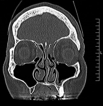
An urgent computed tomography (CT) scan was requested. This showed a soft tissue swelling overlying the left frontal bone and occupying the superior aspect of the left orbit and extending posteriorly (Figure 1). No definitive breach of bone or bony expansion was seen. The appearance of the mass was not in keeping with an orbital abscess.
Coronal computed tomography scan. L = left

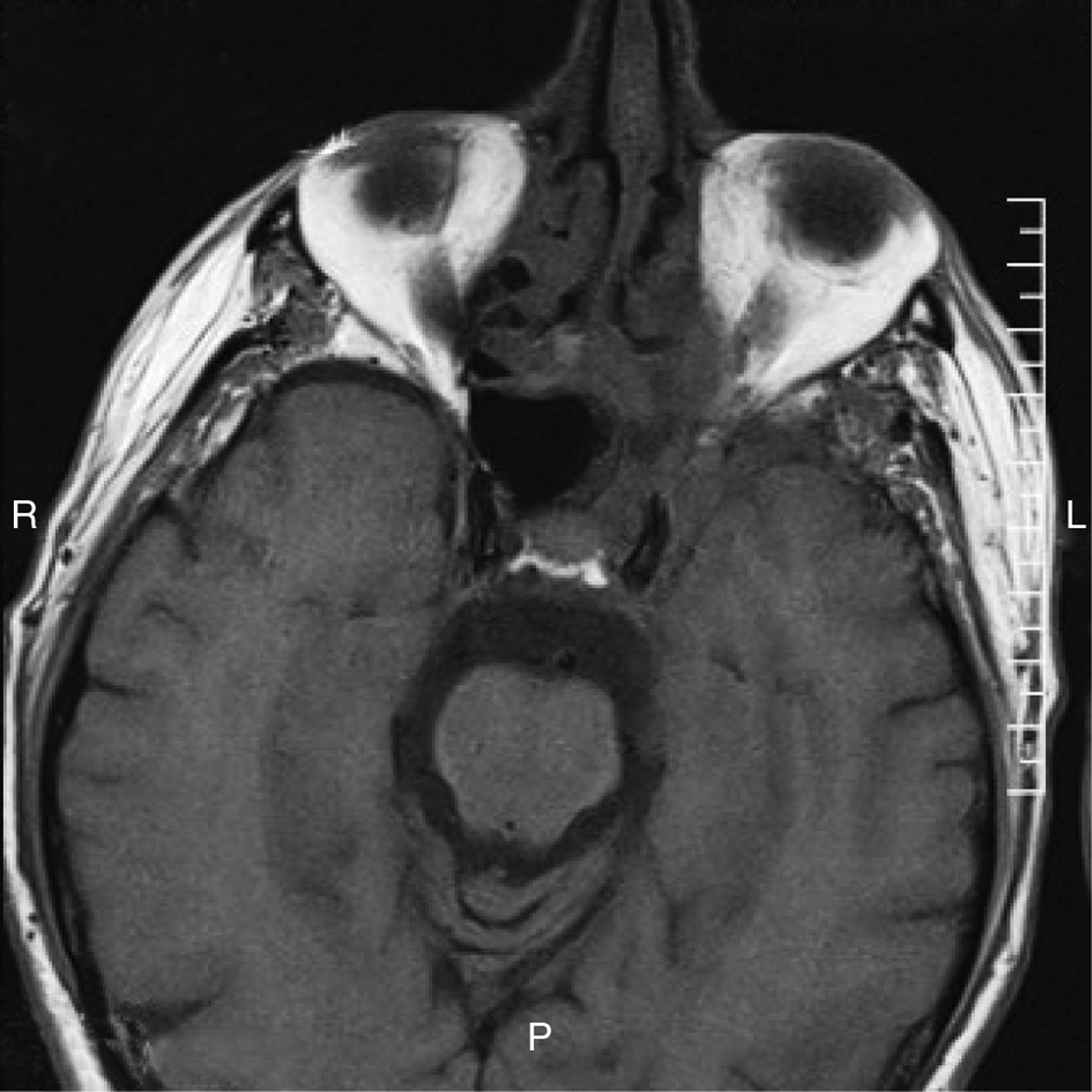
A magnetic resonance imaging scan was requested to assess the extent of the mass. This showed a mass in the inner aspect of the left orbital roof, with low signal change on T1- and T2-weighted images and on short T1 inversion recovery (STIR), and marked post-contrast enhancement (Figure 2). The enhancement extended along the roof and lateral wall of the left orbit, and involved the optic nerve, superior orbital fissure and left cavernous sinus. There was also associated marked meningeal enhancement within the left anterior cranial fossa. The right orbit was normal.
Axial magnetic resonance imaging scan. R = right; L = left; P = posterior
The patient was initially commenced on a course of intravenous ceftriaxone, metronidazole and dexamethasone.
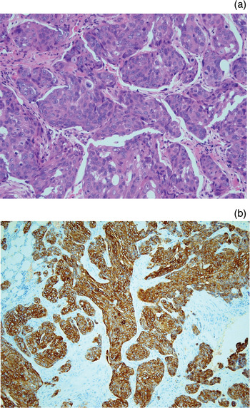
An orbital incisional biopsy was carried out by the orbital surgeon. Histopathological examination of the biopsy specimen revealed poorly differentiated metastatic SCC infiltrating fibrofatty tissue, with focal evidence of squamous differentiation. There was no significant cytoplasmic vacuolisation or nuclear crenation. Immunostaining was positive for the markers cytokeratin 5 and 6 (Figure 3).
Photomicrograph of left orbital biopsy. (a) H&E staining shows a poorly differentiated, metastatic squamous cell carcinoma infiltrating fibrofatty tissue, with focal evidence of squamous differentiation and no significant cytoplasmic vacuolisation or nuclear crenation (×400). (b) Immunostaining shows positivity for the markers cytokeratin 5 and 6 (×100).

Further molecular analysis to identify common patterns of mutation and loss of heterozygosity between the primary tumour and the orbital metastases was not carried out, as our unit did not have the required facilities at that time.
The patient's case was discussed at the multidisciplinary team meeting. Due to the extent of the metastatic tumour, a joint decision was made to pursue palliative treatment including steroids, analgesia and antiemetics.
The patient died on the 22 January 2010, 41 days after presenting with eye symptoms.
Discussion
Orbital metastasis from a primary tumour was first described by Horner in 1864.Reference Horner8 Since then, there have been many other reported cases. Reported primary sources of orbital metastasis have included hepatocellular carcinoma,Reference Srinivasan and Krishnanand9 malignant melanoma,Reference Rosenberg and Finger10 renal cell carcinoma,Reference Shome, Honavar, Gupta, Vemuganti and Reddy11 breast carcinomaReference Goldberg, Rootman and Cline12 and prostatic carcinoma.Reference Saleh13 Breast cancer metastases have been reported to account for the majority of orbital metastasis cases (55–60 per cent).Reference Aspraudis, Garezis, Konstantines and Stefaniotau14
The incidence of orbital metastasis is equal in male and females, with primary tumours of the breast being more common in females and primary tumours of the lung and liver more common in males.Reference Wang, Yang, Li, Hei and Xiao15
• This case represents the first report of laryngeal squamous cell carcinoma (SCC) metastasis to the orbit
• Previously reported unusual sites of laryngeal SCC metastasis have included the forearm and various muscles
• Common clinical signs and symptoms of orbital metastasis include exophthalmos, orbital pain and reduced vision
• Early ophthalmology review is needed, followed by incisional biopsy
• Treatment is palliative, requiring a multidisciplinary approach
• Palliative management may include surgery, chemotherapy, radiotherapy and hormonal treatment
The clinical presentation of orbital metastasis may be broken down into four generalised syndromes: infiltrative (the commonest), mass, inflammatory and functional.Reference Goldberg, Rootman and Cline12
The most common signs and symptoms of orbital metastasis include exophthalmos, orbital pain, reduced vision, periorbital swelling and diplopia.Reference Font and Ferry16 Symptoms may mimic orbital cellulitis.
A study of 28 cases of orbital metastasis found that the most common sign was exophthalmos (occurring in over 75 per cent of patients), followed by pain (29 per cent) and decreased vision (29 per cent).Reference Font and Ferry16
It is important that patients presenting with a possible diagnosis of orbital metastasis receive an early ophthalmology review, to aid early diagnosis and palliative intervention. Diagnosis is made histopathologically from tissue obtained from an incisional biopsy of the orbital mass.Reference Goldberg, Rootman and Cline12
Management of orbital metastasis is often multidisciplinary, using a combination of palliative treatments to achieve symptom control. Treatments may include surgery, radiotherapy, hormone therapy and chemotherapy.Reference Valenzuela, Archibald, Fleming, Ong, O'Donnell and Crompton17
Palliative radiotherapy has been shown to improve the quality of life of patients with orbital metastasis, irrelevant of the primary tumour type. Huh et al. Reference Huh, Nisce and Simpson18 found that symptoms such as pain, proptosis and reduced vision were either partially or completely relieved by palliative radiotherapy. Radiotherapy to orbital metastasis needs to be individualised according to the location and extent of disease.
However, the mean overall survival of patients with orbital metastasis has been reported to be just over one year. Our patient survived for only 41 days from presentation with eye symptoms.
Conclusion
Orbital metastasis from laryngeal SCC is extremely rare. To the best of our knowledge, the presented patient represents the first reported case in the English literature. Further reports are needed to more clearly define the clinical manifestations and management of laryngeal metastasis to the orbit. Facilities to identify common molecular features shared by both the primary laryngeal tumour and the orbital metastasis are currently mainly confined to research units. However, once such techniques become routine pathology practice, further information may be obtained which may improve prognosis.
Despite the poor prognosis of patients with orbital metastasis, early diagnosis is paramount to enable optimal palliative intervention, in order to relieve patient symptoms and thus improve quality of life.
Acknowledgement
We would like to thank Dr Muc for providing pathological slides and interpretation, and Dr Pittaway and Mr Reuser for their input in writing the manuscript.