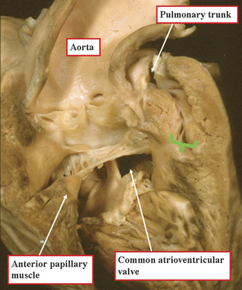
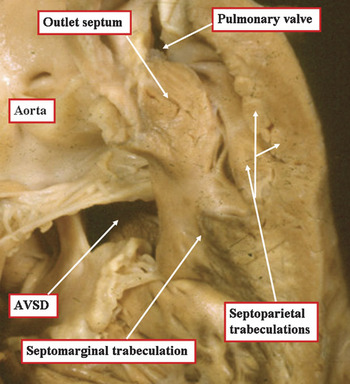
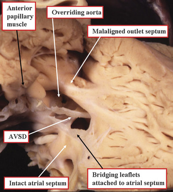
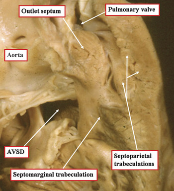
Although usually found in the setting of separate atrioventricular junctions, some patients with tetralogy of Fallot can be found in association with a common atrioventricular junction and an atrioventricular septal defect. Previous reports reveal that up to one-tenth of children with atrioventricular septal defect have associated tetralogy of Fallot,Reference Guo-Wei and Mee1–Reference Uretzky, Puga and Danielson3 while up to 3% of those with tetralogy have a concurrent atrioventricular septal defect co-existing with a common atrioventricular junction.Reference Arciniegas, Hakimi and Farooki2 Up to four-fifths of patients with these combined lesions also exhibit Down’s syndrome.Reference Najm, Van Aesdell and Watzka4 The combination of lesions entails the presence of a common atrioventricular junction, usually guarded by a common atrioventricular valve, and with both atrial and non-restrictive ventricular components of the atrioventricular septal defect (Fig. 1). The additional distinctive feature of the combination relates to the antero-cephalad deviation of the muscular outlet septum. This feature, in combination with the abnormal location of the septo-parietal muscular trabeculations (Fig. 2), produces varying degrees of obstruction of the right ventricular outflow tract. As in patients having tetralogy of Fallot with separate atrioventricular junctions, the presence of variable degrees of pulmonary stenosis at subvalvar and valvar levels, coupled with supravalvar narrowing of the pulmonary trunk and its branches, can further contribute to the obstruction of flow into the lungs. While a common atrioventricular valve is the norm in the setting of Tetralogy of Fallot with atrioventricular septal defect, there may be rare cases in which, notwithstanding the presence of a common atrioventricular junction, there are two identifiable atrioventricular valvar orifices,Reference Bharati, Kirklin, McAllister and Lev5, Reference Tchervenkov, Hill, Del Duca and Korkola6 (Fig. 3) as can be found in the overall spectrum of anatomic variants seen in patients with atrioventricular septal defects. When seen in the setting of separate valvar orifices for the right and left ventricles, it is usually the case that shunting is possible only at ventricular level, the bridging leaflets being firmly attached to the underside of the leading edge of the atrial septum (Fig. 3). This anatomic variant may alter the surgical management at the time of repair. When there is a common atrioventricular valve, then the superior bridging leaflet, which is free-floating, extends to be attached to an apically located papillary muscle within the right ventricle, the so-called Rastelli type C arrangement (Figs 1–3). As all these morphologic variants fall within the spectrum of anatomical heterogeneity characteristic of patients having atrioventricular septal defect with common atrioventricular junction, they will be included in our discussion regarding the surgical management. With regards to the distinction between those having atrioventricular septal defect with tetralogy of Fallot as opposed to those with double outlet right ventricle, it is now accepted that this distinction is primarily based on the degree of aortic override, which can be accurately estimated by echocardiographic techniques. A detailed description of the echocardiographic findings of patients with both tetralogy of Fallot and those with double outlet right ventricle is found elsewhere in this supplement. In this discussion, we confine ourselves to considering patients with fibrous continuity between the leaflets of the aortic valve and the common atrioventricular valve (Fig. 2). As in those with tetralogy of Fallot and separate right and left atrioventricular junctions, a spectrum is seen relating to the progressive commitment of the aortic valve to the right ventricle. Almost always in such cases there will be fibrous continuity between the leaflets of the aortic and common atrioventricular valves, although this is not an essential feature. In over half of the patients with double outlet right ventricle and common atrioventricular junction when the arterial valves are supported by extensive infundibulums,Reference Bharati, Kirklin, McAllister and Lev5 the interventricular communication, although non-committed, extends towards the subarterial areas, as is the case in tetralogy of Fallot with atrioventricular septal defect. This has important surgical implications, as it implies that in the majority of these hearts, similar principles of repair may be applicable to those we will describe later in this review. In patients with the most complex forms of double outlet right ventricle, nonetheless, such as those with isomerism of the atrial appendages and so-called visceral heterotaxy, the surgical management may differ significantly. These details will not be addressed.
The heart has been opened through an anterior incision in the right ventricle to reveal the combined features of tetralogy of Fallot and an atrioventricular septal defect with a common atrioventricular valve. Note the muscular obstruction at the mouth of the narrowed subpulmonary infundibulum (green bracket) and the overriding aorta. There is fibrous continuity between the leaflets of the aortic and common atrioventricular valves.

The ventricular outflow tracts from the heart shown in Figure 1 have been enlarged to show the phenotypic features of tetralogy of Fallot. The muscular outlet septum is deviated, and malaligned relative to the septomarginal trabeculation. There are also anomalous septoparietal trabeculations which complete the muscular obstruction at the mouth of the narrowed and elongated subpulmonary infundibulum. Note that the superior bridging leaflet, in fibrous continuity with the overriding aortic valve, bridges extensively into the right ventricle, being attached to the anterior papillary muscle of the right ventricle (see also Fig. 1).
In this heart with tetralogy of Fallot combined with a common atrioventricular junction, the bridging leaflets of the common valve are firmly attached to the leading edge of the atrial septum so that shunting through the atrioventricular septal defect is possible only at ventricular level. There are also separate valvar orifices within the common junction for the right and left ventricles, the left atrioventricular valvar orifice having the anticipated trifoliate arrangement (also called trileaflet arrangement), albeit not seen in this figure.

Historical perspectives
In the past, the management of patients having tetralogy of Fallot associated with atrioventricular septal defect usually involved initial palliation by construction of a systemic-to-pulmonary arterial shunt, followed by delayed complete repair at the age of 2 to 6 years.Reference D’Allaines, Colvez and Fevre7–Reference Pacifico, Kirklin and Bargeron10 Over the last decade or two, there has been a steady trend towards managing this lesion by early repair in infancy. Despite the recent advances, some controversy still exists surrounding the ideal surgical strategy, the timing of surgical repair, and the role of palliation.
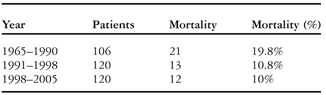
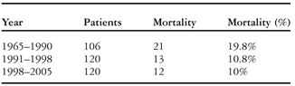
In Table 1, we have summarized the experience with surgical management from the 1960s to 2005.Reference D’Allaines, Colvez and Fevre7–Reference Prifiti, Bonacchi and Bernabei52 Those performing surgery during the 1970s and 1980s generally reported poor outcomes. Rates of mortality after primary repair, whether or not preceded by palliation, were in the range from 10% to 60%, with an average of 20%. Over the subsequent decades, improved diagnostic accuracy and perioperative approaches resulted in improved surgical outcomes. Surgical mortality, nonetheless, has remained relatively constant, at about 10%, over the last 15 or so years (Table 1). Among the factors recognized as playing a significant role for morbidity and mortality following surgical correction are lesions such as insufficiency of the left atrioventricular valve, residual shunting at the ventricular level, residual obstruction of the right ventricular outflow tract, particularly when combined with residual tricuspid valvar regurgitation, or more rarely obstruction of the left ventricular outflow tract. These alterations, alone or in combination, contribute to the greater surgical risk and poorer long-term surgical outcomes in general associated with the repair of tetralogy of Fallot with atrioventricular septal defect as compared to the repair of either lesion in isolation.
Review of historical series from 1965 to 2005 as shown in references #2 through #52. The data has been divided in 3 groups to evaluate the trend in mortality.
Factors affecting outcome
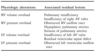
Understanding the pathophysiologic alterations associated with residual lesions is important in delineating their impact on long-term outcomes. The potential occurrence of postoperative multi-valvar pathology substantially increases the risk of early death and subsequent cardiac failure. In Table 2, we have summarized the potential postoperative pathophysiologic changes. Postoperative right or left atrioventricular valvar insufficiency, pulmonary insufficiency, residual obstruction in the right ventricular outflow tract, and residual intracardiac shunts may produce, alone or in combination, a variety of physiologic alterations on the right ventricle and left ventricle.
Physiologic alterations affecting short- and long term outcomes after surgical correction.

Abbreviations: RV – right ventricular; LV – left ventricular; AV – atrioventricular.
Primary repair versus initial palliation
Arguments put forward in the past in favour of palliation as the first line of treatment have been that reconstruction of the atrioventricular valve is technically easier and more successful in older children, and that a larger conduit can be placed from the right ventricle to the pulmonary arteries should this be deemed necessary as part of the repair, as might be required should an anomalous coronary artery be found crossing the right ventricular outflow tract.Reference Najm, Van Aesdell and Watzka4 Subsequent arguments advanced against palliation are the prolonged cyanosis to which children are exposed prior to correction, along with the ongoing stimulus for right ventricular hypertrophy, progression of atrioventricular valvar regurgitation, and possibly distortion of the pulmonary arteries produced by construction of a shunt.Reference Najm, Van Aesdell and Watzka4
Nowadays, the timing of surgery depends largely on the severity of the clinical manifestations, such as cyanosis, and on the philosophy adopted by the individual surgical team. While cyanosis can be severe at birth in some neonates, mandating early surgical intervention, in most children cyanosis is mild, and surgical intervention can be postponed. In general, as for those with atrioventricular septal defect or tetralogy of Fallot in isolation, the current practice at most centres is to undertake primary repair when the infant is aged from 3 to 6 months, depending on the clinical circumstances, seeking to avoid palliation in the most instances. With increasing surgical experience in treating this combination of lesions, a modified Blalock-Taussig shunt or a central shunt is rarely used, mostly being reserved for those infants who present early with profound cyanosis, and have associated co-morbidities such as sepsis, prematurity, low birth weight, or major associated anomalies. Although complete repair in the neonatal period has been proposed by some surgical groups,Reference McElhinney, Reddy and Silverman27 the risk of early complete repair in these high-risk patients may be prohibitive, and palliation still remains a valid option.
Surgical repair
The main goals of surgical correction are to close the interatrial and interventricular communication, to reconstruct the right and left atrioventricular valves, and to relieve the obstructed right ventricular outflow tract.Reference Tchervenkov, Hill, Del Duca and Korkola6 As in isolated atrioventricular septal defect, the common atrioventricular junction can be addressed by using either one-patch,Reference McElhinney, Reddy and Silverman27, Reference Castaneda, Jonas, Mayer and Hanley53 or two patches.Reference Karl22, Reference O’Blenes, Ross and Nanton24, Reference Schmid, Kampmann and Hake26, Reference Ilbawi, Cua and Deleon51 In contrast to atrioventricular septal defect seen in isolation, however, the superiority of one technique versus another has never been addressed, mostly as a result of the relative small number of patients reported in most series. When atrioventricular septal defect has been repaired as an isolated lesion, a higher incidence of left atrioventricular valvar insufficiency and valvar dehiscence has been reported when using the one-patch technique.Reference O’Blenes, Ross and Nanton24
In contrast to atrioventricular septal defect seen in isolation, the coexistence with tetralogy involves aortic override to varying degrees (Fig. 2), such that the antero-superior extent of the ventricular component of the defect may be obscured when approaching from the transatrial route.Reference Tchervenkov, Hill, Del Duca and Korkola6, Reference Schmid, Kampmann and Hake26 This problem may be enhanced in cases with significant aortic override, and especially in those who fall within the spectrum of double outlet right ventricle, when more than half of the aortic valve is supported by the right ventricle. As a result, it may be necessary to make additional modifications of surgical technique.
As in the isolated defect, the operation entails a median sternotomy, standard aortic and bi-caval cannulation, and is undertaken using cardiopulmonary bypass with mild or moderate hypothermia. After the aorta is cross-clamped, and antegrade cardioplegic solution is delivered into the aortic root, an oblique incision is made in the right atrial free wall so that the intra-cardiac anatomy can be inspected. At this time, the morphologic characteristics of the atrial and ventricular components of the defect, and the morphology of the common atrioventricular valve, are established. The right ventricular outflow tract is inspected to determine the surgical approach needed to relieve the obstruction within the right ventricular outflow tract. The septoparietal trabeculations may be divided at this time to expose the extent of the infundibulun and the pulmonary valve. In some cases, the obstruction caused by a narrowed infundibulum can be satisfactorily relieved simply by dividing the obstructing muscle bundles through the trans-atrial route, without the need for a patch or ventricular incision. In others, for example when the infundibulum is markedly narrowed, a small and carefully tailored ventriculotomy may be made completely to relieve the subvalvar obstruction, alone or in combination with an incision across the ventriculo-pulmonary junction should the valvar diameter be particularly small. The decision whether or not to incise the ventricular wall varies greatly among surgeons. If a ventriculotomy is made, the incision is extended proximally just sufficiently to relieve the obstruction caused by the narrowed infundibulum, allowing accurate resection of the septoparietal trabeculations. In rare cases, a ventriculotomy may be useful to expose the anterior rim of the ventricular component of the defect when this is obscured by the superior bridging leaflet, as might occur in patients with significant aortic override or double outlet right ventricle.
When supravalvar pulmonary stenosis is present, the pulmonary trunk is exposed, and a longitudinal incision made in the trunk and extended proximally to the level of the ventriculo-pulmonary junction. At this stage, it is convenient to inspect and size the pulmonary valve to establish whether a transjunctional patch will be required. (A transjunctional patch is often called a “transannular patch”.)
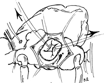
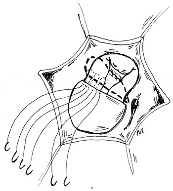
After the initial inspection of the right ventricular outflow tract is completed, the common atrioventricular valve is exposed and tested with cold saline. The point of coaptation of the superior and inferior bridging leaflets is determined, and marked with fine prolene sutures (Fig. 4). This facilitates subsequent repair, serving as a landmark for the closure of the ventricular component of the defect and reconstruction of the atrioventricular valves. Gentle traction applied to the marking sutures placed at the free edge of the superior and inferior bridging leaflets may also aid in exposing the ventricular component of the defect for closure.
The cartoon shows the view obtained by the surgeon of the common atrioventricular valve subsequent to instillation of saline and placement of marking sutures at the zone of apposition between the the superior and inferior bridging leaflets. The arrows show the gentle traction placed on the marking sutures placed in the bridging leaflets to expose the ventricular component of the defect.

Management of the ventricular component
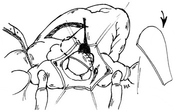
Regardless of the type of material used, the ventricular patch is tailored to the ventricular component of the defect, ensuring anterior redundancy.Reference Tchervenkov, Hill, Del Duca and Korkola6, Reference Schmid, Kampmann and Hake26 The patch needs to be wider anteriorly for the area that corresponds to the subaortic region, thus avoiding the risk of creating obstruction in the newly created outflow tract from the left ventricleReference Karl22, Reference Delius, Kumar and Elliott23, Reference Castaneda, Jonas, Mayer and Hanley53 (Fig. 5). When using this technique, obstruction with the left ventricular outflow tract is uncommon.Reference Najm, Van Aesdell and Watzka4, Reference Delius, Kumar and Elliott23
The cartoon shows the shape of the patch required for closure of the ventricular component, with redundancy in its anterior margin so as to ensure an unobstructed outflow tract from the left ventricle.

The patch can be sutured to the edges of the ventricular component with either a continuous suture or interrupted sutures, taking care to avoid the conduction system along the posterior aspect of the defect, as in repair of the atrioventricular septal defect in isolation. Division of secondary cords may improve visualization of the defect, especially posteriorly, without compromising valvar function. In rare cases where the posterior component of the ventricular septal defect is very shallow, a hybrid approach may be used, employing a patch to close the superior component, but closing the inferior component by attaching the bridging leaflet to the muscular septum with pledgeted sutures. Adequate exposure of the antero-superior quadrant of the defect is important, as residual shunts are more common in this area, and have been recognized as a common cause of reintervention.Reference Najm, Van Aesdell and Watzka4
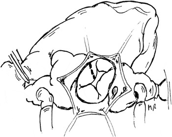
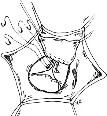
Division of the superior bridging leaflet can also improve exposure of the superior rim of the ventricular component. Division of this leaflet is, of course, part of the repair when using a one-patch technique, but may also be advantageous when using the two-patch repair (see below). In patients with significant aortic override, the superior bridging leaflet should be divided obliquely and towards the right side of the atrioventricular valve, so that the ventricular patch is positioned in an oblique rightward fashion to surround the aortaReference Tchervenkov, Hill, Del Duca and Korkola6 (Fig. 6). A short right ventricular incision may be needed to obtain an unobstructed right ventricular outflow tract in patients with a narrowed infundibulum and pulmonary valvar orifice. The limited number of patients included in most surgical series does not permit definitive conclusions to be made on the effects of a ventriculotomy on surgical outcomes, although a ventriculotomy was said not to affect early outcomes in one series.Reference Najm, Van Aesdell and Watzka4 The long-term effects of a ventricular incision on right ventricular function and arrythmias are well known, nonetheless, at least in the setting of tetralogy in isolation.Reference Gatzoulis, Till and Somerville50 One limitation of most studies performed in patients with tetralogy in isolation is that a ventricular incision is often combined with a transjunctional patch, with consequent loss of pulmonary valvar function. As a result, a confounding factor is introduced when the effects of the ventricular incision are studied in relation to late right ventricular function and arrythmias.
The cartoon shows the incision that can be made in the superior bridging leaflet. This is part and parcel of the one-patch technique, but can also be used to advantage when inserting two patches. See text for further discussion.

In those rare cases in which separate valvar orifices are found in the common junction for the right and left ventricles (Fig. 3), management may be modified to proceed with closure of the ventricular component, if present, by using a patch, or by achieving direct closure by placing pledgeted horizontal mattress sutures through the ventricular septum and through the point of separation of the left and right atrioventricular valves. Repair of the left atrioventricular valve proceeds as described below, albeit that in some patients seen with this combination, the bridging leaflets may be firmly attached to the leading edge of the atrial septum, so that there is no atrial component within the atrioventricular septal defect (Fig. 3). In these instances, it will be necessary to open the atrial septum so as to repair the zone of apposition between the bridging leaflets of the left atrioventricular valve, proceeding with closure of the atrial septum after satisfactory repair of the left valve.
Management of the common atrioventricular valve
In nearly all patients, the superior bridging leaflet is free-floating. In one study,Reference Prifiti, Bonacchi and Bernabei52 Rastelli type C morphology was found in four-fifths of patients, and type B morphology in the remainder. Similar findings were reported in another study,Reference Delius, Kumar and Elliott23 whilst in yet another relatively large series, all but one patient had type C morphology.Reference Najm, Van Aesdell and Watzka4 In general, the principles for management of the left atrioventricular valve follow those used when atrioventricular septal defect is repaired in isolation. The superior and inferior bridging leaflets are secured to the ventricular patch using interrupted horizontal mattress sutures. An autologous pericardial patch is used most often to close the atrial component of the defect, and is incorporated in the same line of sutures (Fig. 7). The reconstructed left atrioventricular valve is then inspected, and tested with cold saline. It is important adequately to close the zone of apposition between the left ventricular components of the bridging leaflets, the so-called cleft, so as to prevent residual or recurrent left atrioventricular valvar insufficiency, and reduce the rate of re-interventions on the left atrioventricular valve.Reference Capouya, Laks and Drinkwater54, Reference Alexi-Meskishvili, Ishno and Dahnert55 The dimensions of the new left valve can be measured with Hegar dilators placed after insertion of the first suture through the edges of the valvar leaflets. In certain cases, it may be necessary to avoid complete closure of the zone of apposition so as to prevent orificial stenosis. From a technical point of view, the zone of apposition can be closed either using simple interrupted sutures, or horizontal pledgeted mattress sutures reinforced with autologous or bovine pericardium (Fig. 8). Continuous sutures should be avoided so as to minimize the risk of dehiscence and the risk of shortening the reconstructed valvar leaflet, which could possibly result in valvar insufficiency. Subsequent to closure of the zone of apposition, the newly restored left valve is tested to determine whether additional maneuvers, such as annuloplasty or commissuroplasty, are needed to obtain valvar competency. These techniques are similar to those used in correction of atrioventricular septal defects when encountered in isolation. Competency of right atrioventricular valve is then tested, and any insufficiency corrected as necessary. In contrast to repair of the isolated defect, however, where it is the rule to have a competent pulmonary valve, and any residual insufficiency of the right atrioventricular valve may better be tolerated, when associated with tetralogy it is wise to spend additional time repairing the right atrioventricular valve as necessary. The combination of pulmonary valvar insufficiency, in cases where a transjunctional patch is needed, and right atrioventricular valvar insufficiency, may result in significant right ventricular volume overload and right-sided failure. This may be further exacerbated by the presence of residual regurgitation across the left atrioventricular valve. Competency of the right atrioventricular valve may be improved by a combination of annuloplasty or cordal reconstruction, in addition to other well-described techniques as in repair of atrioventricular septal defect in isolation. Should patients have a double orifice in the left atrioventricular valve, or deficient valvar leaflet tissue, the valve can be repaired using a modified one-patch technique.Reference Prifiti, Bonacchi and Bernabei52
The cartoon shows how the superior and inferior bridging leaflets are secured to the ventricular and atrial patches using interrupted sutures.

The cartoon shows the technique used to close the zone of apposition between the left ventricular components of the bridging leaflets.

Management of the atrial component
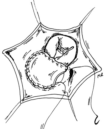
Whether using one or two patches, the atrial component is closed as for the atrioventricular septal defect in isolation (Fig. 9), occasionally leaving a small atrial communication which may be helpful in the postoperative period by allowing a controlled right-to-left shunt. The suture line is brought around the orifice of the coronary sinus postero-inferiorly, which may be left draining to the left or right atrium depending on the preference of the surgeon. Our preference is to leave the coronary sinus draining to the right atrium, carrying the suture line far posteriorly and into the opening of the coronary sinus itself. This is important to avoid injury to the conduction system.
The cartoon shows the completion of the repair by suturing the patch to close the atrial component of the defect.

Management of the right ventricular outflow tract and pulmonary valve
The pulmonary valve is most often exposed with a longitudinal incision along the pulmonary trunk, which is then extended proximally to the attachment of the valvar leaflets without initially compromising the integrity of the valve. The pulmonary valve is inspected, establishing whether it possesses 2 or 3 leaflets, and a pulmonary valvotomy is performed if indicated. The size of the pulmonary valvar orifice is determined by using calibrated Hegar dilators, which can be introduced through either the transatrial or transpulmonary route. In cases where the pulmonary valve can be preserved, a transatrial and transpulmonary repair is favoured by some surgeons, and has been reported with favourable outcomes.Reference Karl22 Conversely, others use a transventricular approach more liberally to relieve the infundibular obstruction completely.Reference Najm, Van Aesdell and Watzka4, Reference Delius, Kumar and Elliott23, Reference Prifiti, Bonacchi and Bernabei52
As when repairing tetralogy in isolation, the goal is to preserve the integrity of the pulmonary valve whenever possible, but significant residual obstruction across the right ventricular outflow tract can have adverse consequences on long-term right ventricular function. Preservation of the pulmonary valve is usually possible in patients presenting with no or minimal narrowing of the valvar orifice, with Z scores of 0 or −1. In contrast, it is generally not feasible in patients with Z values lower than −3 or −4. The optimal management in the presence of mild-to-moderate hypoplasia, with Z values of −2 or −3, remains uncertain. Regardless of the approach used, echocardiographic and haemodynamic assessment of the right ventricular outflow tract at the end of the procedure is important to confirm the adequacy of the repair and appropriate relief of obstruction. Although functional assessment of the right ventricular outflow tract early after discontinuation of cardiopulmonary bypass may be biased by a number of physiologic changes occurring simultaneously, including variations in cardiac output and ventricular function, in general the systolic right ventricular pressure as assessed by direct measurement should be less than three-fifths of the systemic pressure. This finding should be consistent with the echocardiographic estimate of the gradient across the right ventricular outflow tract. In contrast to repair of tetralogy in isolation, an important consideration when associated with atrioventricular septal defect relates to the presence of concomitant insufficiency of the right atrioventricular valve. As we have discussed, right ventricular volume overload resulting from pulmonary valvar regurgitation may adversely affect ventricular function and long-term outcome.
As in surgical correction of tetralogy in isolation, when a transjunctional patch is required because of hypoplasia of the pulmonary valvar orifice, the pulmonary valve may be reconstructed by insertion of a monocusp leaflet or a valved conduit. Although valved conduits are rarely used when repairing tetralogy in isolation, the additional haemodynamic burden of right or left atrioventricular valve insufficiency when associated with atrioventricular septal defect may result in biventricular dysfunction. In these situations, it may be an option to place a valved conduit from the right ventricle to the pulmonary arteries, although this exposes the patient to the need to change the conduit. Monocusp pulmonary valves have also been used to reconstruct the outflow tract in this setting,Reference Najm, Van Aesdell and Watzka4 although reports are conflicting, and generally limited by the small number of patients studied.Reference Gundry, Razzouk, Boskind, Bansal and Bailey56–Reference Brown, Ruzmetov, Vijay, Rodefeld and Turrentine58 In one series,Reference Najm, Van Aesdell and Watzka4 a pericardial monocusp pulmonary valve was constructed in over three-fifths of patients who required a transjunctional patch. No improvement was seen, however, compared to children who were left with pulmonary insufficiency.
Reoperations
Reoperations after repair are common, being reported by some in two-fifths of patients, with a mean interval from complete repair to reoperation of almost 2 years.Reference Najm, Van Aesdell and Watzka4 Freedom from reoperation at 4 years was found to be higher in the patients undergoing primary repair compared to those in whom repair was preceded by palliation. The most frequent indications for reoperation were reconstruction of the right ventricular outflow tract and pulmonary arteries, repair of the left atrioventricular valve, and closure of residual septal defects, findings consistent with those reported by others.Reference Prifiti, Bonacchi and Bernabei52
Conclusions
Over the last two decades, the most common strategy for management of patients with tetralogy of Fallot combined with atrioventricular septal defect has become complete repair in infancy. Palliative procedures, nonetheless, still have a role in the management of newborns or small infants presenting with severe cyanosis and associated medical conditions. Despite the general improvement in outcomes, the mortality associated with complete repair remains significant. The complexity of the surgical approach, combined with the relatively high incidence of residual multi-valvar pathology and other residual lesions, have a significant impact on outcomes over both the short and long terms. Due to the impact of multi-valvar pathology, effort needs to be directed to accomplish the best possible repair of the newly created right and left atrioventricular valves, and to preserving the integrity of the pulmonary valve whenever possible. In patients requiring a transjunctional patch, reconstruction of the pulmonary valve with a monocusp leaflet or valved conduit is an attractive option, although its efficacy in reducing the risk of right ventricular failure remains to be defined.