INTRODUCTION
Today, parasitic organisms are ubiquitous and occur in hosts ranging from bacteria to whales. While in most cases, parasites are tolerated and do not affect the well-being of their hosts, when new strains appear or established parasites are suddenly transferred to new hosts, the results can be devastating. Parasites residing in the tissues or alimentary tract of their hosts are soft bodied and their chances of fossilization are poor. However, most gastro-intestinal parasites produce a resistant stage that aids them in the transfer from host to host, usually via fecal material. Thus by collecting and identifying these resistant stages (cysts, eggs, etc) the parasites can be identified.
Dinosaur coprolites have been used in the past to supply information on the diet of their producers (Bertrand, 1903; Chin and Gill, 1996; Chin, 1997; Chin et al. 1998; Poinar et al. 1998; Prasad et al. 2005). While fossilized dung samples from humans has provided evidence of intestinal parasites (Gonçalves et al. 2003; Greenblatt and Spigelman, 2002), there are no records of parasites from dinosaur coprolites or any other coprolites from Mesozoic terrestrial deposits.
We report here the presence of resistant stages of protozoan and helminth parasites in coprolites from the Early Cretaceous Bernissart Iguanodon shaft in Belgium, showing that dinosaurs apparently suffered from intestinal parasites and establishing the first Cretaceous record of these parasitic groups in terrestrial vertebrates.
MATERIALS AND METHODS
Coprolites from the Bernissart Iguanodon shaft are elongate in shape, ranging from 110 to 130 mm in length and from 23 to 48 mm in diameter (Bertrand, 1903). A single coprolite was used in this study. Before extraction procedures, the coprolite was scrubbed thoroughly with a toothbrush and abrasive soap powder to remove any superficial material. It was then broken up into 1–2 mm size grains with a sterile cast iron mortar and pestle in a sterile clean room. The particles were then treated with 10% hydrochloric acid (HCl) to remove the carbonates. After several water rinses and centrifugation to concentrate the residue, the mixture was resuspended in hydrous hydrofluoric acid (HF) to remove silicates, clay and quartz. After further washings and concentration by centrifugation, the residue was mounted on microscope slides for examination. Additional details on the extraction process have been presented by Gray (1965). Observations and photographs were made with a Nikon Optiphot optical microscope with magnifications up to 1000×.
RESULTS
The extract contained a wide range of well-preserved plant microfossils, such as pollen grains, spores and miscellaneous animal microfossils. Among the latter were cysts of a protozoan pathogen and eggs of 3 helminth parasites. These are described below.
Systematics
Protozoan cysts
Several small, thin-walled, refractive spheres were very similar to cysts of the extant genus Entamoeba Casagrandi and Barbagallo (Patterson et al. 2000). They are described below.
Phylum Protozoa
Family Entamoebidae Chatton
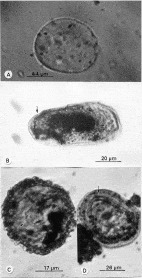
Entamoebites antiquus new genus and species (Fig. 1A).

Fig. 1. Parasite stages in a coprolite from the Bernissart Iguanodon shaft in Belgium. (A) Protozoan cyst of Entamoebites antiquus. (B) Egg of the trematode Digenites proterus. Note operculum (arrow). (C) Egg of Ascarites priscus. Note the mammillated surface and portions of the developing nematode inside (arrow). (D) Egg of Ascarites gerus. Note developing nematode inside (arrow).
Mature cysts (N=3): length, 10–12 μm; thickness of cyst wall, 0·02 μm; nuclei present, spherical to slightly irregular, greatest diameter, 1·5–2·0 μm; diameter of nucleolus (karyosome), 0·9–1·1 μm; cytoplasm filled with coarse, evenly distributed cytoplasmic granules; granules subspherical to oval in shape, with rounded ends, ranging from 0·88 to 1·32 μm in length; vacuoles and chromatoidal bars not present.
Diagnosis: Occurs in Early Cretaceous coprolites.
Etymology: “Antiquus” means old or ancient in Latin.
Type locality: The Early Cretaceous Bernissart Iguanodon shaft, Belgium.
Type specimen: Deposited in the Department of Zoology, Oregon State University, Corvallis, OR; slide # Y2 852 (2).
Trematode egg
An oval-elliptical egg with a smooth wall of medium thickness, an operculum at one end and containing an undeveloped embryo. These characters most closely resemble extant eggs of digenetic trematodes. A description follows.
Phylum Platyhelminthes
Class Trematoda
Digenites proterus, new genus and species (Fig. 1B).
Egg (N=1). Broadly oval in outline, 72 μm in length and 33 μm in greatest width; with an operculum at one end; egg wall 1·7 μm thick at opercular end and 2·4 μm at abopercular side; embryo 36 μm in length and 14 μm in width undeveloped.
Diagnosis: Occurs in Early Cretaceous coprolites.
Etymology: “proterus” is from the Greek word “proteros” meaning early.
Type locality: Coprolite from the Early Cretaceous Bernissart Iguanodon shaft, Belgium.
Type specimen: Deposited in the Department of Zoology, Oregon State University, Corvallis, OR; slide # Y2 852 (2).
Nematode eggs
Several spherical eggs containing juvenile nematodes were identified as ascarid eggs on the basis of their size and morphology. Two types were present. One type had a mammillated surface (covered with small protuberances) which is characteristic of members of the family Ascarididae. It is described below.
Phylum Nematoda
Order Ascarididea Yamaguti
Family Ascarididae Blanchard
Ascarites priscus, new genus and species (Fig. 1C).
Eggs (N=2). Spherical eggs from 42 to 46 μm in greatest diameter; egg shell mammillated, 4·2 μm in thickness; coils of developing juvenile nematode inside egg, with body thickness from 8·1 to 8·4 μm.
Diagnosis: Occurs in Early Cretaceous coprolites.
Etymology: “Priscus” is Latin for ancient.
Type locality: Coprolite from the Early Cretaceous Bernissart Iguanodon shaft, Belgium.
Type specimen: Deposited in the Department of Zoology, Oregon State University, Corvallis, OR; slide # Y2 852 (2).
The second nematode egg type consisted of small, smooth-shelled, spherical eggs. One of these contained a developing juvenile nematode. This type is described below, also in the family Ascarididae.
Ascarites gerus, new species (Fig. 1D).
Egg (N=1). Spherical egg ranging from 49 μm to 52 μm in diameter; egg shell smooth, ranging from 3·4 to 3·6 μm in thickness; enclosed juvenile nematode narrow, 5·4 μm in maximum width; coiled at least twice inside egg.
Diagnosis: Occurs in Early Cretaceous coprolites.
Etymology: “Gerus” is from the Greek word “geras” meaning old age.
Type locality: Coprolite from the Early Cretaceous Bernissart Iguanodon shaft, Belgium.
Type specimen: Deposited in the Department of Zoology, Oregon State University, Corvallis, OR; slide # Y2 852 (2).
DISCUSSION
Dinosaur coprolites are quite rare (Chin, 1997), mainly because of the uncertainty of the identifications. In providing guidelines for determining dinosaur coprolites, Thulborn (1991) listed a set of criteria. These included: (1) that the coprolites occur during the realm of the dinosaurs; (2) that they occur in appropriate habitats (terrestrial, etc); (3) that they are the right size; (4) that they occur in sediments that contain other types of dinosaur fossils; (5) that the contents are consistent with what dinosaur diets were considered to be. All of these criteria are met with in the Wealden material. They are from a terrestrial, Early Cretaceous site that contains skeletal remains of dinosaurs. The contents of the coprolites contained fragments of bones and striated muscles, indicative of the diet of a small to medium-sized carnivore that stripped the flesh off its victims (Bertrand, 1903).
We accept the conclusion of Bertrand (1903) who, after an extensive study of the morphology and contents of the Wealden coprolites, identified them as definitely dinosaurian, possibly originating from the carnivore, Megalosaurus dunkeri. Vertebrates from the Bernissart locality (a freshwater facies of the Neocomian) consist of the remains of 3 dinosaurs (Iguanodon mantelli von Meyer, I. bernissartensis Boulenger and Megalosaurus dunkeri Dames); 2 crocodilians, (Goniopholis simus Owen [reaching about 1 m in length] and Bernissartia fagesi Dollo [reaching 5–6 m in length]), 2 turtles, (Chitracephalus dumoni Dollo and Peltochelys duchasteli Dollo) and 1 urodele (Hylaeobatrachus croyi Dollo) (Dollo, 1909).
These coprolites have not been studied by anyone since Bertrand. Abel (1935) suggested that they may have originated from crocodiles, but gave no evidence for this statement and Bertrand (1903) had already ruled out that possibility due to the absence of large bone fragments and fish scales in the remains. Also, although the feces of crocodilians are reasonably firm while in the colon and rectum, because crocodilians usually defecate in the water, the feces soon become greatly softened and almost immediately dissipate (Frederic L. Frye, personal communication). Since there were only aquatic crocodilians in Europe at that time, including both species found at the Bernissart site (Carroll, 1988), the chances of obtaining 280 crocodilian coprolites when their fecal matter dissipates soon after deposition would be highly unlikely. Even if the Bernissart coprolites were later determined to be non-dinosaurian, these findings establish parasite groups in Early Cretaceous terrestrial vertebrates. It would be very likely that representatives of these same groups of parasites would have occurred in all the major vertebrate groups present at that period, much as they occur in all vertebrate classes today (Erasmus, 1972; Anderson, 2000; Patterson et al. 2000).
Entamoebites antiquus
Within the family Entamoebidae are several extant genera that form cysts similar to those of E. antiquus. However, the round, smooth cysts of E. antiquus are more similar to members of the genus Entamoeba than to the closely related genera Endolimax Kuenen and Swellengrebel and Iodamoeba Dobell, that normally have ovoid, ellipsoidal or pyriform cysts (Spencer and Monroe, 1961; Patterson et al. 2000). Cysts of the Entamoebidae are characteristically refractile and impart a certain lustrousness to the cytoplasmic contents (Spencer and Monroe, 1961), which was noted on the fossil species. In stained microscopic mounts of extant cysts, the wall is often surrounded by a conspicuous clear zone (halo effect), which is present to some degree in the fossil cyst (Fig. 1). Extant cysts of Entamoeba range in diameter from 5 μm to 20 μm, and contain from 1 to 16 nuclei (Kudo, 1954; Spencer and Monroe, 1961; Sloss and Kemp, 1981).
The actual number of nuclei in the cysts of E. antiquus could not be determined. Three are shown in Fig. 1, but since nuclei of unstained Entamoeba cysts are often not visible and not all nuclei are apparent at the same focal level (Spencer and Monroe, 1961), there are probably more present. The nuclei that are visible are small, spherical and possess a delicate nuclear membrane, which is consistent with the condition in mature cysts of extant Entamoeba species. Cytoplasmic granules, which are typical inclusions of Entamoeba cysts, are quite distinct in the fossil. In some species of Entamoeba (E. coli), the cytoplasmic granules are almost as large as the nuclei (Spencer and Monroe, 1961), which approaches the condition in the fossil. The absence of vacuoles and chromatoidal bars in the fossil cysts could indicate their stage of maturity since these organelles usually disappear as the cysts mature (Spencer and Monroe, 1961).
Entamoeba is widespread today and infects amphibians, reptiles, birds and mammals. The extant E. invadens Rodhain is widespread in reptile populations, especially in carnivorous species (Frank, 1984), which supports Bertrand's conclusion that the coprolites came from carnivorous dinosaurs. Pathogenic strains of E. invadens feed on leucocytes, liver cells, epithelial cells and bacteria and can cause disease outbreaks in times of stress (Kudo, 1954; Frank, 1984). Common symptoms of gastro-intestinal infections in lizards and snakes are vomiting and severe diarrhoea. The parasites may invade the liver and cause hepatitis and nephritis (Frye, 1991).
The cyst of E. invadens is the transmission stage of the parasite and is normally passed out in the feces (Keymer, 1981). Cysts remain in the environment until they are ingested by a susceptible vertebrate host, or are taken up and transported by dung-frequenting insects, such as cockroaches, which were abundant throughout the Cretaceous (Rasnitsyn and Quicke, 2002).
Digenites proterus
The egg of Digenites proterus was identified based on its size, shape and presence of an operculum. Eggs of most digenetic trematodes are ovoid to spindle shaped, have an operculum (lid) at one end and range from 70 μm to 86 μm in length (Spencer and Monroe, 1961; Schell, 1970; Erasmus, 1972). The fossil trematode egg falls within this description.
Many species of Digenea parasitize reptiles and birds. Some 400 adult species parasitize turtles, another 250 live in snakes and 75 are adapted to lizards (Brooks, 1984). Adult trematodes occur in the oral cavity, gall bladder, pancreas and kidneys of reptiles and the eggs are passed out of the body with feces, urine and oral mucus. Each egg develops into a miracidium which, in most species, enters a snail and continues through several additional developmental stages before ending up as an adult in a vertebrate (Schell, 1970; Frank, 1981; Brooks, 1984). Most infections are asymptomatic in reptiles (Brooks, 1984) and only cause problems when the hosts are under severe stress.
While some tapeworm eggs also have an operculum, most are oval to spherical in outline and, even early in development, there is usually some indication of hooks associated with most embryos (Sloss and Kemp, 1981).
Ascarites priscus and Ascarites gerus
The eggs of both A. priscus and A. gerus closely resemble those of members of the family Ascarididae. These nematodes are mainly parasites of terrestrial vertebrates with some 50–100 species of ascarids in reptiles (Sprent, 1984). The spherical to subspherical egg of Ascarites priscus, with its mammillated surface, is similar to eggs of Ascaris L. and Ophidascaris Baylis (Spencer and Monroe, 1961; Sloss and Kemp, 1981; Anderson, 2000). Eggs of the python parasite, Ophidascaris moreliae Sprent, can retain their infectivity for 7 years (Anderson, 2000). The adult worms live in the reptile's alimentary tract and deposit large numbers of eggs that are passed out with the feces. Embryonic development continues in the environment until the nematode juvenile is completely formed and infective.
Ascarites gerus has thick-shelled eggs resembling those found in the extant genera Ascaris, Ascaridia Dujardin and Polydelphis Dujardin. Members of Ascaridia occur mainly in gallinaceous birds and their eggs embryonate outside the host's body. Species of Polydelphis live in snakes and lizards (Anderson, 2000).
Little is known about diseases in the Cretaceous. Stages of trypanosomes and malarial organisms in Early Cretaceous sand flies and biting midges, respectively, show that vector-borne diseases occurred some 100 million years ago (Poinar and Poinar, 2004; Poinar and Telford, 2005). While there are Paleozoic records of platyhelminth infections of marine vertebrates (summarized by Poinar, 2003) and records of parasites in subfossil remains of terrestrial vertebrates (Greenblatt and Spigelman, 2002), the present findings are the earliest fossil records of gastro-intestinal parasites of terrestrial vertebrates.
We thank Dr Paul J. M. J. Sartenaer, Institute Royal des Sciences Naturelles de Belgique, for providing the coprolites used in this study, the late Jane Gray for the extraction of the palynomorphs and Dr Charles Wellman, Department of Animal and Plant Sciences, The University of Sheffield, UK, for his courtesy in returning Jane Gray's slides for study. Grateful appreciation is extended to Roberta Poinar for comments on earlier drafts of this manuscript.