Introduction
Angiostrongylus cantonensis-associated eosinophilic meningitis/meningoencephalitis is a worldwide food-borne helminthic zoonosis. Humans acquire the infection by consuming the infective larval stage in the raw or poorly cooked intermediate snail host or other paratenic animal hosts, e.g. crustaceans, frogs, shrimps and monitor lizards, or ingesting fresh/raw vegetables contaminated with the infective larvae (Eamsobhana, Reference Eamsobhana2006; Cross & Chen, Reference Cross, Chen, Black and Seed2007). The migration of larvae through the brain results in cerebral haemorrhage and eosinophilic meningitis, which can be fatal.
Definitive diagnosis of angiostrongyliasis relies on detection of the fifth-stage or juvenile parasitic worms in the cerebrospinal fluid (CSF), and occasionally from the eyes of patients (Wang et al., Reference Wang, Lai, Zhu, Chen and Lun2008; Graeff-Teixeira et al., Reference Graeff-Teixeira, da Silva and Yoshimura2009). Because of the frequent difficulty in obtaining a parasitological diagnosis, immunological means based on enzyme immunoassay format, namely standard enzyme-linked immunosorbent assay (ELISA) and immunoblotting, to detect the parasite-specific antibodies/antigens, have been used to support clinical diagnosis (Eamsobhana & Yong, Reference Eamsobhana and Yong2009). A simpler version, dot-blot ELISA, has been applied in community-based studies of human angiostrongyliasis in endemic areas of north-east Thailand (Eamsobhana, 2010). However, the currently available enzyme-based tests are time-consuming and the procedure is complicated. A simpler, rapid and non-instrumented test format which is specific and sensitive is needed in a basic diagnostic laboratory or field setting – where proper facilities and highly trained personnel are limited.
A colloid gold dot-immunobinding assay on nitrocellulose membrane (NCM) developed in the late 1980 s is a simple, visually read, rapid test that uses colloidal gold conjugated antibody or antigen, for the detection of corresponding antigen or antibody, instead of enzyme conjugates as in ELISA (Chun & Chu, Reference Chun and Chu1989; Spielberg et al., Reference Spielberg, Ryder and Harris1989). The current version of the dot immunogold filtration assay (DIGFA), or flow-through immunoassay, uses NCM as a solid-phase support for the antigen and colloidal gold-labelled antibody/protein A as a probe. By visual examination, the immobilized colloidal gold particles appear as pinkish dots. The test usually gives a diagnostic result within 3–5 min.
In the past decade, this rapid DIGFA format has been used increasingly in clinical laboratories and field settings to detect pathogenic organisms, including a few parasites. The DIGFA test has been adopted successfully for the serodetection of helminth infections with schistosomiasis (Xiao et al., Reference Xiao, Wang and Tian2003; Wen et al., Reference Wen, Chen, Ding, Zhang, Lu, Yu, Shen, Wu, Zhou and Zheng2005), echinococcosis (Eliades et al., Reference Eliades, Karagouni, Stergiatou and Miras1998; Feng et al., Reference Feng, Wen, Zhang, Chen, Ma, Zhang, Qi, Bradshaw, Vuitton and Craig2010), cysticercosis (Gan et al., Reference Gan, Yue, Xun, Kui and Hua2006), paragonimiasis (Wang et al., Reference Wang, Shi and Gan2007) and clonorchiasis (Liu et al., Reference Liu, Hu and Zhang2001).
DIGFA with crude extracts of A. cantonensis as diagnostic antigens had been applied to detect specific anti-A. cantonensis IgG antibodies from angiostrongyliasis patients and several other parasite-positive cases, with 90.5% sensitivity and 98% specificity. However, cross-reactions were observed in cases with trichinellosis (26.7%) and schistosomiasis (3.3%) (Gan et al., Reference Gan, Wang, Guo, Yang, Zhang and Eamsobhana2007). There was excellent agreement (up to 92.2%) between the results obtained by DIGFA and standard ELISA for the detection of A. cantonensis-specific IgG (Gan et al., Reference Gan, Wang, Yang, Zhang, Eamsobhan and Eamsobhana2010). As the type of diagnostic antigen could influence the sensitivity and specificity of the test, a single target-specific antigen of A. cantonensis is needed for the DIGFA to enhance its specificity and sensitivity.
The present study reports the diagnostic value of the DIGFA approach utilizing a purified 31-kDa glycoprotein specific to A. cantonensis as the diagnostic antigen, and protein A colloidal gold conjugate as the antigen-antibody detector. The adoption of the DIGFA format for a more rapid and specific immunodiagnosis of cerebral angiostrongyliasis was assessed in the Parasitology Laboratory, Siriraj Hospital, Bangkok.
Materials and methods
Serum samples
A total of 59 serum samples from archived serum panels were assessed using DIGFA with the 31-kDa A. cantonensis specific antigen. Angiostrongyliasis sera were from clinically diagnosed patients with detectable A. cantonensis-specific antibody which revealed a reactive 31-kDa band in immunoblot (n= 11). Antigen cross-reactivity studies were carried out on sera from patients with gnathostomiasis (n= 8), cysticercosis (n= 5), toxocariasis (n= 2), filariasis (n= 4), paragonimiasis (n= 2) and malaria (n= 2). Clinically related parasitic cases were positive by parasitological and/or serological tests for a specific parasite or its products. Patients with gnathostomiasis were serologically positive on immunoblot test showing a 24-kDa band specific for Gnathostoma spinigerum. Cases of toxocariasis were positive on ELISA using excretory–secretory antigens of the second-stage larvae of Toxocara canis. Patients with cysticerci were diagnosed pathologically as subcutaneous cysticerci. Sera from patients with filariasis, paragonimiasis and malaria were diagnosed parasitologically. A panel of control sera from 25 healthy individuals was negative for any parasitic infection at the time of blood collection.
All serum samples from the reference serum stock collected from the left-over clinical samples were kept at − 80°C at the Department of Parasitology, Siriraj Hospital, Bangkok. The use of stored left-over clinical samples for this study was approved by the Director of Siriraj Hospital, Faculty of Medicine Siriraj Hospital, Mahidol University.
Preparation of diagnostic antigen
Electroeluted purified 31-kDa antigen of A. cantonensis specific to angiostrongyliasis cantonensis was prepared as described previously (Eamsobhana et al., Reference Eamsobhana, Yoolek, Suvouttho and Suvuttho2001). In brief, a 12% sodium dodecyl sulphate–polyacrylamide gel was loaded with 2.0 mg of adult worm extract of A. cantonensis. After electrophoresis, the region containing the 31-kDa targeted antigen was cut into strips and the gel pieces were electroeluted in an electroeluter (Model 422, Bio-Rad, Richmond, Califormia, USA) using a clear membrane cap at a molecular weight cut-off of 12,000–15,000 Da. Elution was done at 10 mA/glass tube constant current for 3 h. Electroeluted protein was desalted and concentrated by ultrafiltration using Centricon YM-10 (Millipore Corporation, Billerica, Massachusetts, USA). The protein content was estimated using a NanoDrop 2000 (Thermo Fisher Scientific, Wilmington, Delaware, USA). The electroeluted fraction containing the 31-kDa specific protein was used in DIGFA for the detection of specific antibody to A. cantonensis.
DIGFA procedure
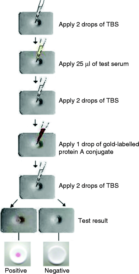
A filtration device to allow rapid antigen–antibody reaction was used as per the original design of the study group at the Institute of Parasitic Diseases, Zhejiang Academy of Medical Sciences, Hangzhou, PR China. The purified antigen (1.0 mg/ml) was dotted (0.5 μl/dot) on to a 0.45-μm pore size nitrocellulose membrane (NCM) (Bio-Rad Laboratories), and laid on the top of water-absorbing pads over the test opening of a flow-through module, comprising a rectangular plastic box (3 cm × 4 cm × 0.6 cm) with a 10 mm test opening cut into the centre of the lid (fig. 1).
(colour online) Schematic diagram of DIGFA procedure to detect human angiostrongyliasis. TBS, Tris buffer solution.

The gold-labelled protein A conjugate was prepared according to Gan et al. (Reference Gan, Wang, Yang, Zhang, Eamsobhan and Eamsobhana2010). Briefly, colloidal gold of 30 nm diameter was prepared by controlled reduction of a boiling solution of 0.2 g/l chloroauric acid (HAuCl4) with 6 g/l sodium citrate. The colloidal gold solution was adjusted to pH 6 with 0.2 m K2CO3. The optimal concentration of protein A for conjugation with colloidal gold was determined by titrating aliquots of diluted protein A with colloidal gold. The pre-determined amount of protein A was added to the colloidal gold solution while stirring, and incubated at room temperature for 30 min. Bovine serum albumin (BSA) was then added to a final concentration of 1% and the mixture was centrifuged at 23,000 rpm for 30 min at 4°C. The supernatant was discarded and the pellet resuspended in 20 mm Tris buffer, pH 7.0, containing 1% BSA. This protein A-coated colloidal gold probe was stored at 4°C.
The DIGFA procedure was performed at room temperature. In the detection test, two drops (about 100 μl) of 2% BSA in Tris buffer (140 mm NaCl, 10 mm Tris-HCl, pH 7.4) were applied to the centre of the device test opening on to the NCM in the well. Then, 25 μl of test serum was dripped slowly into the test opening until completely infiltrated. The membrane was subsequently washed with two drops of Tris buffer. After washing, one drop (about 50 μl) of the gold-labelled protein A conjugate was added and allowed to be absorbed through the membrane, and the final wash was repeated with two drops of Tris washing buffer. A result was considered positive when a well-defined pink dot appeared in the opening. No evidence of such a dot was an indication of a negative result (fig. 1). An inconclusive result was indicated by a very faint pinkish dot. Each serum sample was tested three times to confirm the reproducibility of the results.
Results and discussion
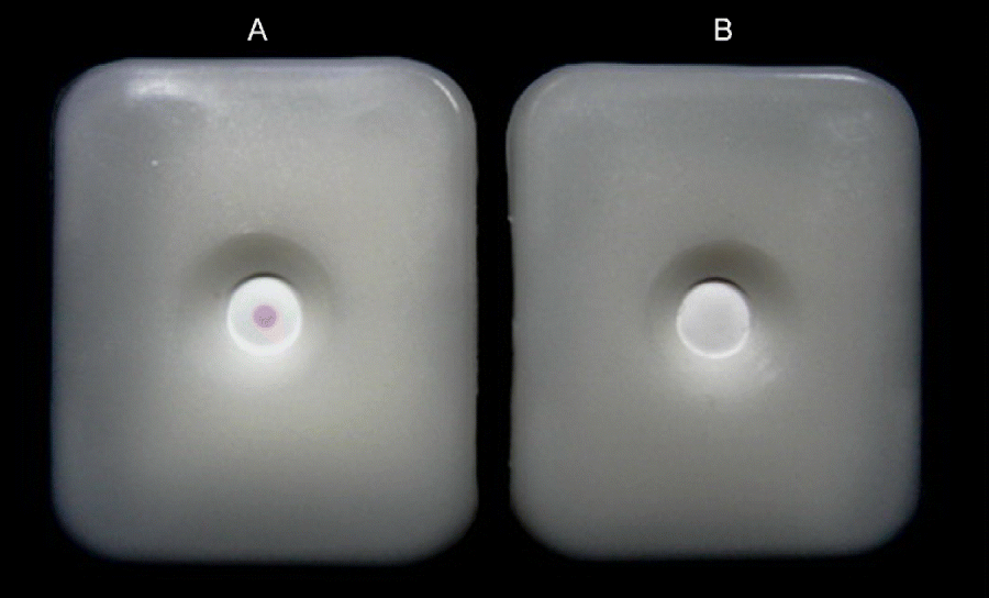
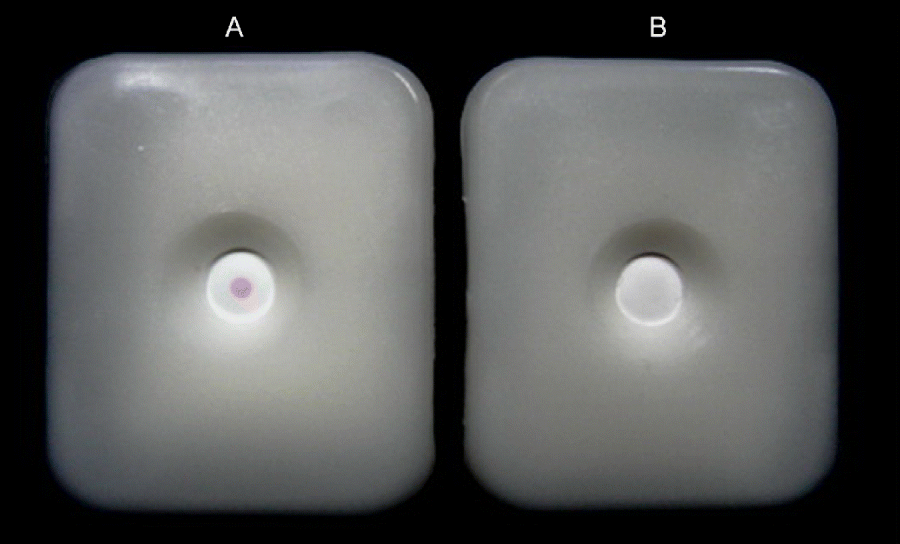
Sera from patients with angiostrongyliasis produced well-defined, positive pink dots with different colour intensities against the purified A. cantonensis antigen. No other sera from patients with gnathostomiasis, cysticercosis, toxocariasis, paragonimiasis, filariasis or malaria, or control sera from normal parasite-free individuals, produced a positive coloured dot. Positive and negative samples in sera of patients for the detection of specific antibodies against the 31-kDa antigen of A. cantonensis using DIGFA are shown in fig. 2.
(colour online) The pink dot in the centre of the opening indicates a positive sample (A) following the application of sera for the detection of specific antibodies against the 31-kDa antigen of Angiostrongylus cantonensis, compared with a negative sample (B) without a pink dot. The diameter of the opening is 10 mm.
The sensitivity and specificity of the DIGFA technique, to detect anti-A. cantonensis specific antibody in angiostrongyliasis cases – previously confirmed by immunoblot test to reveal a 31-kDa reactive band – were both 100%. No cross-reactions in DIGFA were observed with sera from patients with other parasitic diseases, or from healthy control subjects.
The development of a reliable user-friendly test to support clinical diagnosis of eosinophilic meningitis/meningoencephalitis due to A. cantonensis is essential for the improvement of laboratory diagnosis. Currently, immunoblotting is the most commonly utilized immunodiagnostic test for confirmation of infection with this neurotropic nematode (Eamsobhana & Yong, Reference Eamsobhana and Yong2009). In immunoblot analysis, demonstration of a specific antibody against the 31-kDa A. cantonensis antigen in a serum sample indicates infection with angiostrongyliasis (Eamsobhana, Reference Eamsobhana1994). However, although the test provides the advantages of sensitivity and specificity, it requires long reaction times and involves multiple steps. Therefore, the utilization of such a test has been confined to laboratories equipped with tools and devices for performing analyses.
The gold-based DIGFA, which differentiates positive from negative results by the development of a coloured dot, is being used increasingly to support clinical diagnosis of a variety of human diseases. DIGFA is more rapid and easier to perform than the traditional enzyme-coupled ELISA and the whole assay can be completed in about 3–5 min with visual interpretation of results, and without the need for expensive or special equipment.
The DIGFA approach using a specific 31-kDa glycoprotein of A. cantonensis presented here was easily adapted from the DIGFA method using crude extracts of A. cantonensis (Gan et al., Reference Gan, Wang, Guo, Yang, Zhang and Eamsobhana2007). In comparison to DIGFA with crude parasite antigens, results with better sensitivity and specificity were obtained with the use of a specific diagnostic antigen (31-kDa glycoprotein of A. cantonensis). This antigen has been shown earlier to be specific for angiostrongyliasis cantonensis patients (Eamsobhana et al., Reference Eamsobhana, Yoolek and Punthuprapasa2003, Reference Eamsobhana, Yoolek, Punthuprapasa and Suvouttho2004). The performance using purified 31-kDa A. cantonensis antigen to detect a corresponding specific anti-A. cantonensis antibody in infected sera is in good agreement with that of immunoblot tests, which revealed a 31-kDa reactive/diagnostic band. No cross-reaction was observed in cases with other parasitic diseases or normal healthy controls, thus attaining 100% specificity and sensitivity. Nonetheless, further studies to determine its use in other Angiostrongylus spp. infection in humans and animals, as well as further testing to determine possible cross-reactivity with other nematode infections (e.g. trichinellosis, schistosomiasis), are warranted.
The pore size of the nitrocellulose membrane, a solid-phase support for antigen, will directly affect the filtration rate of the serum sample. In a flow-through format, serum sample and reagents are drawn quickly through the porous NCM, hence reducing low-affinity, non-specific binding and minimizing chemical interference from any incubation steps (Wu et al., Reference Wu, Xu, Yan, Zhang, Liu and Li1999). Many investigators have reported 0.45 μm pore size of NCM as the optimal size to be used in DIGFA procedures (Huang et al., 1996, 1998; Mansour et al., Reference Mansour, El-Assa, El-Mahdy, Hendawy, Salem and Demerdash2009). In our DIGFA test with 0.45 μm pore size of NCM, each reagent was added after the previous one was totally absorbed, generally 1 min per step. The result could be observed and recorded immediately after the last washing buffer had filtrated and the whole assay took about 3 min. However, fibrin clot or high blood lipid in the serum will interfere with the drawing rate of the NCM, and with reaction between antigen and antibody, which will lead to false-positive or false-negative reactions. Such sera must be centrifuged to get rid of fibrin clot or blood lipid before testing (Wu et al.,Reference Wu, Xu, Yan, Zhang, Liu and Li1999; Mansour et al., Reference Mansour, El-Assa, El-Mahdy, Hendawy, Salem and Demerdash2009).
Although gold-labelled antibody/immunoglobin probes have been widely applied for immunodiagnostic assays related to human infections, the application of protein A could replace antibody/immunoglobulins to be labelled with colloidal gold, due to its very large surface area and good biocompatibility and stability (Horisberger & Clerr, Reference Horisberger and Clerr1985; Eliades et al., Reference Eliades, Karagouni, Stergiatou and Miras1998). Protein A is capable of binding to the Fc region of either human immunoglobulins, especially IgGs, or those of other species, including monkey, pig, dog, cat, rabbit, guinea-pig, mouse, rat, etc. (Brada & Roth, Reference Brada and Roth1984; Chandler et al., 2000).
In the present study, protein A, conjugated with colloidal dye particles, was used as a reagent for the detection of specifc IgG molecule against A. cantonensis in DIGFA. The 31-kDa glycoprotein antigen, purified from A. cantonensis adult worms by electrophoresis and electroelution, is among the principal antigens recognized by sera of humans with A. cantonensis infection, as well as by sera of immunized mice, rats and rabbits (Eamsobhana, Reference Eamsobhana1994). As protein A can bind to the Fc region of IgGs from a large number of other animal species, its use as a reporter with this specific antigen will also be applicable for immunological screening of 31-kDa antibody against A. cantonensis in wild animal hosts, for control measures to prevent human infection.
The strength of colour development in DIGFA is closely related to the size and quality of colloidal gold particles. Gold particles measuring 20–30 nm are suitable and are easy to identify in a flow-through format with NCM (Chandler et al., 2000). Particles smaller than 15 nm were too small to produce a strong colour, while particles larger than 60–70 nm self-aggregated after storage at 4°C for several days (Shim et al., 2007; Gui et al., Reference Gui, Wang, Guo and Zhu2008). In order to increase the signal-to-background ratio, colloidal gold with a diameter of 30 nm was chosen. Our test results showed that the use of 30-nm colloidal dye particles, conjugated with protein A, produced a well-defined pink spot with no apparent background staining in all the positive serum samples, while negative serum samples were absolutely clear.
Furthermore, the intensity of colour dots reflected the antibody activity level of immune serum (Ali et al., Reference Ali, Sibel, Salih, Haluk and Hadi2005). In the present study, 4 out of 11 angiostrongyliasis serum samples gave faint but visible pink dots. This could be attributed to the low antibody level in this group of infected patients as the intensities of dots were in good agreement with their corresponding immunoblot results – which also developed a faint-coloured specific band at the position of 31 kDa (data not shown). This indicated that the two methods show good concordance. In addition, colloidal gold-labelled protein A and enzyme-conjugated immunoglobulins show similar sensitivity, as both produce comparable colour signals. It is important to note that the enzyme-coupled method uses poisonous/carcinogenic reagents, such as BCIP (5-bromo-4-chloro-3′-indolyl phosphate p-toluidine salt) and NBT (nitro-blue tetrazolium chloride) as the common colorimetric substrates to generate the visual signal.
In conclusion, the 3-min visually read DIGFA test utilizing purified A. cantonensis antigen, as reported here, is reliable and reproducible for specific immunodiagnosis of human infection with A. cantonensis. It can be applied as an additional routine test in basic medical laboratories for clinical diagnostic support. In addition, the DIGFA serology approach appears to be suitable for practical use under field conditions without instrumentation. The test can be used in epidemiological studies to determine the seroprevalence of angiostrongyliasis. Large-scale sero-epidemiological studies in endemic communities in north-east Thailand are under way to evaluate its usefulness as a field detection method.
Acknowledgements
The authors thank the Director of the Siriraj Hospital, Faculty of Medicine Siriraj Hospital, Mahidol University for permission to carry out this work, and Sudarat Boonyong for technical assistance. We also thank the editor and two anonymous reviewers whose comments and suggestions were very useful in improving the paper. This study was funded in part by the Department of Disease Control, Ministry of Public Health, Thailand, and the University of Malaya (YHS: grants H-5620009 and H-50001-00-A000025).