Non-technical Summary
Shells from marine mollusks offer valuable clues about life in the oceans of the past, including how they interacted with parasites. In this study, we examined shells from a common gastropod, Zidona dufresnii, to see whether any evidence, such as holes, growth patterns, and malformations, suggests that other animals used their shells for shelter or predation. We found that some of these markings formed while the mollusks were still alive, demonstrating ancient parasitic relationships. Furthermore, we discovered that the shells vary greatly in age, ranging from ~3,000 to almost 40,000 years BP. These findings contribute to a better understanding of how parasites and mollusks coexisted and provide new insights into the dynamics of marine ecosystems throughout geological history, illustrating how marine life evolved over time.
Introduction
Marine mollusk shells, both during the animal’s lifetime and after death, are widely used by other organisms as substrate for attachment, habitat, refuge, and feeding (Lopes, Reference Lopes, Netto, Carmona and Tognoli2012; Aguirre et al., Reference Aguirre, Richiano, Álvarez and Farinati2016; Richiano et al., Reference Richiano, Aguirre and Giachetti2021; Marini et al., Reference Marini, Spotorno, Diniz and Dentzien-Dias2022). The primary ecological interactions involve bryozoans, annelids, and other mollusks, with many mollusk species serving as intermediate or permanent hosts for numerous parasite groups (Ituarte et al., Reference Ituarte, Cremonte and Zelaya2005; Huntley and De Baets, Reference Huntley and De Baets2015; Huntley et al., Reference Huntley, De Baets, Scarponi, Linehan, Epa, Jacobs and Todd2021). However, not all of these interactions leave marks on the shells—the only remains of mollusks typically preserved in the fossil record—limiting the identification of traits related to parasitism (Huntley and De Baets, Reference Huntley and De Baets2015). Nevertheless, the study of parasite evidence in fossil shells holds significant potential for understanding variations in marine disease prevalence over time, particularly during periods of elevated extinction rates or environmental changes (De Baets et al., Reference De Baets, Huntley, Klompmaker, Schiffbauer and Muscente2021).
A major challenge is how to determine whether a structure was produced by ante-mortem or postmortem interaction (Huntley, Reference Huntley2007; Huntley et al., Reference Huntley, De Baets, Scarponi, Linehan, Epa, Jacobs and Todd2021). To gain a better understanding, Huntley and De Baets (Reference Huntley and De Baets2015) categorized the traits and malformations found into two groups: (1) interactions where only perforations, excavations, or destruction of the shell by the settling organism occurs, making it impossible to distinguish ante-mortem from postmortem interactions; and (2) interactions that result in a growth response of the mollusk, allowing for the isolation of the invading organism, indicating that the trait or malformation was produced by an ante-mortem interaction. Nevertheless, it is often difficult to differentiate traits related to parasitism from traits that are the result of bioerosion. Most studies conducted on Southern Atlantic mollusks indicate that the traits observed in the shells refer to bioerosions, such as perforations and excavations, which, by themselves, do not allow the determination of whether they were produced while the animal was alive or after it was dead (Lopes and Buchmann, Reference Lopes and Buchmann2008; Lopes, Reference Lopes, Netto, Carmona and Tognoli2012; Richiano et al., Reference Richiano, Aguirre, Farinati, Davies and Castellanos2015; Charó et al., Reference Charó, Cavallotto and Aceñolaza2017; Erthal et al., Reference Erthal, Ritter and Kotzian2017; Erthal and Ritter, Reference Erthal and Ritter2020; Richiano et al., Reference Richiano, Aguirre and Giachetti2021; Verde et al., Reference Verde, Castillo, Martín-González, Cruzado-Caballero, Mayoral and Santos2022; Schmidt-Neto et al., Reference Schmidt-Neto, Horodyski, Ritter and Dasgupta2023; Anzai et al., Reference Anzai, Diniz, Spotorno and Dentzien-Dias2024).
Generally, parasite–host interactions in the fossil record have been studied across a range of different organisms or their remains, including mollusks, mammals, and even coprolites (fossilized feces) (Ruiz and Lindberg, Reference Ruiz and Lindberg1989; Poinar and Boucot, Reference Poinar and Boucot2006; Dentzien-Dias et al., Reference Dentzien-Dias, Poinar, Figueiredo, Pacheco, Horn and Schultz2013; Huntley and De Baets, Reference Huntley and De Baets2015; Huntley et al., Reference Huntley, De Baets, Scarponi, Linehan, Epa, Jacobs and Todd2021; Marini et al., Reference Marini, Spotorno, Diniz and Dentzien-Dias2022). Ruiz and Lindberg (Reference Ruiz and Lindberg1989) identified traces of trematode parasites in bivalves of the family Veneridae, which, in the form of small internal pits in the shells occur at least since the Neogene, documenting interactions that date back at least 5 million years. Ituarte et al. (Reference Ituarte, Cremonte and Deferrari2001, Reference Ituarte, Cremonte and Zelaya2005) described igloo-shaped shell structures, formed by the bivalve’s attempt to isolate the parasite. Other structures that provide evidence of parasitism include fixed and free pearls, irregular calcium deposits (ICD), discoloration, and shell blisters (Stunkard and Uzmann, Reference Stunkard and Uzmann1958; Johannessen, Reference Johannessen1973; Campbell, Reference Campbell1985; Cremonte and Ituarte, Reference Cremonte and Ituarte2003). From Brazil, Marini et al. (Reference Marini, Spotorno, Diniz and Dentzien-Dias2022) observed blisters in the bivalve Mactra isabelleana d’Orbigny, Reference d’Orbigny1846, which are associated with external bioerosions of the ichnogenus Caulostrepsis, and described a new type of malformation—the sheet.
However, despite the growing number of studies on parasite–host interactions in recent years, there are still numerous knowledge gaps to be filled. One of the major gaps, which we address here, concerns parasite–host interactions in marine gastropods, since most previous studies have concentrated on parasitic traits in bivalves (Ituarte et al., Reference Ituarte, Cremonte and Deferrari2001, Reference Ituarte, Cremonte and Zelaya2005; Huntley and De Baets, Reference Huntley and De Baets2015; Scarponi et al., Reference Scarponi, Azzarone, Kowalewski and Huntle2017; De Baets et al., Reference De Baets, Huntley, Klompmaker, Schiffbauer and Muscente2021; Huntley and Scarponi, Reference Huntley and Scarponi2021; Huntley et al., Reference Huntley, De Baets, Scarponi, Linehan, Epa, Jacobs and Todd2021; Marini et al., Reference Marini, Spotorno, Diniz and Dentzien-Dias2022).
Materials and methods
Study area
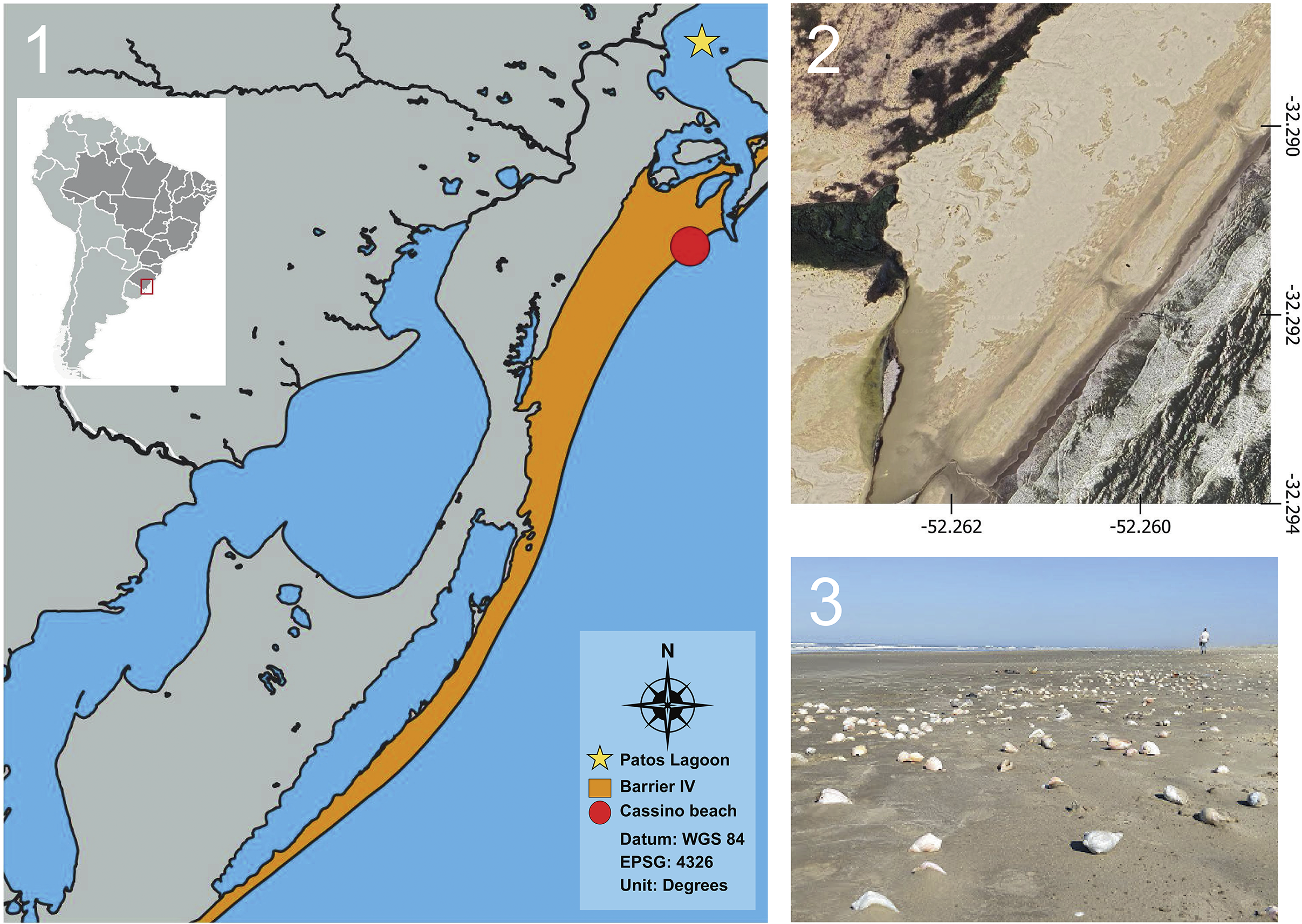
The study area, Cassino Beach (Fig. 1), is located on the southern Rio Grande do Sul Coastal Plain (RSCP), immediately beyond the outflow of the Patos Lagoon, in the state of Rio Grande do Sul, southern Brazil. Cassino Beach is a dissipative beach characterized by fine-grained sediment, a gentle slope, and a wide coastal face (Calliari and Klein, Reference Calliari and Klein1993). Due to the orientation of the coast, the dominant waves are of the swell type, generated in the mid-latitudes. In addition, local waves produced by strong maritime breezes occur, especially during spring and summer (Dillenburg et al., Reference Dillenburg, Tomazelli and Barboza2004). The mean significant wave height is approximately 1.5 m, potentially exceeding 2 m during storm events, which generally occur between autumn and winter. In such cases, the average sea level along the coast may rise by 1.3 m (Barletta and Calliari, Reference Barletta and Calliari2001).
Location map of the study area. (1) Extension of the Rio Grande do Sul Coastal Plain. The positions of Barrier IV and Cassino Beach (red dot) are indicated. (2) Google Earth image of the dune field of Cassino beach. (3) Shell-rich deposits at Cassino Beach.

Figure 1. Long description
Panel 1 is a geographical map of the Rio Grande do Sul Coastal Plain. An inset map of South America at the top-left shows the study area located in Southern Brazil. The main map features a large orange-shaded region along the Atlantic coast labeled as Barrier I V. A red dot marks Cassino beach on the eastern shoreline. A yellow star to the North marks Patos Lagoon. A compass rose points North. Technical data includes Datum W G S 84, E P S G 4326, and Unit Degrees. Panel 2 is a satellite photo of the Cassino beach dune field with a coordinate grid. The Y-axis on the right shows latitudes negative 32.290 to negative 32.294. The X-axis at the bottom shows longitudes negative 52.262 to negative 52.260. The image shows sandy textures and dark vegetation patches. Panel 3 is a ground-level photo of Cassino Beach. In the foreground, numerous white and tan shells are scattered across the flat, damp sand. In the distant background, a single person stands near the shoreline under a clear blue sky.
The sediments on the continental shelf of southern Brazil were accumulated within the various environments of a lagoon–barrier system, which was subject to changing sea levels during the Late Pleistocene (Correa, Reference Correa1996a). Paleocoastlines are known for the period between 17,500 and 6,500 years BP. During the Pleistocene Last Glacial Maximum (LGM), these relict sediments, deposited in an earlier environment but not yet buried on a continental shelf, were largely exposed due to low sea levels (Correa, Reference Correa1996a, Reference Correab).
The results of this dynamic process are frequently observed on Cassino Beach, including accumulations of mollusk shells, as well as vertebrate teeth, and bones that had previously been buried but were subsequently reworked and redistributed by wave action (Lopes and Buchmann, Reference Lopes and Buchmann2008). As demonstrated by Ritter and Erthal (Reference Ritter and Erthal2016), these accumulations may contain shells of different ages, including fossils as well as recent specimens. This is due to the geological evolution of the RSCP, characterized by successive marine regressions and transgressions, which resulted in the formation of a complex lagoon–barrier system (Villwock, Reference Villwock1984; Villwock et al., Reference Villwock, Tomazelli, Loss, Dehnhardt, Bachi and Dehnhardt1986; Calliari and Klein, Reference Calliari and Klein1993; Lopes and Buchmann, Reference Lopes and Buchmann2008). During each regression of the sea, associated with the progradation of the coastal plain, a new coastline was formed where shells accumulated, later to be buried in the sand (Villwock, Reference Villwock1984; Villwock et al., Reference Villwock, Tomazelli, Loss, Dehnhardt, Bachi and Dehnhardt1986; Lopes and Buchmann, Reference Lopes and Buchmann2008; Ritter and Erthal, Reference Ritter and Erthal2016). During transgressions, these old coastlines were submerged, and the sediments were partly remobilized during storm events and redeposited on the new coastal face. Over the formation of the RSCP, at least four major cycles of regression and transgression occurred, resulting in mixed deposits containing shells of different ages (Villwock, Reference Villwock1984; Villwock et al., Reference Villwock, Tomazelli, Loss, Dehnhardt, Bachi and Dehnhardt1986; Calliari and Klein, Reference Calliari and Klein1993).
On the Rio Grande do Sul continental shelf, submerged beaches were identified at current water depths between 20 and 40 meters. These quartzose, well-sorted sandy deposits represent ancient coastal lines formed during phases of stable sea level throughout the Quaternary. Considered relict sediments, these deposits are no longer in equilibrium with current sedimentary dynamics but preserve relevant paleoenvironmental evidence for reconstructing the paleogeographic evolution of the region (Correa, Reference Correa1996a).
Materials and methods
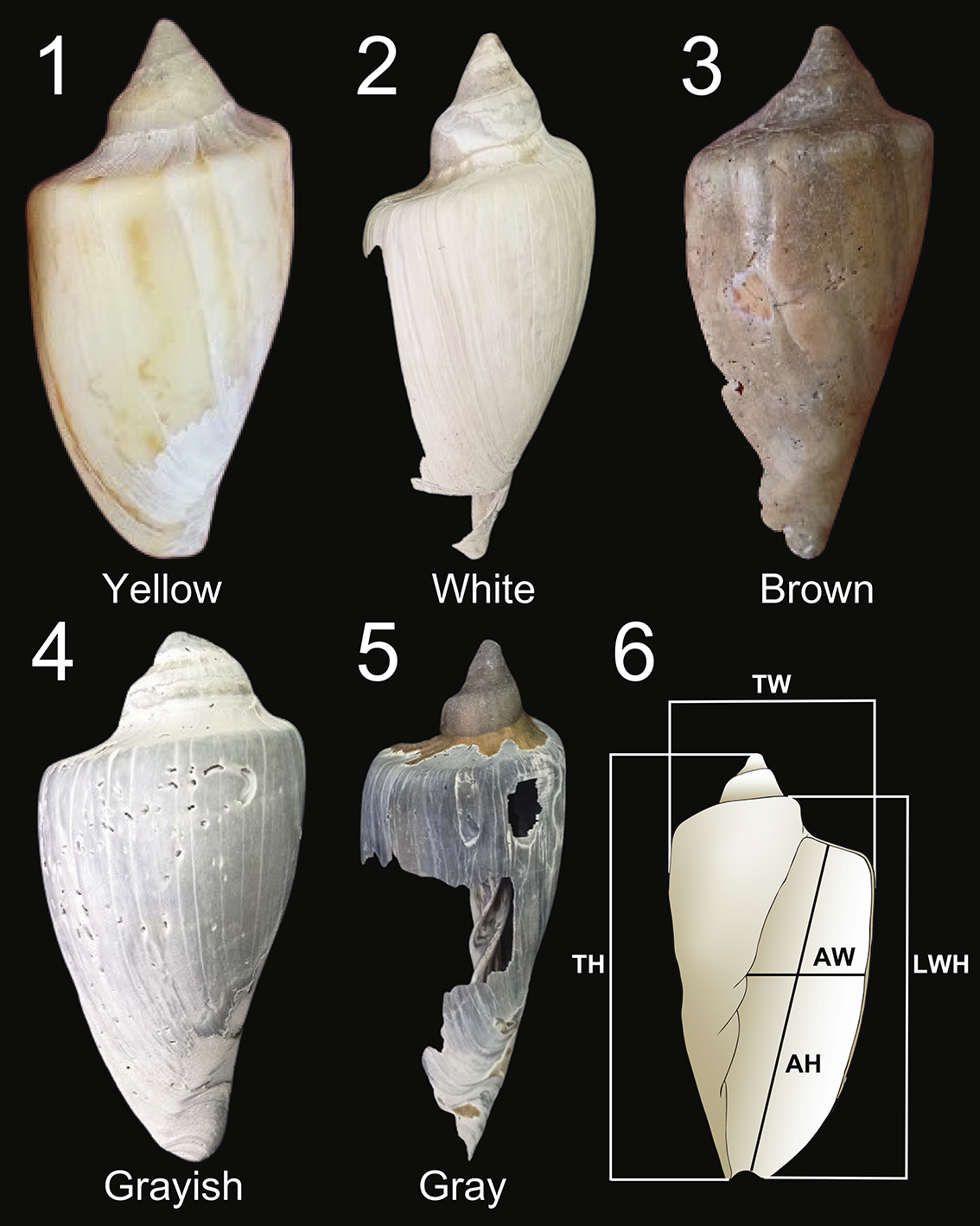
For this study, a death assemblage of shells of the gastropod Zidona dufresnii (Donovan, 1823) (Caenogastropoda, Volutidae) was analyzed. Zidona dufresnii inhabits sandy substrates at water depths between 35 and 60 meters along the western margin of the South Atlantic Ocean in from Rio de Janeiro, Brazil (22°S) southward to San Matias, Patagonia, Argentina (42°S) (Giménez and Penchaszadeh, Reference Giménez and Penchaszadeh2002, Reference Giménez and Penchaszadeh2003; Giménez et al., Reference Giménez, Penchaszadeh, Brey and Mackensen2004, Reference Giménez, Healy, Hermida, Nostro and Penchaszadeh2008; Medina et al., Reference Medina, Romero, Bidau and Narvarte2017, Reference Medina, Romero, Crespi-Abril and Narvarte2020). The species was selected due to its high abundance in this region—being commercially exploited in Uruguay and Argentina (Giménez et al., Reference Giménez, Penchaszadeh, Brey and Mackensen2004; Riestra et al., Reference Riestra, Lozoya, Fabiano and Carrizo2006)—and its variation in coloration (Fig. 2), shape, and abundance in the deposits of Cassino Beach, which may indicate temporal mixing.
(1–5) Color scheme for the shells of Zidona dufresnii. The scale includes the colors yellow, white, brown, grayish, and gray. (6) Diagram of the shell of Z. dufresnii. TH = total height; LWH = last whorl height; TW = total width; AH = aperture height; AW = aperture width.

The specimens were surface collected at Cassino Beach. All shells were collected, irrespective of their preservation state or size. In the laboratory, all shells were washed in flowing water to remove adhering sediment and dried in an oven at 40º C for six hours.
All shells were examined for coloration according to a color scale developed from the specimen themselves, covering the original shell color, bleached and darker shades, as well as the presence or absence of color pattern and luster. Measurements of shell size were also taken, including height, width, and aperture size (Fig. 2).
All Z. dufresnii shells were examined using a stereomicroscope to identify the types of bioerosion and shell malformations present. To complement this analysis, photographs were taken using scanning electron microscopy (SEM) at Centro de Microscopia Eletrônica do Sul (CEME-Sul/FURG), enabling a more detailed visualization of the observed structures. Subsequently, these internal structures were compared with those documented in the literature to identify potential causes of malformation.
To estimate the age of the shells, radiocarbon dating (14C) was performed using accelerator mass spectrometry (AMS) on six specimens exhibiting distinct taphonomic characteristics, such as coloration, presence or absence of luster, and ornamentation. Three of these samples were analyzed at International Chemical Analysis (ICA), North Bethesda, Maryland, USA, while the other three were analyzed at Vilnius Radiocarbon, Vilnius, Lithuania. The samples were processed using standard methods in both laboratories. Calibrations were performed using the software OxCal (version 4.4.4) (Bronk Ramsey, Reference Bronk Ramsey2009). To calculate radiocarbon ages, we used the Marine20 curve of Heaton et al. (Reference Heaton, Köhler, Butzin, Bard and Reimer2020) with an adjustment considering the marine reservoir effect, assuming a local variation (ΔR) of 117 ± 29 (Alves et al., Reference Alves, Macario, Souza, Pimenta, Douka, Oliveira, Chanca and Angulo2015), based on SHCal20 for Southern Hemisphere calibration (Hogg et al., Reference Hogg, Heaton, Hua, Palmer and Turney2020).
To facilitate interpretation of the obtained data, simple statistical analyses were performed, such as measures of mean and dispersion, the chi-square test (x2) to determine whether there is a significant relationship, and logistic regression analysis, integrating taphonomic data with interactions to identify the main factors contributing to the presence or absence of bioerosion and indications of parasitism. For this analysis, shell color, the presence or absence of color pattern, and luster were considered as independent variables, while internal growth patterns were taken as dependent variables.
Repository and institutional abbreviation
All specimens are deposited in the malacology collection of the Laboratório de Geologia e Paleontologia (LGP) at Universidade Federal do Rio Grande (FURG) under the acronym LGP/W.
Results
Shell preservation and color
The pristine shells of Zidona dufresnii are fusiform, lustrous, and thick-shelled, with pointed apex. The shell is orange-yellow in color, with a pattern of reddish-brown axial zigzag lines. The aperture is high subrectangular, accounting for approximately three-quarters of the total shell height (Rios, Reference Rios1994, Reference Rios2009).
Of the 894 shells of Z. dufresnii analyzed (Supplementary Table 1), 464 were more or less complete, without significant fractures or excessive bioerosion, allowing for accurate measurements. However, none of the beached shells studied herein had the fragile elongated apex preserved. The mean height of the shells, derived from the last whorl since the apex is always incomplete, was 91.80 mm (minimum 50.44 mm, maximum 146.45 mm), and the width was 52.90 mm, varying between 30.37 mm and 76.50 mm (Supplementary Fig. 1).
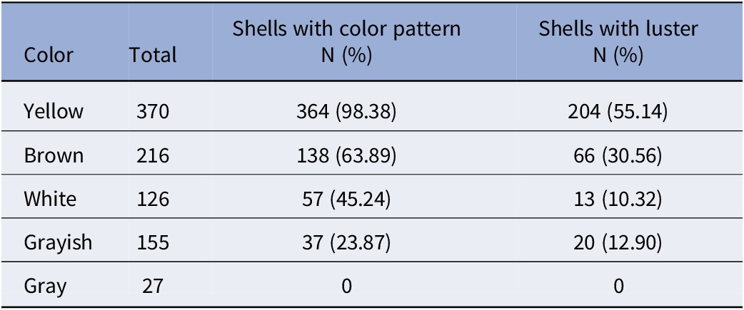
Shell colors range from the original yellow to entirely gray. A total of 310 shells (34.68%) still are lustrous, while 584 shells (65.32%) are dull. In 601 shells (67.23%), the original zig-zag color pattern is present, whereas it has faded in 293 shells (32.77%). As shown in Table 1, overall shell color is related to the presence or absence of luster and zig-zag pattern. Shells in original or near original color also preserve the original zig-zag pattern, and half of them still show luster. Shells that have changed color, particularly those that are gray, show a gradually fading color pattern and, primarily, luster.
Relationship between color and the presence of color pattern and luster in shells of Zidona dufresnii

Table 1. Long description
The table consists of four columns: Color, Total, Shells with color pattern N (percent), and Shells with luster N (percent). The data is organized into five rows by shell color:
* Yellow: Total 370 shells. 364 shells (98.38 percent) have a color pattern. 204 shells (55.14 percent) have luster.
* Brown: Total 216 shells. 138 shells (63.89 percent) have a color pattern. 66 shells (30.56 percent) have luster.
* White: Total 126 shells. 57 shells (45.24 percent) have a color pattern. 13 shells (10.32 percent) have luster.
* Grayish: Total 155 shells. 37 shells (23.87 percent) have a color pattern. 20 shells (12.90 percent) have luster.
* Gray: Total 27 shells. 0 shells have a color pattern. 0 shells have luster.
Bioerosion traces
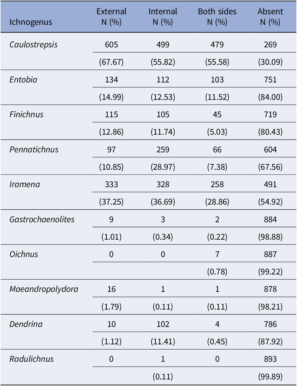
Bioerosion traces were identified in 709 shells, representing 79.3% of the sample. In many specimens, these perforations are restricted to the outside or the inside (aperture); in some shells, they are present on both sides (Table 2).
Distribution of bioerosion traces of different ichnogenera in shells of Zidona dufresnii

Table 2. Long description
The table consists of five columns: Ichnogenus, External N percent, Internal N percent, Both sides N percent, and Absent N percent.
* Caulostrepsis: External 605 67.67 percent, Internal 499 55.82 percent, Both sides 479 55.58 percent, Absent 269 30.09 percent.
* Entobia: External 134 14.99 percent, Internal 112 12.53 percent, Both sides 103 11.52 percent, Absent 751 84.00 percent.
* Finichnus: External 115 12.86 percent, Internal 105 11.74 percent, Both sides 45 5.03 percent, Absent 719 80.43 percent.
* Pennatichnus: External 97 10.85 percent, Internal 259 28.97 percent, Both sides 66 7.38 percent, Absent 604 67.56 percent.
* Iramena: External 333 37.25 percent, Internal 328 36.69 percent, Both sides 258 28.86 percent, Absent 491 54.92 percent.
* Gastrochaenolites: External 9 1.01 percent, Internal 3 0.34 percent, Both sides 2 0.22 percent, Absent 884 98.88 percent.
* Oichnus: External 0, Internal 0, Both sides 7 0.78 percent, Absent 887 99.22 percent.
* Maeandropolydora: External 16 1.79 percent, Internal 1 0.11 percent, Both sides 1 0.11 percent, Absent 878 98.21 percent.
* Dendrina: External 10 1.12 percent, Internal 102 11.41 percent, Both sides 4 0.45 percent, Absent 786 87.92 percent.
* Radulichnus: External 0, Internal 1 0.11 percent, Both sides 0, Absent 893 99.89 percent.
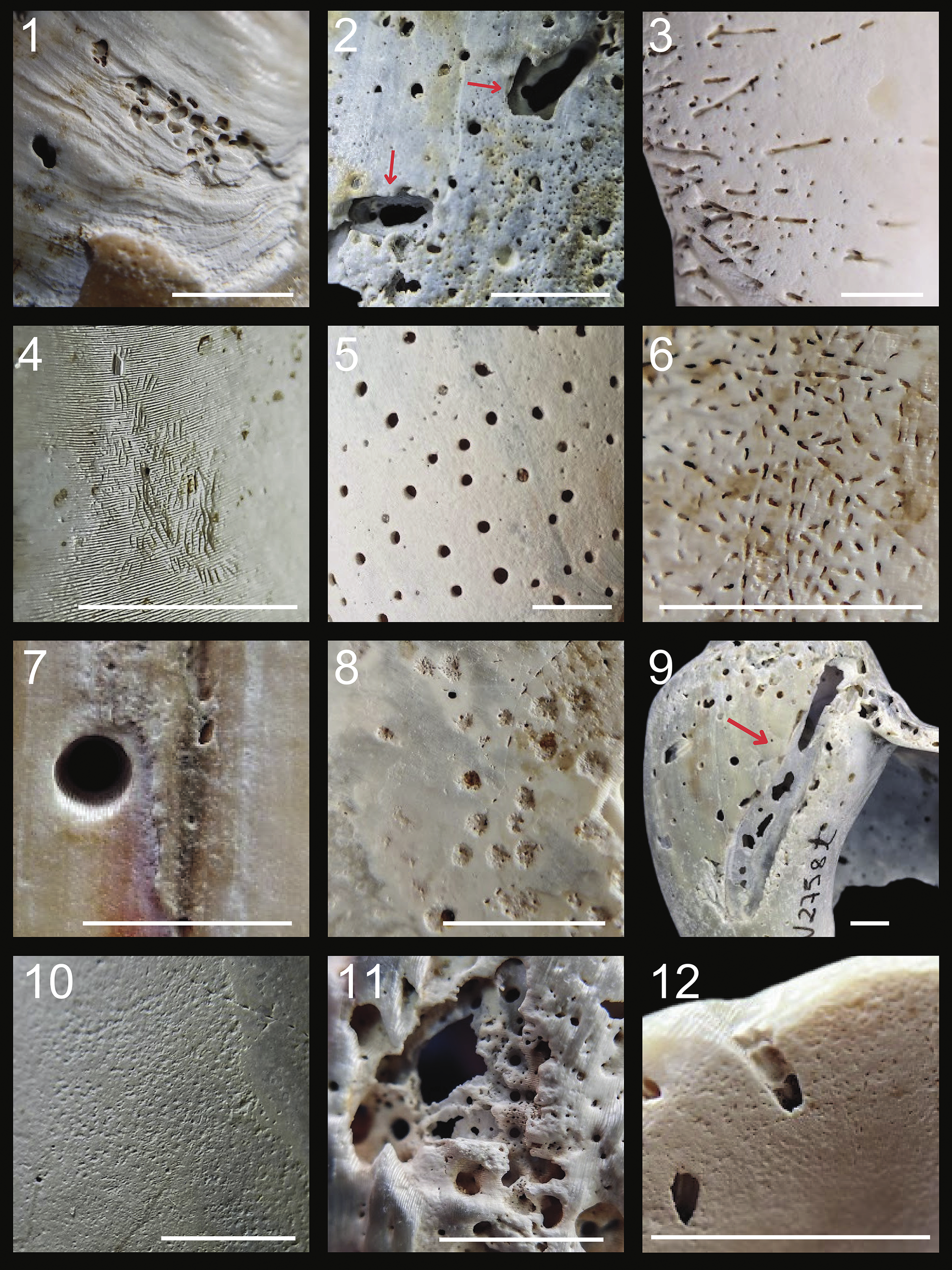
From the group of bioeroded shells, 10 distinct ichnogenera are recorded, with often more than one type of bioerosion occurring in a single shell (Fig. 3). The most common ichnogenus, on both the outer and inner sides, is Caulostrepsis, which is characterized by a U-shaped tunnel excavated by polychaetes (Charó et al., Reference Charó, Cavallotto and Aceñolaza2017). Iramena, which is produced by ctenostome bryozoans and characterized by a system of tunnels with an irregular pattern of openings to the shell surface, is the second-most common taxon (Pereira et al., Reference Pereira, Mocho and Lourenço2009; Charó et al., Reference Charó, Cavallotto and Aceñolaza2017), followed by Finichnus, formed of groups of shallow elliptical or pear-shaped pits produced by cheilostome bryozoans (Taylor et al., Reference Taylor, Wilson and Bromley2013). Entobia, produced by clionid sponges, consists of interconnected galleries parallel to the surface, with perforations in the form of a network often covering the entire shell. Pennatichnus, which is also produced by cheilostome bryozoans, has axial openings leading to the surface in a feather-like pattern (Charó et al., Reference Charó, Cavallotto and Aceñolaza2017; Taylor et al., Reference Taylor, Wilson and Bromley2013). Gastrochaenolites is characterized by perforations that are wedge shaped in longitudinal cross section, with a circular or oval surface opening distinctly narrower than the main chamber. This structure is produced by boring bivalves (Gibert et al., Reference Gibert, Domènech and Martinell2007; Charó et al., Reference Charó, Cavallotto and Aceñolaza2017), which use the shell of Z. dufresnii as a substrate and permanent habitat. Oichnus, characterized by circular or oval perforations perpendicular to the shell surface, which may or may not penetrate the shell (Charó et al., Reference Charó, Cavallotto and Aceñolaza2017), represent drill holes made by carnivorous gastropods. Maeandropolydora is formed of long cylindrical galleries of sinuous, irregular, or almost rectilinear shape (Bromley and D’Allessandro, Reference Bromley and D’Allessandro1983), which are excavated by polychaetes. Dendrina consists of a dendritic bioerosion pattern, forming a prostrate system of roofless channels that bifurcate dichotomously (Wisshak et al., Reference Wisshak, Titschack, Kahl and Girod2017; Wisshak and Neumann, Reference Wisshak and Neumann2018). Radulichnus consists of short, parallel rows of shallow grooves occurring in small clusters, made by mollusk-grazing gastropods (Gibert et al., Reference Gibert, Domènech and Martinell2007; Lopes and Pereira, Reference Lopes and Pereira2018).
Bioerosion traces found in shells of Z. dufresnii. (1) Caulostrepis (D376). (2) Gastrochaenolites (LGP-W2758t). (3) Iramena (D349). (4) Radulichnus (D428). (5) Entobia (LGP-W2755o). (6) Pennatichnus (D444). (7) Oichnus (D515). (8) Dendrina (D444). (9) Maeandropolydora (LGP-W2758t). (10) Finichnus (D444). (11, 12) Shells showing distinct overlapping bioerosion traces of two or several ichnogenera: (11) LGP-W2757t; (12) D239. Scale bars = 0.5 cm.

Figure 3. Long description
A collection of twelve macro photographs arranged in a three-column by four-row grid. Each panel includes a white scale bar representing 0.5 cm.
* Panel 1 shows Caulostrepis D 3 7 6, featuring a series of small, interconnected pits forming a linear track on a light-colored shell surface.
* Panel 2 shows Gastrochaenolites L G P dash W 2 7 5 8 t, with two red arrows pointing to large, elongated oval-shaped boreholes.
* Panel 3 shows Iramena D 3 4 9, displaying numerous thin, dark, branching or straight tunnel-like traces across a smooth white surface.
* Panel 4 shows Radulichnus D 4 2 8, consisting of fine, parallel scraping marks clustered together on a textured shell surface.
* Panel 5 shows Entobia L G P dash W 2 7 5 5 o, characterized by a dense distribution of small, circular, dark holes of varying sizes.
* Panel 6 shows Pennatichnus D 4 4 4, featuring short, dash-like indentations arranged in a somewhat organized, radiating pattern.
* Panel 7 shows Oichnus D 5 1 5, highlighting a single, perfectly circular, deep drill hole with smooth edges.
* Panel 8 shows Dendrina D 4 4 4, showing shallow, rosette-shaped or star-shaped erosional pits.
* Panel 9 shows Maeandropolydora L G P dash W 2 7 5 8 t, where a red arrow points to a long, winding, and irregular gallery or tunnel system.
* Panel 10 shows Finichnus D 4 4 4, displaying a very fine, granular texture of tiny, shallow depressions.
* Panel 11 shows shell L G P dash W 2 7 5 7 t, exhibiting a complex, overlapping network of multiple ichnogenera resulting in a highly porous, sponge-like appearance.
* Panel 12 shows shell D 2 3 9, featuring distinct, larger overlapping bioerosion traces that have breached the shell surface in several places.
Live–live interactions
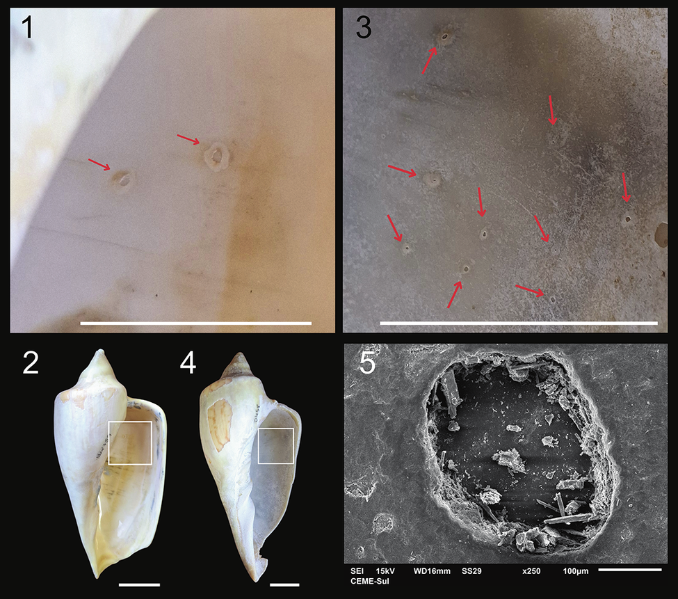
Several types of abnormal internal shell growth (i.e., malformations or pathologies) were identified on the inside of Z. dufresnii shells. These include pits in 170 shells (19.02%), verrucous structures in 115 shells (12.86%), attached pearls in 112 shells (12.53%), and blisters and needles in 47 shells (5.26%). Sheets occur in only 12 shells (4.70%) and igloo-like structures in two (0.22%). Pits (Fig. 4) are small, oval cavities with raised edges that may occur all over the inside of the shell. In bivalves, such pits are known to be induced by parasitic trematodes and are regarded as a defensive mechanism to encompass the parasite (Ruiz and Lindberg, Reference Ruiz and Lindberg1989).
Pit-type malformations in shells of Z. dufresnii. (1–4) Shells with pit malformation on the inside: (1, 3) LGP-W2759; (2, 4) D458. Scale bars = 2 cm. (5) SEM image showing detail of a pit structure (LGP-W2756j).

Figure 4. Long description
Panel 1 at the top-left shows a close-up of the smooth interior of shell L G P dash W 2 7 5 9 with two red arrows pointing to small circular pit malformations. Panel 3 at the top-right shows a wider view of the interior of shell D 4 5 8 with nine red arrows indicating a higher density of similar pits. Panel 2 at the bottom-left displays the full ventral view of shell L G P dash W 2 7 5 9 with a white square highlighting the area shown in panel 1. Panel 4 in the bottom-middle displays the full ventral view of shell D 4 5 8 with a white square highlighting the area shown in panel 3. Panel 5 at the bottom-right is a grayscale S E M micrograph of a single pit from shell L G P dash W 2 7 5 6 j. The pit is an irregular oval depression with rough, fragmented edges and debris inside. Technical text at the bottom of panel 5 reads S E I 15 k V, W D 16 m m, S S 2 9, times 2 5 0, and 100 micrometers next to a scale bar.
Pits were never described from marine gastropods, but terrestrial gastropods are known to encapsulate trematodes (Gerard et al., Reference Gerard, Tombeur, Dahirel and Ansart2023) and may form pits during the encapsulation process. In some Z. dufresnii shells, up to 16 pits occur, indicating high levels of infestation.
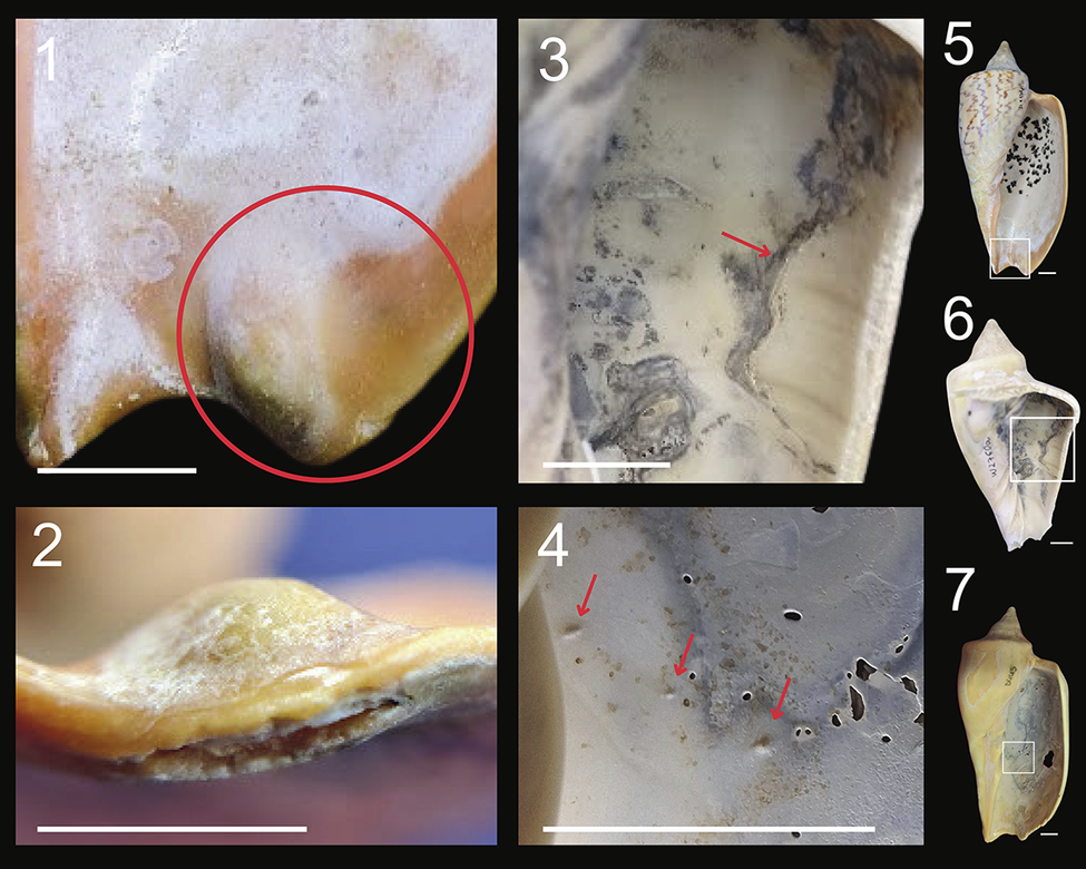
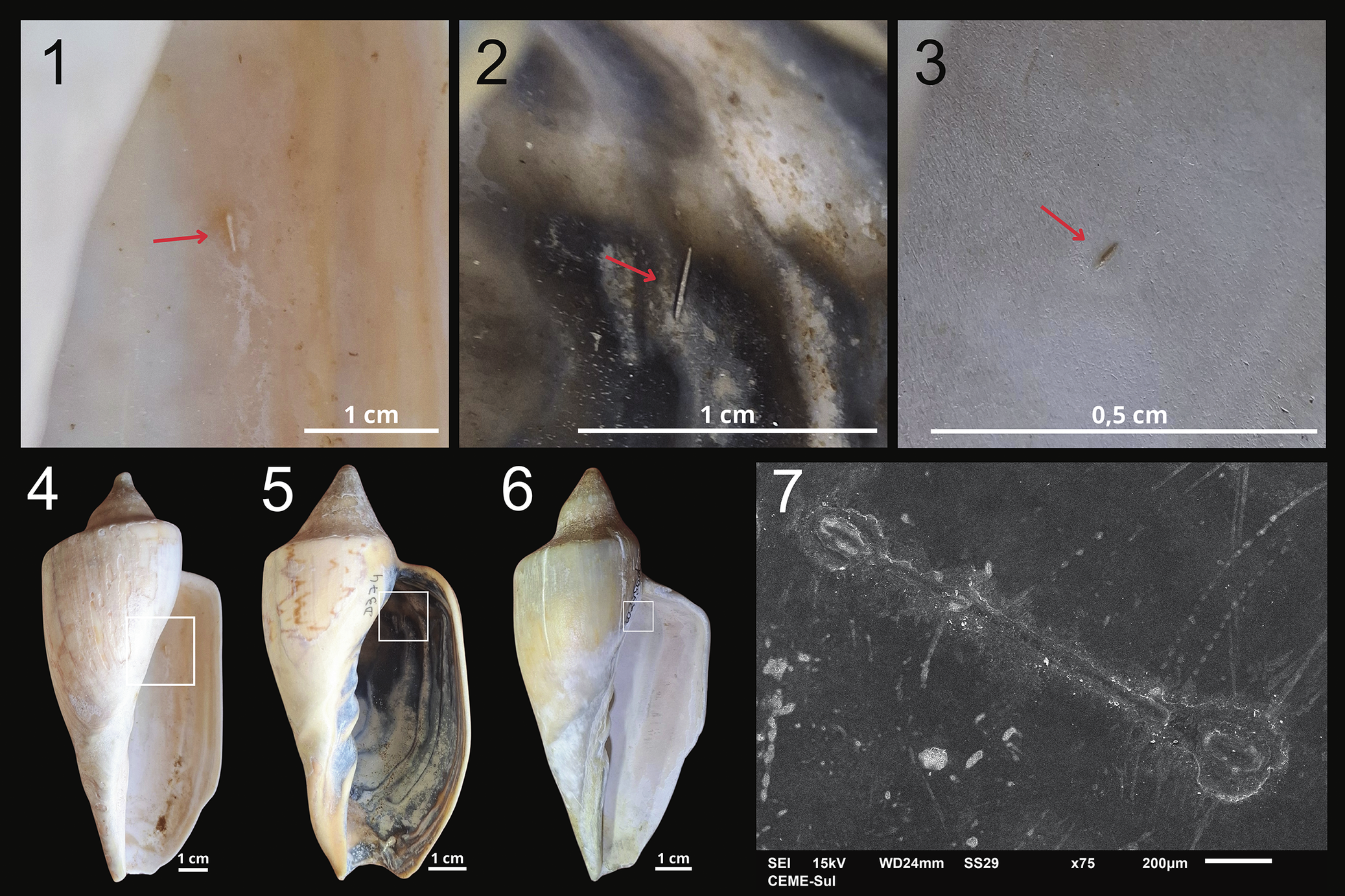
Attached pearls (Fig. 5.4) are large, solid structures, usually rounded, sometimes elongated, and fixed to the inner wall of the shell. While they are generally produced by the gastropod to encapsulate small particles, such as sand grains, which get trapped in the space between the shell and the visceral mass, they may also be associated with the presence of trematode parasites (Taylor et al., Reference Taylor, Wilson and Bromley2013; De Baets et al., Reference De Baets, Dentzien-Dias, Upeniece, Verneau and Donoghue2015). Shell blisters (Fig. 5.1, 5.2) are elevated, hollow structures inside the shell that are formed by the gastropod when a foreign body is penetrating the shell. They are generally associated with bioerosions from the outside of the shell (Huntley and Scarponi, Reference Huntley and Scarponi2012; De Baets et al., Reference De Baets, Dentzien-Dias, Upeniece, Verneau and Donoghue2015; Huntley and De Baets, Reference Huntley and De Baets2015; Marini et al., Reference Marini, Spotorno, Diniz and Dentzien-Dias2022). Sheets (Fig. 5.3) are shell layers that also enclose a void but cover a significant portion of the inner surface of the shell and have no defined shape (Marini et al., Reference Marini, Spotorno, Diniz and Dentzien-Dias2022). They are larger than blisters but not as elevated and may also be associated with bioerosion on the outer face (Marini et al., Reference Marini, Spotorno, Diniz and Dentzien-Dias2022). Verrucous structures (Fig. 6) are often spherical or elongated in outline, with a rough texture, and may also show variation in color (da Rosa et al., Reference da Rosa, Marini, Spotorno and Dentzien-Dias2026). Needles (Fig. 7) are elongated structures approximately 1.5 mm long, which suggest that small, straight foreign bodies were encapsulated in the shell (da Rosa et al., Reference da Rosa, Marini, Spotorno and Dentzien-Dias2026). The SEM images (Fig. 7) show that some of these structures have the shape of rods with rounded ends.
Blister, sheet, and attached pearl malformations in shells of Z. dufresnii. (1, 2, 5) Blister (D194). (3, 6) Sheet with the internal layer of the structure broken (LGP-W2760a). (4, 7) Attached pearls (D445). Scale bars = 1 cm.

Figure 5. Long description
The photo is organized into two columns of large macro images and a right-hand column of full-shell views.
* Panel 1 at the top left shows a macro view of a blister malformation D 1 9 4, with a red circle highlighting a raised, rounded protrusion on the shell surface.
* Panel 2 below it shows a cross-sectional side view of the same blister, revealing its thickness and elevation from the shell plane.
* Panel 3 in the center top shows a macro view of a sheet malformation L G P-W 2 7 6 0 a, where a red arrow points to a jagged, broken edge of an internal structural layer.
* Panel 4 in the center bottom shows a macro view of attached pearls D 4 4 5, with three red arrows pointing to small, distinct calcified nodules fused to the interior shell wall.
* Panels 5, 6, and 7 on the right show the complete shells for specimens D 1 9 4, L G P-W 2 7 6 0 a, and D 4 4 5 respectively. Each full shell contains a small white square indicating the anatomical location of the malformations shown in the macro panels.
White scale bars representing 1 cm are present in the bottom left of panels 1 through 4 and to the bottom right of panels 5 through 7.
Verrucous malformation in shells of Z. dufresnii. (1–4) Shells with the malformation on the inside: (1, 3) D549; (2, 4) LGP-WW2761g. Scale bars = 1 cm. (5) SEM image showing detail of verrucous structure (LGP-W2760a).

Needle-type malformations in shells of Z. dufresnii. (1–6) Shells with needle malformation on the inside: (1, 4) D372; (2, 5) D374; (3, 6) LGP-W2756d. (7) SEM image showing detail of a needle structure, where it is possible to perceive its rod-like shape (B70).

Figure 7. Long description
The figure consists of seven panels.
Panels 1, 2, and 3 are close-up photos of the interior shell surfaces of specimens D 3 7 2, D 3 7 4, and L G P-W 2 7 5 6 d respectively. Each panel features a red arrow pointing to a small, thin, calcified needle-like structure protruding from the smooth nacreous surface. Scale bars indicate 1 centimeter for panels 1 and 2, and 0.5 centimeters for panel 3.
Panels 4, 5, and 6 show the full ventral view of the same three shells. Each shell has a white square box highlighting the specific region on the inner lip or columella where the malformations are located. Scale bars at the bottom right of each shell indicate 1 centimeter.
Panel 7 is a grayscale S E M micrograph of specimen B 7 0 at 75 times magnification. It shows a detailed view of a single rod-like needle structure lying across the shell surface. The needle has a slightly bulbous base and a long, straight shaft. Technical data at the bottom of the panel includes S E I, 15 k V, W D 24 millimeters, S S 29, and a 200 micrometer scale bar.
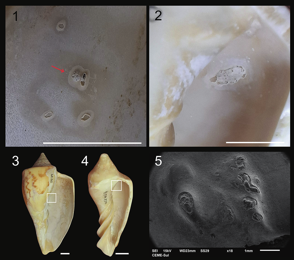
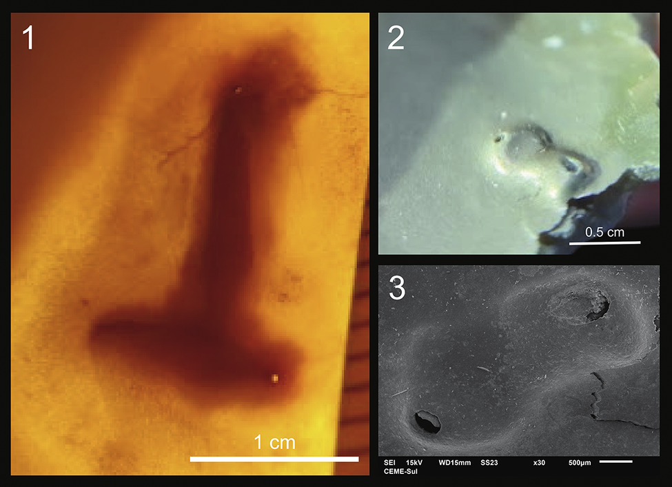
Igloo-like structures, as described by Ituarte et al. (Reference Ituarte, Cremonte and Deferrari2001), are dome shaped, with a lateral aperture. The igloo-like structures found in Z. dufresnii shells are semi-elliptical domes with an aperture (Fig. 8), measuring approximately 1 cm. According to Huntley et al. (Reference Huntley, De Baets, Scarponi, Linehan, Epa, Jacobs and Todd2021), igloos are formed when the walls of a pit structure grow over the pit and fuse, creating a hollow blister-like structure with a tiny opening at the anterior side of a metacercaria. This means that the igloo structures represent unsuccessful attempts to isolate the parasite. Their construction is more intricate than that in other structures since the parasite may stimulate or modify the activity of the host. Besides the aperture, there may or may not be an erosional hollow or divot in the shell surface below the igloo (Huntley et al., Reference Huntley, De Baets, Scarponi, Linehan, Epa, Jacobs and Todd2021).
Igloo-like malformations in shells of Z. dufresnii. (1, 2) Shells showing igloo-like malformations on the inside: (1) D190; (2) B46. (3) SEM image showing detail of an igloo-like structure (B46).

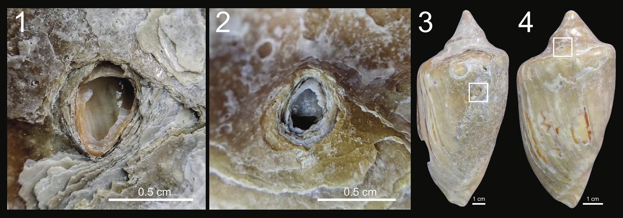
Rounded malformations observed on the outer shell surface of Z. dufresnii shells formed around barnacles that attached to the shell (Fig. 9). These structures are known as bioclaustrations and are located in the posterior region of the shells, particularly near the apex. In total, eight specimens exhibited this structure, with two of them retaining barnacle remains.
Bioclaustration in Z. dufresnii shells. (1, 3) D496. (2, 4) D460.

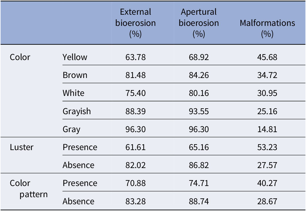
Shell color, as an index of alteration, is correlated with the presence of bioerosion on both inner and outer shell surfaces, as well as with malformations (Table 3). A statistically significant association of coloration and malformations was confirmed by the chi-square test (x2 = 29.495, df = 4, p = 6.2 × 10⁻⁶). Statistically significant correlation was also confirmed for coloration and occurrence of bioerosions on the outer shell surface (x2 = 50.944, df = 4, p = 2.293 × 10⁻¹⁰) and on the inner shell surface (x2 = 51.517, df = 4, p = 1.741 × 10⁻¹⁰).
Relationships between color, luster, and color pattern, in shells of Zidona dufresnii showing external, internal, and malformation bioerosions

Table 3. Long description
The table is organized into three main categories: Color, Luster, and Color pattern. Each category is evaluated against three percentage-based metrics: External bioerosion, Apertural bioerosion, and Malformations.
* Color category:
- Yellow: 63.78 percent external, 68.92 percent apertural, 45.68 percent malformations.
- Brown: 81.48 percent external, 84.26 percent apertural, 34.72 percent malformations.
- White: 75.40 percent external, 80.16 percent apertural, 30.95 percent malformations.
- Grayish: 88.39 percent external, 93.55 percent apertural, 25.16 percent malformations.
- Gray: 96.30 percent external, 96.30 percent apertural, 14.81 percent malformations.
* Luster category:
- Presence: 61.61 percent external, 65.16 percent apertural, 53.23 percent malformations.
- Absence: 82.02 percent external, 86.82 percent apertural, 27.57 percent malformations.
* Color pattern category:
- Presence: 70.88 percent external, 74.71 percent apertural, 40.27 percent malformations.
- Absence: 83.28 percent external, 88.74 percent apertural, 28.67 percent malformations.
Logistic regression analysis indicated that the only taphonomic characteristic of the shells representing a robust predictor is the presence of luster. This suggests that lustrous shells are more likely to show malformations (Supplementary Appendix 1).
Age of the shells
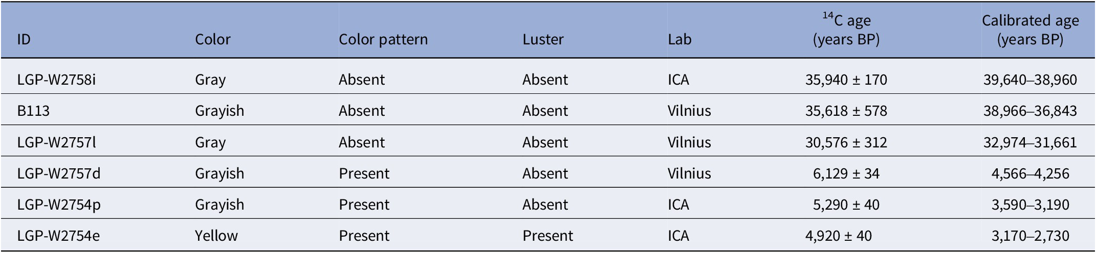
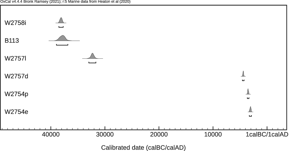
The six shells analyzed yielded ages between approximately 2,730 and 39,640 years BP (Table 4). Three shells are less than 7,000 years old and are thus attributed to the Holocene. The remaining three shells are older than 30,000 years and thus are Late Pleistocene in age (Fig. 10; Supplementary Appendix 2).
Calibrated 14C model ages (95.4%) for the shell samples, using the Marine20 curve (Heaton et al., Reference Heaton, Köhler, Butzin, Bard and Reimer2020)

Table 4. Long description
The table contains seven columns: I D, Color, Color pattern, Luster, Lab, C 14 age in years B P, and Calibrated age in years B P.
Row 1: I D L G P dash W 2 7 5 8 i, Gray, Color pattern Absent, Luster Absent, Lab I C A, C 14 age 35,940 plus or minus 170, Calibrated age 39,640 to 38,960.
Row 2: I D B 1 1 3, Grayish, Color pattern Absent, Luster Absent, Lab Vilnius, C 14 age 35,618 plus or minus 578, Calibrated age 38,966 to 36,843.
Row 3: I D L G P dash W 2 7 5 7 l, Gray, Color pattern Absent, Luster Absent, Lab Vilnius, C 14 age 30,576 plus or minus 312, Calibrated age 32,974 to 31,661.
Row 4: I D L G P dash W 2 7 5 7 d, Grayish, Color pattern Present, Luster Absent, Lab Vilnius, C 14 age 6,129 plus or minus 34, Calibrated age 4,566 to 4,256.
Row 5: I D L G P dash W 2 7 5 4 p, Grayish, Color pattern Present, Luster Absent, Lab I C A, C 14 age 5,290 plus or minus 40, Calibrated age 3,590 to 3,190.
Row 6: I D L G P dash W 2 7 5 4 e, Yellow, Color pattern Present, Luster Present, Lab I C A, C 14 age 4,920 plus or minus 40, Calibrated age 3,170 to 2,730.
Modeled dating of the samples LGP-W2758i, B113, LGP-W2757l, LGP-W2757d, LGP-W2754p, and LGP-W2754e using OxCal (v4.4.4).

Figure 10. Long description
The horizontal X-axis is labeled Calibrated date cal B C forward slash cal A D, with a scale ranging from 40000 on the left to 1 cal B C forward slash 1 cal A D on the right. Six samples are listed vertically on the left.
* L G P dash W 2 7 5 8 i shows a narrow probability peak centered around 38000.
* B 1 1 3 shows a wider probability distribution also centered near 38000.
* L G P dash W 2 7 5 7 l shows a peak centered around 32000.
* L G P dash W 2 7 5 7 d shows a sharp, narrow peak located near 4000.
* L G P dash W 2 7 5 4 p shows a sharp peak located near 3000.
* L G P dash W 2 7 5 4 e shows a sharp peak located near 2000.
Each probability curve includes a horizontal bracket underneath indicating the 95.4 percent confidence interval. The top three samples represent an older cluster between 40000 and 30000, while the bottom three represent a much younger cluster between 5000 and 1.
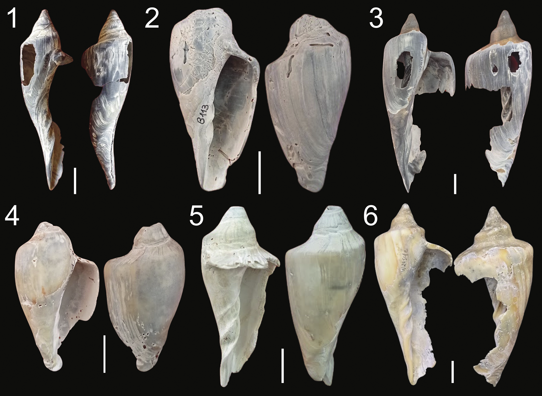
The dated specimens exhibited distinct taphonomic characteristics (Fig. 11). Holocene shells retain the original zig-zag color pattern, and the youngest one also preserves luster. By contrast, luster or color pattern are absent in the Late Pleistocene shells, and overall shell color shows different shades of gray.
Dated shells. (1) LGP-W2758i. (2) B113. (3) LGP-W2757l. (4) LGP-W2757d. (5) LGP-W2754p. (6) LGP-W2754e. Scale bars = 2cm.

Figure 11. Long description
A collection of six numbered panels against a black background, each displaying two views of a fossilized shell. A white vertical scale bar representing 2 centimeters is placed at the bottom right of each panel.
* Panel 1: Two views of a slender, elongated shell labeled L G P dash W 2 7 5 8 i. The shell is dark gray with white striations and significant breakage on the body whorl.
* Panel 2: Two views of a broader, conical shell labeled B 1 1 3. It has a light gray, weathered surface with visible growth lines.
* Panel 3: Two views of a shell labeled L G P dash W 2 7 5 7 l. The shell is dark gray with large holes and jagged edges indicating heavy erosion.
* Panel 4: Two views of a robust, light-colored shell labeled L G P dash W 2 7 5 7 d. It features a smooth, cream-colored surface with some pitting near the base.
* Panel 5: Two views of a pale, greenish-gray shell labeled L G P dash W 2 7 5 4 p. The shell has a distinct flared lip and a pointed spire.
* Panel 6: Two views of a yellowish-tan shell labeled L G P dash W 2 7 5 4 e. The shell shows significant fragmentation along the aperture and outer lip.
Discussion
The occurrence of bioerosion in shells of Zidona dufresnii from the Rio Grande do Sul Coastal Plain has rarely been reported in the scientific literature. Lopes (Reference Lopes, Netto, Carmona and Tognoli2012, Reference Lopes2013) identified bioerosion traces attributed to the ichnogenera Entobia and Caulostrepsis. Subsequently, Schmidt-Neto et al. (Reference Schmidt-Neto, Horodyski, Ritter and Dasgupta2023) documented Gastrochaenolites and Rogerella from shells of the same species. Additional types of bioerosion traces occur in other gastropods and in bivalves from the same shell-rich deposits (Lopes, Reference Lopes, Netto, Carmona and Tognoli2012; Schmidt-Neto et al., Reference Schmidt-Neto, Horodyski, Ritter and Dasgupta2023; Anzai et al., Reference Anzai, Diniz, Spotorno and Dentzien-Dias2024; da Rosa et al., Reference da Rosa, Marini, Spotorno and Dentzien-Dias2026).
According to previous studies, Entobia is the predominant ichnogenus documented from mollusk shells found at Cassino Beach, followed by Maeandropolydora and Pennatichnus (Lopes and Buchmann, Reference Lopes and Buchmann2008; Lopes, Reference Lopes, Netto, Carmona and Tognoli2012). These structures are indicative of deep-layer perforation, suggesting that the respective shells remained exposed to seawater for extended periods, sufficient for the development of a mature community of bioeroders (Bromley and Asgaard, Reference Bromley and Asgaard1993). According to Lopes (Reference Lopes, Netto, Carmona and Tognoli2012), although Entobia remains the most abundant, some shells of bivalves and gastropods also exhibited the presence of Caulostrepsis, indicating a change in the fauna of bioeroding mollusks. However, in more recent studies, other ichnogenera have begun to be identified more frequently, such as Iramena and Finichnus (Lopes and Buchmann, Reference Lopes and Buchmann2008; Lopes, Reference Lopes, Netto, Carmona and Tognoli2012; Anzai et al., Reference Anzai, Diniz, Spotorno and Dentzien-Dias2024). These traces are shallower than those quoted in the preceding and are usually rapidly obliterated when exposed to erosive agents, which may indicate that this type of bioerosion occurred relatively recently (Bromley and Asgaard, Reference Bromley and Asgaard1993). The respective shells, once reworked, must have remained available to the bioeroders for some time before being transported to the beach.
The ichnogenera Gastrochaenolites, Oichnus, and Maeandropolydora are rare (Lopes and Buchmann, Reference Lopes and Buchmann2008; Lopes, Reference Lopes2011, Reference Lopes, Netto, Carmona and Tognoli2012; da Rosa et al., Reference da Rosa, Marini, Spotorno and Dentzien-Dias2026), and Radulichnus was documented only in samples from barrier III (Lopes and Pereira, Reference Lopes and Pereira2018) but not from Cassino Beach or the shallow platform.
It is important to emphasize that the majority of the bioerosion traces found only on the outside of the shells have no direct association with shell growth. Therefore, it is impossible to determine whether these structures were produced before or after the death of the mollusk. As an exception, Oichnus, which is a predation trace, implies that the mollusk was usually still alive when the bioerosion occurred (Charó et al., Reference Charó, Cavallotto and Aceñolaza2017). All Oichnus traces observed in the study material perforated the shell from outside, indicating successful predation. Numerous shells not only display different types of bioerosion but also preserve an ecological succession, as evidenced by the superposition of different traces. Together, these traces indicate that the respective shells experienced multiple distinct episodes of bioerosion during the period they remained exposed as a substratum.
All malformations found in the shells of Z. dufresnii occurred while the gastropods were still alive, representing reactions involving calcium carbonate production mainly to isolate foreign bodies that had entered the shell. However, not all of these malformations are necessarily results of parasitism, in which one species (the parasites) benefits at the expense of the other (the host). Some structures previously described from mollusks, particularly bivalves, such as pits and igloos are known to represent host response to the presence of trematode parasites that reached the visceral mass (Ituarte et al., Reference Ituarte, Cremonte and Zelaya2005; Huntley and De Baets., Reference Huntley and De Baets2015; Huntley et al., Reference Huntley, De Baets, Scarponi, Linehan, Epa, Jacobs and Todd2021). Blister and sheet structures are usually associated with external bioerosion of Caulostrepsis, caused by spionid polychaetes that perforate the shell from the outside, causing a protective response of the mollusk (Huntley and Scarponi, Reference Huntley and Scarponi2012; Huntley et al., Reference Huntley, De Baets, Scarponi, Linehan, Epa, Jacobs and Todd2021; Marini et al., Reference Marini, Spotorno, Diniz and Dentzien-Dias2022).
Similarly, the structures observed in the shells of Z. dufresnii may indicate evidence of infestation by both trematodes and boring polychaetes. The pits identified in the specimens exhibit shapes and sizes consistent with those previously described in the literature for bivalves, characterized by shallow rounded depressions with elevated margins. Their morphology corresponds to the body plan of the parasites, which range from rounded to oval in outline shape and are dorsoventrally flattened. SEM images reveal that the deposited material and the growth pattern of the margins surrounding the pits differ from the normal mineral structure of a healthy shell. Furthermore, the clustered localization of these pits in specific shell regions, combined with a uniform size class, suggests a mass infestation process. Given that only two specimens with structures resembling igloos—albeit more elongated than those known from bivalves and occurring in joined pairs—further studies are required on these structures. Nevertheless, their shape, position, and size provide strong evidence of a trematode-induced origin. Last, the blisters and sheets, associated with external Caulostrepsis-type borings, can be attributed to polychaete parasites, as observed in bivalves.
Many other cases of malformations found in the shells may simply indicate that foreign particles, such as sand grains, spicules, or other bioclasts, caused discomfort to the mollusk. Malformations of the pearl, needle, and verrucous types may occur either due to the entry of an external object between the shell and the visceral mass (da Rosa et al., Reference da Rosa, Marini, Spotorno and Dentzien-Dias2026) or due to parasite infestation, but the actual cause cannot be distinguished without additional evidence (Huntley and De Baets, Reference Huntley and De Baets2015). Recent studies provide evidence that gymnophallid trematodes are directly involved not only in the formation of igloos and pits but also in the development of pearls in bivalves (Benito et al., Reference Benito, Izagirre, Lekube, Zaldibar, Villalba, Montaudouin, Daffe, Soto and Cerio2023; Marchiori et al., Reference Marchiori, Quaglio, Franzo, Brocca, Aleksi, Cerchier, Cassini and Marcer2023).
In the analyzed shells of Z. dufresnii, there are indications that the verrucous malformations may also be related to trematode parasites. SEM analysis of these structures reveals an irregularly shaped center covered with a layer of shell and surrounded by a gentle depression. The size and shape of this type of malformation are compatible with clusters of metacercariae, commonly found in live tissues of mollusks and occasionally causing changes in the formation of their shells (Cremonte and Ituarte, Reference Cremonte and Ituarte2003; Averbuj and Cremonte, Reference Averbuj and Cremonte2010; Cremonte et al., Reference Cremonte, Pina, Gilardoni, Rodrigues, Chai and Ituarte2013; Di Giorgio et al., Reference Di Giorgio, Gilardoni and Ituarte2014; Ribeiro et al., Reference Ribeiro, Coaglio, Oliveira, Pereira and Lima2017). In 53.04% of shells exhibiting verrucous malformations, pits were also present, indicating that these specimens were infested by trematode parasites. Pearl structures, indicative of the occurrence of foreign bodies within the mollusk, occur in 35.65% of the shells with verrucous malformations.
Needle-type malformations may have been caused by the presence of sponge spicules inside the shells (da Rosa et al., Reference da Rosa, Marini, Spotorno and Dentzien-Dias2026). This hypothesis was proposed after observing needle-shaped structures in SEM images, where the two ends of the needle form minute ring-shaped structures. Although considerable variation in shape and size exists, some types of sponge spicules exhibit the same kind of structure (Łukowiak, Reference Łukowiak2016, Reference Łukowiak2020; Rasbold et al., Reference Rasbold, McGlue, Stevaux, Parolin, Silva and Bergier2019; Łukowiak et al., Reference Łukowiak, Van Soest, Klautau, Pérez, Pisera and Tabachnick2022).
Similar morphological malformations to those associated with the presence of barnacles in the studied gastropod shells were already documented by da Rosa et al. (Reference da Rosa, Marini, Spotorno and Dentzien-Dias2026). According to the authors, due to the large size of the gastropods and the cirripedes lodging only in the apical region, parasitism is excluded in this case since the host organism is not affected. Furthermore, the shells of Z. dufresnii show no internal alterations resulting from this interaction. Consequently, bioclaustration by barnacles can be considered a commensal or neutral interaction. Although gastropod shell growth normally occurs on the inner surface and along the margin of the aperture, some species, such as those belonging to the family Volutidae, which have large shells and a well-developed mantle, may partially extend the mantle over the external surface of the shell, allowing the secretion of additional calcium carbonate to cover damaged or perforated areas in a process known as shell repair (Giménez and Penchaszadeh, Reference Giménez and Penchaszadeh2002; Giménez et al., Reference Giménez, Penchaszadeh, Brey and Mackensen2004, Reference Giménez, Ojeda and Boy2024; Checa, Reference Checa2018; Medina et al., Reference Medina, Romero, Crespi-Abril and Narvarte2020).
The rising sea level over the past several thousand years may be a factor contributing to an increase in the prevalence of trematode parasites in mollusks. Secondary factors, such as temperature, productivity, salinity, and taxon abundance, associated with the rise in sea level can make this increase favorable (Huntley et al., Reference Huntley, Fürsich, Alberti, Hethke and Liu2014). Thus, the greater abundance of parasitism indicators in recent shells in the study material, which are characterized by the persistence of luster, may indicate that the infestation rate of trematode parasites in Z. dufresnii is increasing. Another factor that might influence the greater number of malformations in shells with luster compared with older shells is that these young shells, due to a shorter exposure to reworking and bioerosion, have preserved these structures more effectively.
Several authors have claimed that shell color changes with age (Wehmiller et al., Reference Wehmiller, York and Bart1995; Erthal et al., Reference Erthal, Ritter and Kotzian2017; Martinez and Rojas, Reference Martínez and Rojas2023), indicating that the darker the shell, the older the sample is. However, as observed in this study, shell color is not necessarily related to age, with specimens of similar color dated as Holocene or Late Pleistocene. Consequently, it is not possible to assert age using only this parameter. The same effect was observed by da Rosa et al. (Reference da Rosa, Marini, Spotorno and Dentzien-Dias2026), who documented that darker shells of Pachycymbiola brasiliana (Lamarck, Reference Lamarck1811) showed younger ages compared with those of lighter coloration. Nevertheless, luster and color patterns are preserved only in shells from the Holocene, potentially serving as an indicator of age. The presence of luster indicates that the shells have not been exposed long enough to the environment to lose this outermost shell layer. By contrast, the zig-zag color pattern is embedded in the deeper shell layers and persists for a longer period before being lost due to erosion or the degradation of the pigments. It is also observed that bioerosion in the studied shells increased over time (see Table 3), and pristine shells are less bioeroded than older ones.
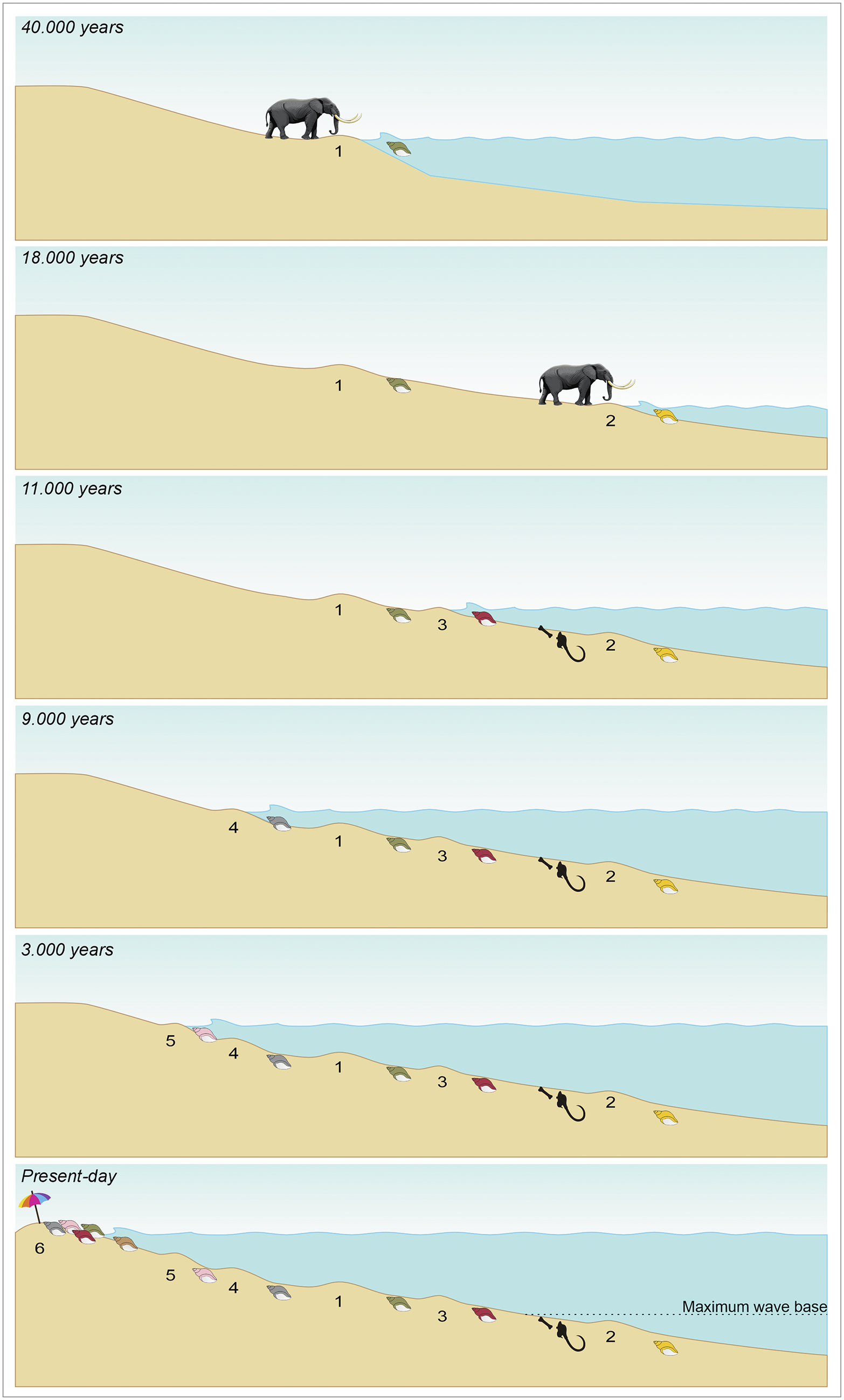
One very interesting aspect that draws attention is related to the ages of the material collected. So far, all shells collected from beaches and the shallow platform in southern Brazil that were dated fall into two intervals, from the present to 8,615 years BP and from 31,661 BP to 41,167 years BP (Lopes et al., Reference Lopes, Oliveira, Figueiredo, Kinoshita, Baffa and Buchmann2010; Ritter et al., Reference Ritter, Erthal, Kosnik, Coimbra and Kaufman2017; Tâmega et al., Reference Tâmega, Spotorno-Oliveira, Dentzien-Dias, Buchmann, Vieira, Macario, Nash, Guimarães, Francischini and Bassi2019; Anzai et al., Reference Anzai, Diniz, Spotorno and Dentzien-Dias2024; da Rosa et al., Reference da Rosa, Marini, Spotorno and Dentzien-Dias2026) (Supplementary Appendix 3), indicating a hiatus from approximately 9,000 to 30,000 years BP, including the LGM. A possible explanation for the absence of shells from this period is that they are preserved in sediment layers currently located at depths greater than 80 meters, thus not undergoing reworking. By contrast, between 30,000 and 40,000 years BP, the coastline was at a depth of approximately 60 meters below the present sea level (Spratt and Lisieck, Reference Spratt and Lisiecki2016), meaning that these sediments are positioned close to those of the Holocene (Fig. 12). This proximity facilitates simultaneous reworking and transport of fossil materials of both ages, contributing to the formation of significantly time-averaged associations.
Reconstruction of coastal ridges 1 to 6 in relation to Late Pleistocene to recent variations in sea level, illustrating potential areas of subaerial and submarine deposition and/or reworking.

Figure 12. Long description
The diagram consists of six stacked panels showing a coastal slope and sea level at different time intervals.
* 40,000 years. Sea level is high. An elephant stands on a ridge labeled 1 at the shoreline. A green deposit is visible just below the water line.
* 18,000 years. Sea level has dropped significantly. Ridge 1 is now far inland. The elephant stands on a new, lower ridge labeled 2 at the new shoreline. A yellow deposit is at the water line.
* 11,000 years. Sea level is rising. Ridge 2 is submerged. A new ridge labeled 3 is forming between ridges 1 and 2. A red deposit appears at ridge 3. A black hook and bone icon is located underwater near ridge 2.
* 9,000 years. Sea level continues to rise. Ridge 3 is submerged. A new ridge labeled 4 forms landward of ridge 1. A grey deposit is at the shoreline.
* 3,000 years. Sea level rises further. Ridge 4 is submerged. A new ridge labeled 5 forms further up the slope. A pink deposit is at the shoreline.
* Present-day. Sea level is at its highest point in the sequence. Ridges 1, 3, 4, and 5 are all submerged. A final ridge labeled 6 is at the current shoreline, marked by a beach umbrella and colorful towels. A horizontal dotted line labeled Maximum wave base sits just above the submerged ridge 2 and the hook icon.
In addition, relict sediments are widely distributed on the southern Brazilian shelf, where they are extensively reworked by the combined effects of the modern hydrodynamic regime, terrestrial sediment input, and post-glacial sea-level rise (Silva et al., Reference Silva, Barboza, Rosa and Dillenburg2014; Carassai et al., Reference Carassai, Lavina, Chemale and Girelli2019; Barboza et al., Reference Barboza, Dillenburg, Ritter, Angulo, Silva, Rosa, Caron and Souza2021).
Conclusion
Pathologies in gastropod shells are generally poorly documented. We surveyed a total of 894 shells of the volutid gastropod Zidona dufresnii from Cassino Beach, Rio Grande do Sul, southern Brazil, for malformations, and 332 of these show pathologies. Various types of malformations were identified, the most common being pits, followed by verrucous structures and attached pearls. Four of these malformation types are associated with parasitism. Pits, verrucous, and igloo-like structures commonly are induced by parasitic trematodes, while shell blisters are generally caused by spionid polychaetes. So far, evidence of parasitism in shells of marine gastropods is rare in the fossil record, and, as far as we know, has been documented only from the South Atlantic. Our results indicate that the present-day beach deposits contain shells from periods both preceding and following the LGM, reflecting a large degree of time averaging. These findings suggest that further detailed analysis of fossil gastropod shells may reveal additional, more diversified biological interactions in the paleontological record.
Acknowledgments
We are grateful to the Coordenação de Aperfeiçoamento de Pessoal de Nível Superior Financing code 001, through the Programa de Pós-Graduação em Oceanologia of Universidade Federal do Rio Grande (PPGO-FURG), and Conselho Nacional de Desenvolvimento Científico e Tecnológico (CNPq) for funding this research. We thank Vilnius Radiocarbon (Vilnius, Lithuania) for the academic support to students, which made radiocarbon dating accessible. We thank the Centro de Microscopia Eletrônica do Sul (FURG) team that helped with the SEM analyses. We thank J. Huntley (University of Missouri, Columbia, U.S.A) for his prior assistance with the structures found and to K. de Baets (University of Warsaw, Poland) for the discussions we had at the beginning of the analysis of the material. We thank D. Fonseca (FURG) for the statistical analysis and S. Medeiros (FURG) for the illustration prepared for this work. We are indebted to A. Santos (University of Oviedo, Spain) for helping us identify the bioerosion traces. We also thank the entire LGP team for their help with the collections and preparation of the studied materials. We are also thankful to the editor S. Schneider and anonymous reviewers who greatly improved the text. An artificial intelligence–based language model (Qwen) was used solely to assist with the translation and linguistic editing of the manuscript. All scientific content and interpretations are the responsibility of the authors.
Competing interests
The authors declare no competing interests.
Data availability statement
Data available from the Dryad Digital Repository: https://doi.org/10.5061/dryad.j3tx95xwg.