INTRODUCTION
Trypanosoma cruzi, the agent of Chagas disease, is a flagellate protozoan parasite of major medical importance, since more than 10 million people in Central and South America carry the parasite, with many more at risk of infection (WHO, 1991; Dias, Reference Dias, Wendel, Brener, Camargo and Rassi1992). Human infection occurs by contact with the contaminated faeces of domestic blood-sucking reduviid bugs, blood transfusion or congenital transmission. In recent years, the incidence of T. cruzi infection has decreased thanks to control of the domestic vectors and screening of blood banks.
T. cruzi is associated with a very high clinical and epidemiological pleomorphism. Clinically, Chagas disease presents a short acute phase with abundant parasites in the bloodstream, followed by a life-long chronic phase maintained with scarce parasitaemia. The chronic disease presents different clinical manifestations, ranking from an absence of symptoms to severe disease. The outcome of infection in a particular individual is the result of a set of complex interactions among the host genetic background, environmental and social factors, and the genetic composition of the parasite (Campbell et al. Reference Campbell, Westenberger and Sturm2004). Epidemiologically, the parasites are associated with 2 main transmission cycles, sylvatic and domestic, involving many different mammals and insect vectors, and in some places domestic and wild vectors may co-exist; in these regions, the parasite population structure is complex, and it is common to find humans and animals infected with T. cruzi I and T. cruzi II mixtures (Coronado et al. Reference Coronado, Zulantay, Albrecht, Rozas, Apt, Ortiz, Rodriguez, Sanchez and Solari2006; Rozas et al. Reference Rozas, De Doncker, Adaui, Coronado, Barnabé, Tibyarenc, Solari and Dujardin2007). The evolutionary history of T. cruzi might contribute to a better understanding of this pleomorphism. Numerical taxonomic analysis suggested the existence of 2 major, highly heterogeneous, phylogenetic lineages of T. cruzi differing in several biological properties (Tibayrenc, Reference Tibayrenc1995). These lineages are called T. cruzi I and T. cruzi II (Zingales et al. Reference Zingales, Stolf, Souto, Fernandes and Briones1999; Brisse et al. Reference Brisse, Dujardin and Tibayrenc2000a; Mendonca et al. Reference Mendonca, Nehme, Santos, Cupolillo, Vargas, Junqueira, Naiff, Barrett, Coura, Zingales and Fernandes2002). The classification of T. cruzi into 2 lineages is supported by biochemical and molecular data obtained using multilocus enzyme electrophoresis (Tibayrenc et al. Reference Tibayrenc, Neubauer, Barnabe, Guerrini, Skarecky and Ayala1993), RAPDs (Tibayrenc et al. Reference Tibayrenc, Neubauer, Barnabe, Guerrini, Skarecky and Ayala1993; Tibayrenc, Reference Tibayrenc1995), mini exon genes and 24SαLSU rRNA (Souto et al. Reference Souto, Fernandes, Macedo, Campbell and Zingales1996; Nunes et al. Reference Nunes, de Carvalho and Buck1997). Using RAPDs and MLEE, the second lineage was further divided into 5 phylogenetic subdivisions: IIa, IIb, IIc, IId, IIe. The term DTU ‘Discrete Typing Unit’ was adopted to designate a set of stocks that are genetically more similar to each other than to any other stock; the existence of 6 DTUs in total was proposed (Brisse et al. Reference Brisse, Dujardin and Tibayrenc2000a, Reference Brisse, Barnabe and Tibayrencb, Reference Brisse, Henriksson, Barnabe, Douzery, Berkvens, Serrano, De Carvalho, Buck, Dujardin and Tibayrenc2003). The presence of these stable lineages is hypothesized to be the result of predominant clonal evolution (Tibayrenc et al. Reference Tibayrenc, Ward, Moya and Ayala1986), inter-dispersed with hybridization events. Indeed, phylogenetic analysis based on DNA sequence data has confirmed that T. cruzi strains belonging to DTU IId and DTU IIe are derived from the hybridization of strains similar to DTU IIb with DTU IIa or with DTU IIc, respectively (Machado and Ayala, Reference Machado and Ayala2001). Using limited data with 9 loci as markers, a model was proposed in which a fusion between the ancestral DTU I and IIb strains resulted in the DTU IIa and IIc strains. Later on, a second hybridization event between DTU IIb and DTU IIc resulted in the hybrids DTU IId/IIe, which are extensively heterozygous (Westenberger et al. Reference Westenberger, Barnabe, Campbell and Sturm2005).
Obviously, the choice of the genetic markers selected for phylogenetic studies may play a major role in the resulting hypothesis. In this context, antigen genes represent an interesting source of information. Indeed, they may be prone to polymorphism as an adaptive answer to the immune selective pressure (Victoir and Dujardin, Reference Victoir and Dujardin2002). Analysis of their polymorphism may thus give a unique perspective on the population structure of pathogens, which is shaped among others by the host's immune response (Gupta and Anderson, Reference Gupta and Anderson1999). This approach was used here on a representative panel of T. cruzi isolates: 12 loci of antigen genes were analysed by multilocus PCR-RFLP and evolutionary history was inferred by phenetic analysis. Results were compared with those obtained using the gold standard MLEE as well as RAPD markers.
MATERIALS AND METHODS
Strains
Twenty T. cruzi strains were used here: all of them were previously typed by MLEE (multilocus enzyme electrophoresis). A T. cruzi marinkellei strain was used as outgroup. The complete description of each stock used in the study has been described elsewhere (Rozas et al. Reference Rozas, De Doncker, Adaui, Coronado, Barnabé, Tibyarenc, Solari and Dujardin2007).
PCR analysis
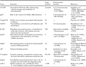
Eleven antigen-encoding genes with different chromosomal location, function and copy number were used for the analysis (Table 1). Our study was focused in the coding region of the markers; except in kmp11 and 1f8 where we targeted the whole sequence (coding and non-coding), and in hsp70 where we studied coding and the non-coding region separately. Twelve loci were PCR amplified and digested with restriction enzymes as described previously (Rozas et al. Reference Rozas, De Doncker, Adaui, Coronado, Barnabé, Tibyarenc, Solari and Dujardin2007). The selected enzymes (given in parenthesis) for each gene were: cruzipain (HpaII), Tcgp63-II (TaiI), 1f8 (Alw21I), kmp11 (HpaII), hsp60 (Hae III), hsp70 intragenic (Cac8I), hsp70 intergenic (Csp6I), SAPA (HpaII), gp72 (TaqI), sa85-1 (BsaJI), Tc-CRT (HaeIII) and Fl-160 (HpaII).
Antigen genes studied to assess genetic polymorphism

Phenetic analysis
After PCR-RFLP analysis, a character matrix was created. The presence of a band was given a score of 1, and the absence was scored 0, no matter if the band was strong or weak. The matrix obtained was analysed using the PHYLIP package, version 3.6 (Felsenstein, University of Washington, 2002; available at http://evolution.genetics.washington.edu/phylip.html); RESTDIST (restriction fragments distance, modification of Nei and Li restriction fragments distance method (Nei and Li, Reference Nei and Li1979); UPGMA (unweighted pair group method using arithmetic averaging); and CONSENSE (majority rule consensus). The robustness of the branching nodes was evaluated by SEQBOOT (bootstrap analysis with 1000 replications). Dendograms were done using the program DRAWGRAM (tree plotting). Three types of dendrograms were built-up: (i) using 7 reference strains and each of the 12 amplicons cleaved with all restriction enzymes (further called 7-strains gene-specific analysis), (ii) using 7 reference strains and a combination of results obtained with all amplicons and all restriction enzymes (further called 7-strains combined analysis) and (iii) using 21 strains, all amplicons but 1 restriction enzyme only – the most discriminant – (further called the 21-strains combined analysis).
RESULTS
Individual phenetic analysis
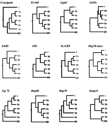
The different restriction patterns obtained for each antigen gene were submitted to individual phenetic analyses (7-strains gene-specific analysis). Comparative analysis of the 12 phenograms revealed the following results (Fig. 1). (1) T. c. marinkellei branched outside the T. cruzi group in all trees, except that based on cruzipain and Kmp11 characters. (2) T. cruzi I branched separately from lineage II in 4 trees, based respectively on Fl-160, sa85, Tc-CRT and hsp60 genes. (3) DTUs IId and IIe clustered together in 9 trees: (i) in 3 cases, together with IIb (fI-160, Tcgp63-II and SAPA), (ii) in 2 cases, together with IIc (SA85 and Hsp60), (iii) in 2 cases, together with IIb and IIc (hsp70 inter and gp72), (iv) once together with IIa and IIb (cruzipain) and (v) once separately from other DTUs of lineage II (Tc-CRT). (4) DTUs IIa and IIc clustered together in 5 phenograms: (i) once closer to DTU I (Tcgp63-II), (ii) once closer to IIb (Tc-CRT) and (iii) 3 times in variable positions (FI-160, 1F8 and SAPA).
Genetic polymorphism in the target antigen-encoding genes for 7 reference strains of Trypanosoma cruzi (7-strains gene-specific analysis). The dendograms shown were constructed from PCR-RFLP data using the UPGMA algorithm.

Combined phenetic analysis
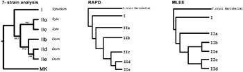
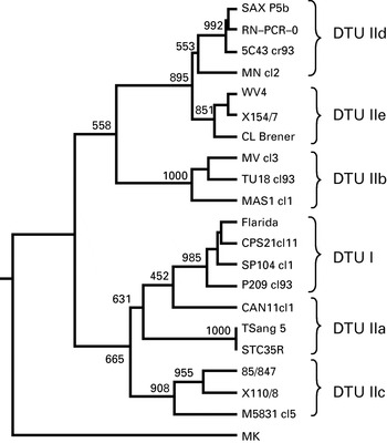
In the next phase, data obtained from the 12 different markers were combined for a global phenetic analysis. This was firstly done for the 7 reference strains, with all restriction enzymes used in this study (7-strains combined analysis) and secondly for 21 strains, with 1 restriction enzyme only by marker (21-strains combined analysis), selected on the basis of its highest discriminatory power. The combined 7-strains analysis (Fig. 2) showed that T. cruzi marinkellei branched outside the T. cruzi cluster. Within, T. cruzi, the 2 lineages I and II were clearly distinguished. Within lineage II, DTUs IIa and IIc clustered together, separately from the IId-e cluster; IIb branched closer to IId-e. The whole structure of the tree was supported by bootstrap values ranging between 88·2 and 100. The combined 21-strains analysis (Fig. 3) revealed 2 pieces of information. First, strains from the same DTU clustered together, with 1 exception, CANIIIcl1 (DTU IIa), which branched between I and IIa, but closer to I, but with a very low bootstrap value (close to 46%). Second, the general structure of the phenogram was similar to that resulting from the combined 7-strains analysis, with the expection that DTU I did not branch separately from lineage II, but clustered together with DTUs IIa and IIc.
Genetic polymorphism using UPGMA for the 7-strain analysis of Trypanosoma cruzi combining PCR-RFLP data for 11 markers and several restriction enzymes (7-strains combined analysis). The dendogram shows the relationships among the different T. cruzi DTUs. For a comparative display we included previously obtained results using MLEE and RAPDS. The bootstrap values (given as percentage from 1000 replicates) are indicated in the branches.

Genetic polymorphism in Trypanosoma cruzi after PCR-RFLP analysis using 11 antigen-encoding genes on 21 reference strains and 1 restriction enzyme per gene (21-strains combined analysis). Bootstrap values (% from 1000 replicates) shown above the dendogram branches were performed with UPGMA.

DISCUSSION
Here the value of antigen gene-unique interfaces in the host-parasite relationships has been explored for the understanding of the evolution within T. cruzi. We selected 11 genes and 12 loci associated that have different functions and are considered to be involved in host-parasite interaction (cell adhesion, infection, molecular mimicry). The polymorphism of the respective genes in a representative sample of the diversity of T. cruzi was screened by PCR-RFLP and evolutionary relationships were inferred by phenetic analysis. Our results supported the current classification (Brisse et al. Reference Brisse, Barnabe and Tibayrenc2000b) of T. cruzi into 6 DTUs. Indeed, on the 21-strains phenogram and despite the fact that only 1 restriction enzyme was used for each antigen gene, all strains clustered together according to their DTU, except for CANIIIcl1 (IIa) which was closer to DTU I than to IIa. This result suggests that the evolution of antigen genes, despite being under immune selection pressure, reflects to a certain extent the same evolutionary history of T. cruzi as the housekeeping genes considered, for instance in MLEE studies. It has also practical implications as it indicates that the markers supporting the different clusters can be used as a DTU-diagnostic. This was highlighted in a previous study, and an algorithm for the unambiguous identification of the 6 DTUs by successive PCR-RFLP assays was proposed (Rozas et al. Reference Rozas, De Doncker, Adaui, Coronado, Barnabé, Tibyarenc, Solari and Dujardin2007), hereby providing a typing alternative for laboratories not equipped for MLEE or sequencing.
In the study of 7-strains individual markers the hybrids DTU IIa/IIc were more related to DTU I or DTU II using different markers, this observation is in agreement with previous studies, where the zymodeme III is a hybridization event result between T. cruzi I and T. cruzi II (Sturm et al. Reference Sturm, Vargas, Westenberger, Zingales and Campbell2003; Westenberger et al. Reference Westenberger, Barnabe, Campbell and Sturm2005); and the hybrids DTUs IId and IIe fell in the same cluster indicating a relationship with DTU IIb or DTU IIc. This result agrees with the hypothesis that DTU IIb and IIc may be the hybrid IId/IIe parentals (Machado and Ayala, Reference Machado and Ayala2001; Gaunt et al. Reference Gaunt, Yeo, Frame, Stothard, Carrasco, Taylor, Mena, Veazey, Miles, Acosta, de Arias and Miles2003; Brisse et al. Reference Brisse, Henriksson, Barnabe, Douzery, Berkvens, Serrano, De Carvalho, Buck, Dujardin and Tibayrenc2003; Elias et al. Reference Elias, Vargas, Tomazi, Pedroso, Zingales, Schenkman and Briones2005; Westenberger et al. Reference Westenberger, Barnabe, Campbell and Sturm2005). Recently, using 5S rRNA analysis it was confirmed that the hybrids IId/IIe are heterozygous and have sequences from DTU IIb and IIc (Westenberger et al. Reference Westenberger, Sturm and Campbell2006).
The combined 7-strains analysis (that integrates more genetic characters for each marker) permitted a new perception of the relationships between the DTUs. Comparison with phenograms built-up from MLEE and RAPD (Brisse et al. Reference Brisse, Dujardin and Tibayrenc2000a, Reference Brisse, Barnabe and Tibayrencb, Reference Brisse, Henriksson, Barnabe, Douzery, Berkvens, Serrano, De Carvalho, Buck, Dujardin and Tibayrenc2003) data revealed similarities and differences. With the three methods, T. c. marinkellei branched outside the T. cruzi cluster, and the two lineages I and II were clearly differentiated. Within lineage II, the only similarity concerned the clustering of DTUs IId and IIe, which was shown by PCR-RFLP and RAPD. On the PCR-RFLP tree, DTUs IIa and IIc clustered together, and IIb was closer to the IId/e cluster. Interestingly, this structure was the one that fitted better with epidemiological features of the different DTUs: (i) lineage I, being encountered across the entire Chagas endemic area in sylvatic as well as domestic transmission cycles, (ii) IIa/c being associated with a sylvatic transmission cycle and (iii) IIb/d/e being associated with a domestic transmission cycle and only collected South of the Amazon basin (Brisse et al. Reference Brisse, Dujardin and Tibayrenc2000b).
The 7-strains individual trees antigen genes that better supported the discrimination of sylvatic and domestic DTUs in lineage II were fl-160 and Tcgp63-II. The former antigen harbours a series of CD8+ T cells that may have a pathogenic or protective role in chronic Chagas disease (Fonseca et al. Reference Fonseca, Moins-Teisserenc, Clave, Ianni, Nunes, Mady, Iwai, Sette, Sidney, Marin, Goldberg, Guilherme, Charron, Toubert, Kalil and Cunha-Neto2005), while the latter is a surface antigen with metalloprotease activity and a possible role in host cell infection (Cuevas et al. Reference Cuevas, Cazzulo and Sanchez2003). Considering that these 2 genes encode important players in the host-parasite interactions, our findings could open two ways of further exploration. On one hand, it would be worthwhile to check if selection of specific variants of these antigens (or others) by the hosts encountered in the sylvatic and domestic transmission cycles could explain the obtained phenetic structure. On the other hand, it would be relevant to revisit the clinical pleomorphism of Chagas disease in humans in the light of antigen gene polymorphism. T. cruzi presents a predominantly clonal evolution (Tibayrenc et al. Reference Tibayrenc, Ward, Moya and Ayala1986), but its evolutionary history is hypothesized to have been shaped by a series of recombination events (Westenberger et al. Reference Westenberger, Barnabe, Campbell and Sturm2005, Reference Westenberger, Sturm and Campbell2006). Two observations made in the present study support this. First, we observed differences in the topology of the 7-strains individual trees according to the targeted antigen gene. This figure is classically observed in populations undergoing recombination events, where the genealogical relationships between sequences vary with position (Nordborg et al. Reference Nordborg2000). Alternatively, this result could reflect the existence of a different selection pressure on the different antigen genes here selected, an explanation proposed for similar observations in Leishmania (Garcia et al. Reference Garcia, Kindt, Quispe-Tintaya, Bermudez, Llanos, Arevalo, Banuls, De Doncker, Le Ray and Dujardin2005). This could theoretically be verified by measuring the ratio of synonymous to non-synonymous changes in the corresponding sequences. Secondly, the clustering in the 7-strains combined tree of (i) IIa with IIc and (ii) IId with IIe indicates a shared history of the respective DTUs within both clusters. As mentioned previously, it was hypothesized that IIa/c would result from a fusion between I and IIb and that IId/e would result from a hybridization between IIb and IIc (Sturm et al. Reference Sturm, Vargas, Westenberger, Zingales and Campbell2003; Westenberger et al. Reference Westenberger, Barnabe, Campbell and Sturm2005). The evolutionary link with the putative parents of these DTUs is still visible in the 7-strains individual trees: (i) IIa/c were associated with I in 1 tree and with IIb in another one, while (ii) IId/e were associated with IIb in 3 trees and with IIc in 2 trees. Furthermore, the number of parental characters still associated with the hybrid DTUs (1+1 for IIa/c and 3+2 for IId/e) fits with the postulated more ancient character of the fusion event leading to IIa/c.
In conclusion, our results show that antigen genes can indeed provide relevant and original information for the understanding of the evolutionary history of T. cruzi. The PCR-RFLP approach is ideal for a first screening, and has the advantage over MLEE, for instance, that it can be applied on a small number of parasites. However, it should be completed by a more extensive study of the respective genes, as we addressed a fraction only of the genetic sequence, by the use of restriction enzymes. Whole sequencing might be the second step, even if it would be complex for interpretation, especially for highly repeated genes. These studies should be coupled with clinical studies, as antigen genes might give clues for the understanding of the clinical pleomorphism characterizing T. cruzi.
Support for this work was obtained from the programme of Bilateral Scientific Collaboration Flanders – Chile 2004–2005.