Introduction
Compression of the spinal cord due to metastases occurs in 5–10% of cancer patients during the course of their disease.Reference Rades, Stalpers and Veninga1 Radiotherapy is a key treatment for such compression, as well as for painful bone metastases.
Primary tumours of the lung, breast and prostate commonly lead to bone metastases with spinal cord compression. The most frequent area is at the dorsal–lumbar level.
Anterior–posterior fields are usually generated to treat the patient. The advantage of this technique is that planning and treatment can be rapid.
Improving planning in three-dimensional (3D) conformal radiotherapy using imaging computed tomography (CT) would be helpful because evaluation of dose distribution in each part of the patient is possible. Field parameters are selected to reduce the dose reaching the organ-at-risk using these images.
Field, wedge, gantry angle and weight were selected for each patient to obtain optimal uniformity for the dose within the treatment volume. Reducing the dose for the rest of the volume was also possible, in contrast with conventional planning.
CT was carried out within a few minutes when the physician decides that treatment was indicated. Imaging, contouring, planning, data input into the accelerator and treatment could be completed in 2 hours if the case is urgent. This technique therefore does not involve treatment delay.
The aim of this study was to compare conventional dosimetry (posterior–anterior fields) with an optimized 3D conformal plan (using oblique fields) to treat the vertebral column.
MATERIAL AND METHODS
CT Somaton Emotion 6 (Siemens, Erlangen, Germany) was used for imaging. The table of the system was identical to the accelerator, producing good accuracy in each fraction. Patients were treated in the supine position with an appropriate headrest; a knee immobilization system was employed for comfort. The patient's arms were placed outside the irradiation area. To treat the cervical spine, arms were placed at the side of the body; they were placed over the head to treat the dorso–lumbar spine. We used a sagital plane defined by the nose, chin and hip to align the patient. Reference points were visualized using radio-opaque marks. A CT of 5-mm slice thickness was done and registered in the treatment planning system.
Treatment plans were generated for a Varian Clinac 600C (Varian Inc., San Jose, CA) with multileaves (width, 1 cm) collimator. The fields were verified with a CR Orex ACL 1417 (Orex Computed Radiography Ltd, Yokneam, Israel) computed radiography system. A Pinnacle3 version 7.4f (Philips/ADAC, Milpitas, CA) was used for calculation of treatment planning with a convolution-superposition algorithm. The calculation grid was 4 mm.
Fifteen patients were selected, and split into three groups of five based on tumour localization: cervical, dorsal and lumbar. The planning target volume (PTV) was generated with a 3D margin of 1 cm from the clinical target volume (CTV). The patient received 8 Gy in one fraction; retreatment with an additional fraction was feasible, safe and effective if persistent or recurrent bone pain was present. Five fractions of 4 Gy were used for cervical cases.Reference Manzano, Bellavita and Rossi2,Reference Agarwal, Baum and Hoefnagel3
Depending on tumour position, the oesophagus, lung, heart, trachea, intestine and kidneys were delineated as organs-at-risk. We evaluated a set of variables in each organ: mean dose, volume of organ that received a dose >70% (V70) and >90% (V90) of the prescription dose.
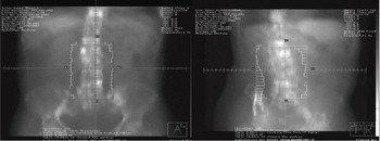
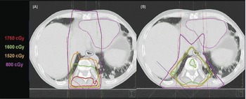
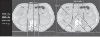
The reference plan (“conventional plan”) consisted of two fields in the anterior–posterior configuration (Figures 1a, 2a and 3a). The PTV was covered with 90% of the prescription dose, minimizing the anterior field weight with 120% of overdose as a limit. Mean weight of the anterior field was (26 ± 6)%. Statistical uncertainties were shown at the 2σ level. The treatment plan (“optimized plan”) consisted of two oblique wedged posterior fields and an anterior field (Figures 1b, 2b and 3b). Inverse planning was not necessary to optimize field weights. The PTV was covered with 95% of the prescription dose, maximizing the oblique field weight, with 107% of overdose as a limit. Mean weight of the anterior field was (23 ± 6)%.
(a) Anterior field. (b) Oblique posterior field.

(a) Conventional planning (dorsal area). (b) Optimized planning (dorsal area).

(a) Conventional planning (lumbar area). (b) Optimized planning (lumbar area).

In some cases of cervical treatment, we chose two opposite fields to keep the dose in the oral cavity as low as possible, or in four fields in an X configuration to avoid irradiating the shoulders.Reference Agarwal, Baum and Hoefnagel3,Reference Khan4
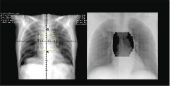
Repeatability of patient positioning treatment was checked with two perpendicular images using the digital Orex CR system (Figure 4).
(a) Digital reconstruction radiograph. (b) Portal image verification.

RESULTS
With regard to the conventional plan, the average volume with a dose >110% was (18 ± 8)%. The PTV in the optimized plan never has exceeded 107% (Figures 2 and 3).
Wide-field radiotherapy is inevitably associated with greater toxicity. The field size average is shown in Table 1, specifying for each location. The average number of vertebras irradiated is 5 ± 3.
Average field size

Statistical uncertainties are shown at the 2σ level.
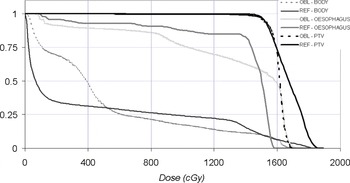
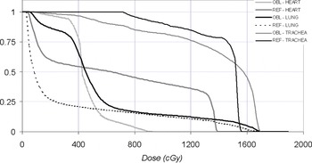
In the dorsal localization, we evaluated a set of dose parameters to the lung. Dose increase in the optimized plan was irrelevant because the volume of irradiated lung was too low. Differences in the irradiation of the oesophagus and trachea between both types of plans were not observed (Figures 5 and 6). Identical results were observed in the cervical localization.
Dose–volume – histogram comparing both plans for the PTV, body and oesophagus.

Dose–Volume – Histogram comparing both plans for the heart, lung and trachea.

The advantage of the optimized plan was better conformity to the PTV, producing a reduction of body volume at high dose (Figures 2 and 3). In the dorsal localization, mean of V70 was (1,300 ± 1,600) cm3 with the optimized plan, better than the (1,800 ± 2,600) cm3 obtained in the reference plan. We also improved V90 from (900 ± 1,200) cm3 to (1,200 ± 2,000) cm3.
In the lumbar localization, we studied dose distribution in the kidneys and intestine, obtaining a higher mean dose and irradiated volume in the optimized plan (although these values were not relevant when taking into account total volume of the kidneys). The volume of irradiated intestine with a high dose was reduced with the optimized plan. In the dorsal localization, the optimized plan achieved better conformity of the high dose to the PTV, reducing the dose to the tissue irradiated in the dorsal area.
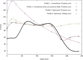
Radiation techniques and energies in relation to depth dose distribution are shown in Figure 7. The curves represent two anterior–posterior fields with 6-MV photons (profile 1), a single posterior filed with 6-MV photons (profile 2) and two wedged oblique and anterior fields (profile 3). If the depth dose distribution of this configuration to treat a vertebral body to an extreme depth of 11 cm is evaluated, a single-field technique is not recommended to avoid a maximal dose in the spinal cord of >115%. Optimized planning profile 3 reduced the dose to 60% in the anterior part of the body. To evaluate the dose contribution of each oblique field, we represented the depth dose distribution in the axis of this field in optimized planning profile 4. Direction of these profiles is clarified in Figure 3. The oblique fields irradiated sane tissue too but, outside the PTV, the slope of the depth dose was very sharp, decreasing to a low ose.
Depth-dose distribution of various radiation techniques and energies.

Tolerance to treatment was good in all cases. There is no severe toxicity grade >1 (CTCA 3.0) and myelitis was not seen. Pain was controlled in 67% of patients, and motor function in 80%. Eighty-seven percent of patients who could ambulate before treatment retained this capacity.
CONCLUSION
The optimized plan followed ICRU criteria, reaching PTV coverage of 95–107% of the prescription dose. In the conventional plan, high dose had to be accepted to get 90% of the prescription dose in the PTV.
The dose–volume histogram of the organs-at-risk has no relevant clinical differences. In the lumbar localization, a minor intestine-irradiated volume with high dose was obtained. The optimized plan did not lead to patient suffering because it is a simple technique and not time-consuming. We adopted this configuration field as part of the treatment protocol in our department.
In contrast with the anterior–posterior configuration, oblique fields improve dose conformity and limit high dose to all of the body except the spinal cord. Radiation Oncologists may prefer this technique because it has lower toxicity than the conventional approach.