Introduction
Restricting contact with helminths can disrupt the microbiome and imbalance immune reactivity (Leung et al., Reference Leung, Graham and Knowles2018). Consequently, controlled infection with helminths could provide effective therapy against allergies and autoimmune diseases (Flohr et al., Reference Flohr, Quinnell and Britton2009; Smallwood et al., Reference Smallwood, Giacomin, Loukas, Mulvenna, Clark and Miles2017). Ulcerative colitis is a progressive inflammation of the colon, with the highest prevalence in Europe and North America and increasing number of cases in newly industrialized countries (Molodecky et al., Reference Molodecky, Soon, Rabi, Ghali, Ferris, Chernoff, Benchimol, Panaccione, Ghosh, Barkema and Kaplan2012; Ng et al., Reference Ng, Shi, Hamidi, Underwood, Tang, Benchimol, Panaccione, Ghosh, Wu, Chan, Sung and Kaplan2018). The common symptoms of ulcerative colitis include progressive loosening of bloody stools, rectal bleeding and diarrhoea. Studies in rodents as well as human clinical trials have indicated inhibition of colitis by parasite infection (Elliott and Weinstock, Reference Elliott and Weinstock2012; Heylen et al., Reference Heylen, Ruyssers, Gielis, Vanhomwegen, Pelckmans, Moreels, De Man and De Winter2014; Maruszewska-Cheruiyot et al., Reference Maruszewska-Cheruiyot, Donskow-Łysoniewska and Doligalska2018). Induction of colon inflammation with dextran sulphate sodium (DSS) in mice produces a model of human ulcerative colitis (Okayasu et al., Reference Okayasu, Hatakeyama, Yamada, Ohkusa, Inagaki and Nakaya1990). Our studies showed that in BALB/c mice with DSS-induced colitis infected with Heligmosomoides polygyrus, more nematodes matured to adults, the nematodes were larger and their sex ratio was altered (Donskow-Łysoniewska et al., Reference Donskow-Łysoniewska, Bien, Brodaczewska, Krawczak and Doligalska2013). The different developmental stages of H. polygyrus have distinct proteomic profiles (Hewitson et al., Reference Hewitson, Ivens, Harcus, Filbey, McSorley, Murray, Bridgett, Ashford, Dowle and Maizels2013), which influence the host immune response (Morgan et al., Reference Morgan, LaCourse, Rushbrook, Greetham, Hamilton, Barrett, Bailey and Brophy2006). Immunoglobulin G (IgG1) recognized fewer molecules in fourth-stage larvae (L4) from mice with colitis compared to control mice (Donskow-Łysoniewska et al., Reference Donskow-Łysoniewska, Bien, Brodaczewska, Krawczak and Doligalska2013). The greatest changes in specific antibodies in mice serum were observed 6 days after infection when H. polygyrus developed into L4 stage (Donskow-Łysoniewska et al., Reference Donskow-Łysoniewska, Bien, Brodaczewska, Krawczak and Doligalska2013). Also, male nematodes had higher viability in mice with colitis which is inconsistent with previous observations that molecules from male H. polygyrus are more immunogenic than molecules from female H. polygyrus (Adams et al., Reference Adams, East, Monroy, Washington and Dobson1987). Possibly male and female nematodes in mice with colitis produce different molecules from those developed in control mice. To evaluate the humoral response in mice with colitis against L4 of H. polygyrus; we identified immunogenic proteins of male and female parasites separately using 2D electrophoresis and determined the level of specific antibodies in mucosa and serum by enzyme-linked immunosorbent assay (ELISA).
Materials and methods
Nematode preparation
The experiments were conducted on BALB/c mice. Eight-week old pathogen-free males were allowed to adjust to the laboratory conditions for 7 days before the start of the experiment at the animal-house in the Faculty of Biology. Animals were then placed in groups of five in cages with room temperature maintained at 24–25 °C, 50% humidity under a 14/10 h light and darkness cycle, and allowed ad libitum access to water and commercial pellet food. For the induction of acute colitis, mice received 3% DSS, 35–50 kDa (TdB Consultancy AB, Uppsala, Sweden) in drinking water for 3 days before infection with 300 infective larvae (L3) H. polygyrus. Infected mice without induced colitis were used as control. There were no significant differences in daily consumption of water between mice given DSS and controls. The mice were sacrificed 6 days after infection, when the parasite was in L4. The small intestine of mice was removed, ligated at both ends with cotton twine, to prevent contamination of the medium with digested matter, and then incubated for 2 h at 37 °C in Petri dishes containing RPMI-1640 medium with L-glutamine (2 mm), penicillin (100 U mL−1) and streptomycin (100 μg mL−1) (Gibco, Inchinnan, Scotland, UK). The larvae were harvested and the sex of L4 stage determined by locating the bursa at the caudal end of pre-adult male larvae.
Somatic antigen larvae L4 preparation
Five hundred worms of male or female L4 from control mice and DSS-treated mice were homogenized in a metal hand-held homogenizer in lysis buffer (8 m urea, 40 mm Tris base, 4% CHAPS, Sigma) supplemented with a cocktail of protease inhibitors (Sigma), followed by centrifugation at 13 000 g for 5 min. The supernatant was collected and stored at −80 °C until experimental use. The final protein concentration of L4 homogenate was measured by RC DC™ Protein Assay (Bio-Rad).
Gel electrophoresis
The supernatant from L4 extracts was purified using a Ready-Prep 2-D Clean up Kit (Bio-Rad). Isoelectric focusing (IEF) was performed using immobilized pH gradient (IPG) strips and a Protean IEF Cell. L4 protein in rehydration buffer was loaded onto 17 cm pH 3–10 IPG strips overnight, followed by 3500 V for 36 h at 20 °C and a maximum current setting of 50 μA per strip. Focused strips were reduced and alkylated by 10 min incubation in equilibration buffer (6 m urea, 0.375 m Tris HCl, 2% sodium dodecyl sulphate and bromophenol blue) with 2% 1,4-dithiothreitol (Promega), and then with freshly added 2% iodoacetamide (Sigma). Equilibrated proteins were then separated in the second dimension on sodium dodecyl sulphate-polyacrylamide gel electrophoresis (SDS-PAGE) in a Large Format 1-D Electrophoresis Chamber (Bio-Rad) at 50 V for 10 min, followed by 90 V for 10 min and 120 V for 10 h. Gels were visualized using silver staining (Pierce Silver Staining Kit, Thermo Scientific) or using western blotting. Images were taken by Syngene G:BOX (Synoptics, Cambridge, UK). A minimum of three gels were run for every variation of L4 extracts.
Immune detection
Serum was prepared from blood samples taken after cardiac puncture. Proteins from 2D gels were transferred onto polyvinylidene difluoride (PVDF) membranes (Merck) in 4 °C transfer buffer [25 mm Tris, 192 mm glycine, 20% (v/v) methanol pH 8.3] at 100 V for 30 min using semi-dry blotting apparatus (Bio-Rad). The membranes were blocked overnight in 5% skimmed milk in Tris-buffered saline/0.1% Tween 20 (TBS-T) at 4 °C then exposed to sera (1:100) from H. polygyrus-infected mice with induced colitis or mice without colitis followed by goat anti-mouse IgG1 or goat anti-mouse IgA conjugated to horseradish peroxidase (HRP) (Santa Cruz Biotechnology, 1:20 000). To confirm the reproducibility of immune recognition, analysis was repeated at least twice. Samples without serum were used as negative controls. The 2-DE blots were visualized by enhanced chemiluminescence (SuperSignal West Pico Chemiluminescent Substrate, Pierce, Thermo Scientific).
Mass spectrometry and bio-informatics
Spots of interest that were recognized by IgG1 or IgA were excised from the 2D gels using sterile disposable scalpel blades then subjected to trypsin digestion. Samples were analysed at the Mass Spectrometry Laboratory, Institute of Biochemistry and Biophysics, Polish Academy of Science. Gel pieces were washed three times in 100 mL of 50 mm ammonium bicarbonate, 50% (v/v) methanol and then twice in 100 mL of 75% (v/v) acetonitrile, before drying.
Gel pieces were rehydrated with trypsin solution (20 mg trypsin mL−1, 20 mm ammonium bicarbonate), and incubated for 4 h at 37 °C. Peptides were extracted from the gel pieces by washing twice in 100 μL of 50% (v/v) acetonitrile/0.1% (v/v) trifluoroacetic acid, before being transferred into solution to a fresh 96-well plate and dried before mass spectrometry analysis. All peptide samples were separated on an LC system (Famos/Switchos/Ultimate, LC Packings) using water that contained 0.1% TFA as the mobile phase and then transferred to a nano-HPLC RP-18 column (nanoACQUITY UPLC BEHC18; Waters Associates, Milford, MA, USA) using an acetonitrile gradient (0–60% ACN) in the presence of 0.05% formic acid with a flow rate of 150 μL min−1 and analysed by electrospray ionization Orbitrap mass spectrometry. A blank run preceded each analysis. Tandem mass spectral data were analysed by using the MASCOT program (Matrix Science Ltd, v2.1.1, London, UK) against the NCBI/Nematoda and worm-Base databases. For gel spot identifications, a peptide mass tolerance of 0.1 Da was used. Bio-informatic analysis of obtained results was carried out using the Blast2Go program.
Specific antibody in serum and mucosa detection
Mucus and serum IgG1, IgA and IgE levels specific to male or female L4 were measured in individual mice. Maxisorb microtitre plate wells (Nunc, Thermo Fisher Scientific) were coated overnight at 4 °C with 100 μL L4 somatic antigens (5 μg mL−1) in 50 mm carbonate buffer, pH 9.6. The plates were washed and blocked with 5% non-fat milk powder in phosphate-buffered saline pH 7.4 for 1 h at room temperature. After washing, 100 μL of abomasal mucus sample, diluted 1:5 (IgG1 and IgA) or undiluted (IgE) was added and incubated for 2 h at room temperature. Wells were rewashed and 50 μL of goat anti-mouse IgG HRP (Santa Cruz Biotechnology, 1:20 000)/goat anti-mouse IgA (α-chain-specific) – HRP (Sigma, 1:200)/rat anti-mouse IgE (Serotec, Oxford, UK; 1:2000) and samples were incubated for 1 h at room temperature. After the final wash, TMB substrate was added. Reactions were stopped by using 2 m sulphuric acid and the optical density (OD) values were read at 490 nm.
Statistical analyses
The significance of the differences between groups was determined by the one-way analysis of variance (GraphPad Software Inc., La Jolla, USA). When the P value was below 0.05, the multiple-comparison test was used. Data were expressed as mean ± s.e.m. A P value of <0.05 was considered to be statistically significant.
Results
Differences in the production of antibody against male and female H. polygyrus L4 between mice with and without colitis
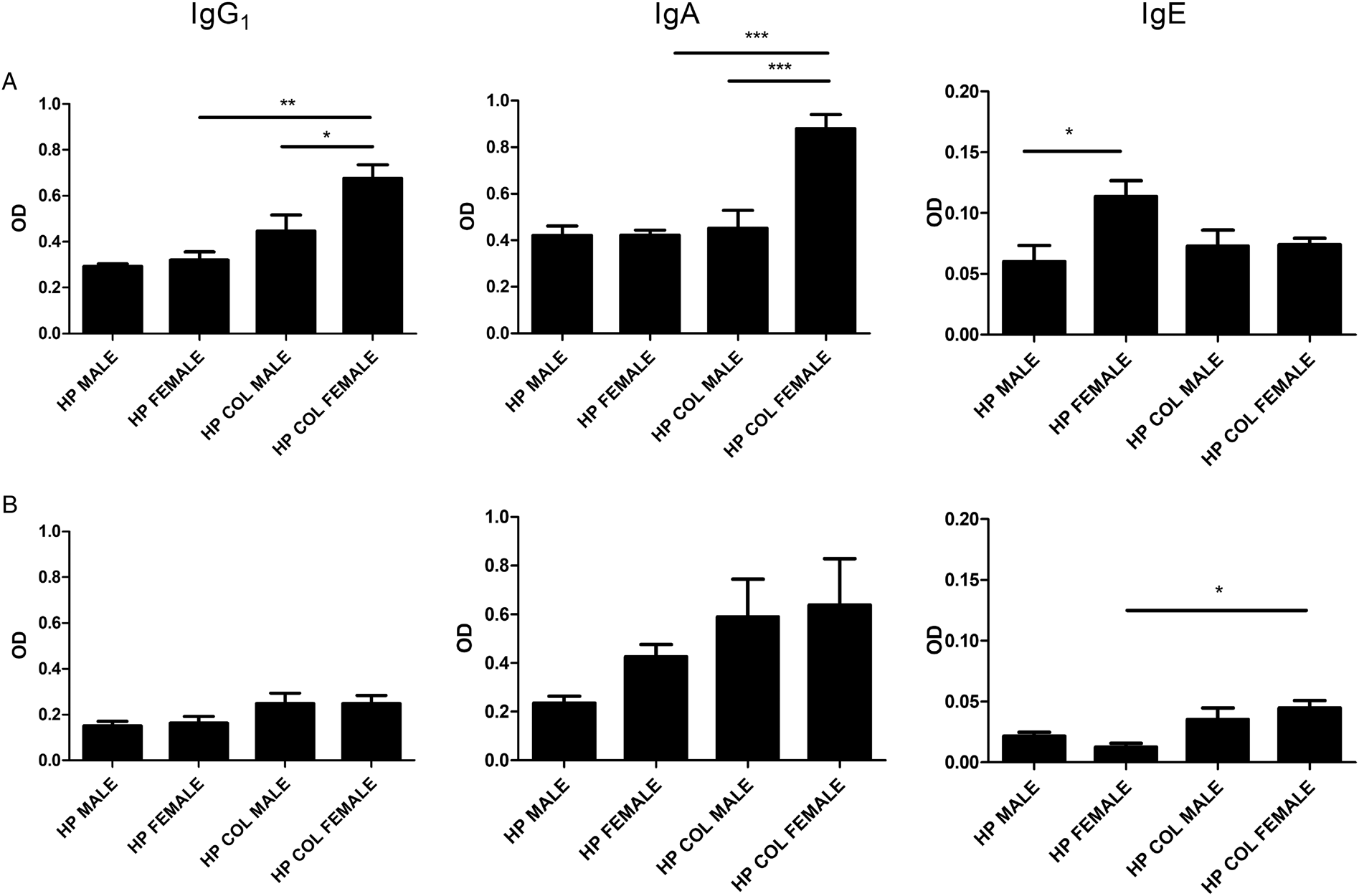
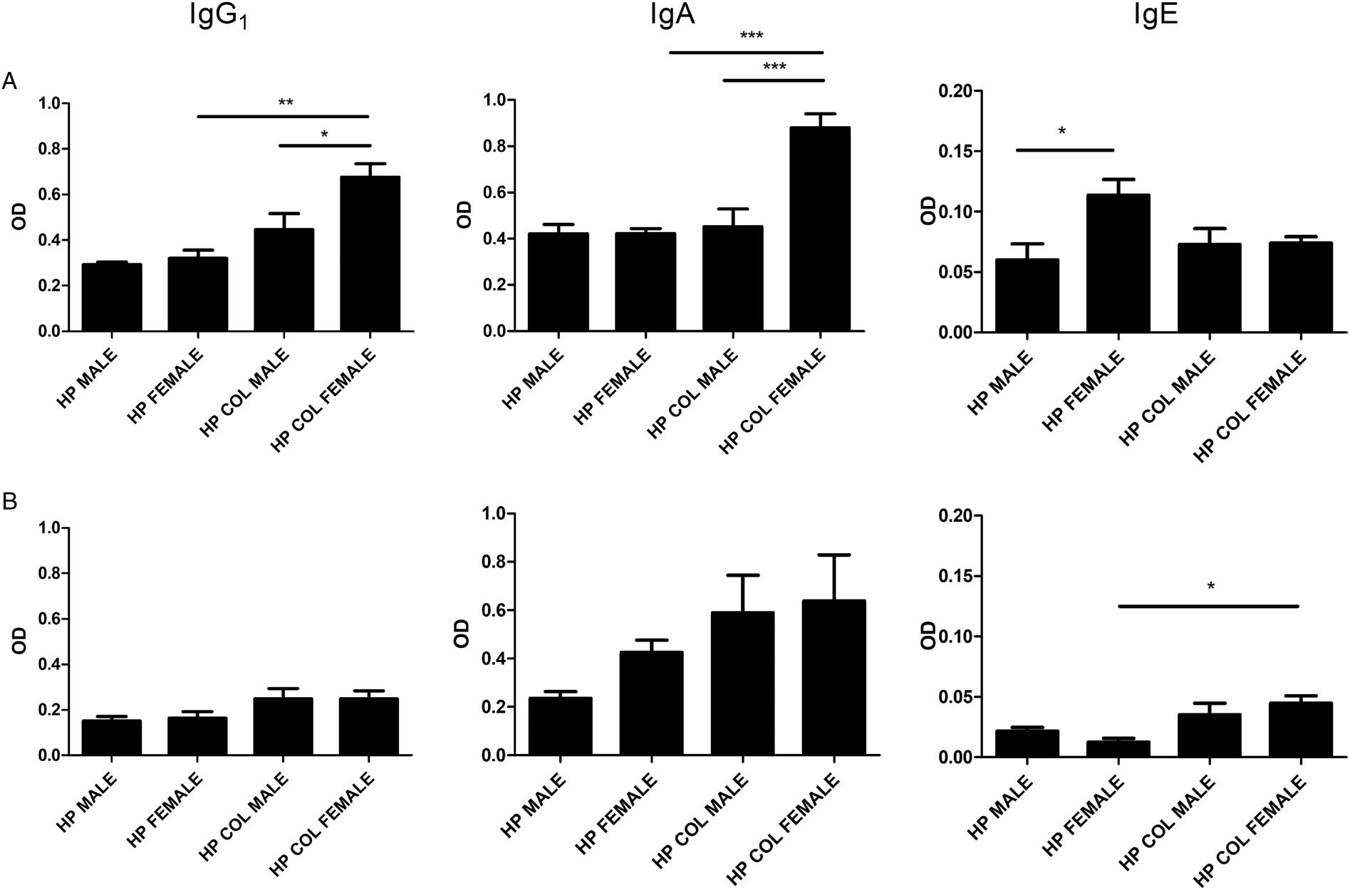
The activity of IgE in serum was higher against somatic antigens from female L4 than male L4 somatic antigens from control mice. There were no differences in the levels of IgG1 or IgA in serum and mucosa as well as mucosal IgE against male or female nematodes from control mice. In mice with colitis, the activity of IgG1 and IgA in serum was higher against female H. polygyrus than against male H. polygyrus. The levels of IgE in serum and IgG1, IgA and IgE in mucosa were similar between male and female L4 stages. Mice with colitis have higher levels of IgG1, IgA in serum and IgE in mucosa against female H. polygyrus compared to control mice (Fig. 1).
Effects of colitis in mice on recognition of male or female L4 H. polygyrus molecules by IgG1, IgA and IgE antibodies in small intestine (A) or in serum (B). Level of antibodies IgG1, IgA and IgE in small intestine and in serum of mice with induced DSS-colitis or without 6 days after infection with H. polygyrus against male or female L4 as measured by ELISA. HP Male, antigen of male L4 H. polygyrus from control mice; HP Female, antigen of female L4 from control mice; HP Col Male, antigen of male L4 from DSS-treated mice; HP Col Female, antigen of female L4 from DSS-treated mice. The results were expressed as the mean absorbance (OD) ± s.e. of five mice. *P < 0.05; **P < 0.005; ***P < 0.0005 compared between groups as highlighted with a line.
Identification of immunogenic proteins of male and female H. polygyrus L4 stage
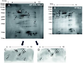
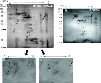
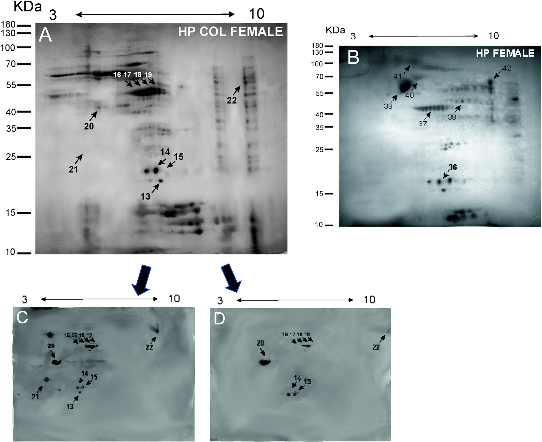
Somatic proteins of male and female L4 from mice with and without colitis were separated by 2D electrophoresis and silver stained. Most of the proteins were concentrated between pH 5 and 7. Their molecular weight varied from 10 to 100 kDa. Around 150 spots for male and female L4 from control mice and 200 spots for male and female L4 from mice with colitis were detected. Results of 2D separation are presented in Figs. 2A, B and 3A, B. Duplicate gels were transferred onto PVDF membranes and proteins were probed with serum from mice with colitis (for somatic antigen of larvae developed during colitis) or with serum from control animals (for material of larvae developed under normal conditions). Most of the proteins were identified as H. polygyrus products although some assignments were based on the closest hits in Nematoda: Teladorsagia circumcintra, Ancylostoma caninum and Dictyocaulus viviparus.
Immunoproteomic analysis of somatic antigens from male H. polygyrus L4 from mice with colitis or controls. 2D silver stained gels of male H. polygyrus from mice with colitis (A) and male L4 from control mice (B). IEF was performed with molecules extracted from L4 with an IPG strip with pH range of 3–10. SDS-PAGE was performed on 12% gel. Western blot analyses of proteins of male H. polygyrus from mice with colitis separated in 2D electrophoresis were transferred to PVDF membrane. The blot was probed with mouse with colitis serum (1:100), followed by HRP-conjugated anti-mouse IgG1 (C) or anti-mouse IgA (D) and visualized by enhanced chemiluminescence. Spots detected by antibodies are indicated by arrows. The numbers refer to the table and western blot results.

Immunoproteomic analysis of somatic antigens from female H. polygyrus L4 from mice with colitis or controls. 2D silver stained gels of female H. polygyrus from mice with colitis (A) and female L4 from control mice (B). IEF was performed with molecules extracted from L4 with an IPG strip with pH range of 3–10. SDS-PAGE was performed on 12% gel. Western blot analyses of proteins of female H. polygyrus from mice with colitis (C, D) separated in 2D electrophoresis were transferred to PVDF membrane. The blot was probed with mouse with colitis serum (1:100), followed by HRP-conjugated anti-mouse IgG1 (C) or anti-mouse IgA (D) and visualized by enhanced chemiluminescence. Spots detected by antibodies are indicated by arrows. The numbers refer to the table and western blot results.
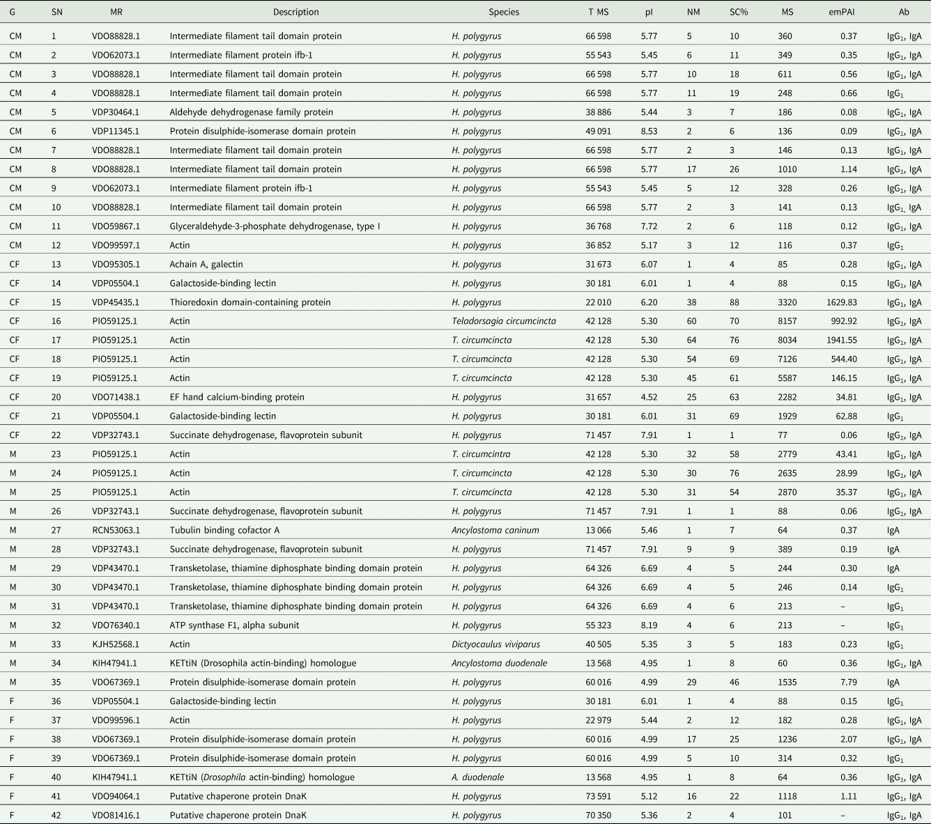
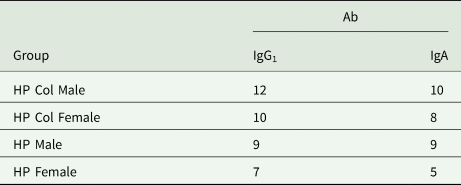
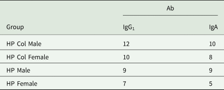
In male L4, serum IgG1 from mice with colitis recognized 12 spots, whereas serum IgA recognized 10 proteins. An intermediate filament protein was the major group of identified spots with most of the spots recognized by both IgG1 and IgA. Aldehyde dehydrogenase family protein, disulphide-isomerase domain protein, glyceraldehyde-3-phosphate dehydrogenase, type I were recognized by both IgG1 and IgA. Actin was recognized only by IgG1. Most of the proteins had stronger signals for IgG1 as spot number 11 (glyceraldehyde-3-phosphate dehydrogenase, type I) for IgA (Fig. 2C and D).
In female L4, serum IgG1 from mice with colitis recognized 10 spots whereas serum IgA recognized eight spots. Most of the recognized proteins were actins (four spots recognized by both IgG1 and IgA) and galectins (three spots; two recognized by both IgG1 and IgA and one by only IgG1). Additionally, thioredoxin domain-containing protein, EF hand and succinate dehydrogenase, flavoprotein subunit were recognized by both IgG1 and IgA serum antibodies (Fig. 3C and D).
Serum IgG1 from control mice recognized nine spots whereas serum IgA also recognized nine spots in male L4; five spots were recognized by both IgG1 and IgA making 13 recognized spots; four of these spots were actin. Three other spots were thiamine diphosphate binding domain protein and one was transketolase. The remaining immunogenic proteins were succinate dehydrogenase, flavoprotein subunit, tubulin binding cofactor A, ATP synthase F1 α subunit, kettin and protein disulphide-isomerase domain protein.
Serum IgG1 from control mice recognized seven spots in female L4 whereas serum IgA recognized five spots. The identified molecules were galectin-binding protein, actin, protein disulphide isomerase domain protein, kettin and putative chaperone protein DNA K (Tables 1 and 2).
Immunogenic protein spots identified by mass spectrometry
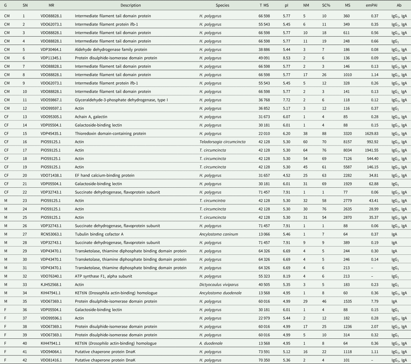
SN, spot number; MR, MASCOT results (NCBI); T MS, theoretical monoisotopic mass (Da); pI, calculated isoelectronic point; MP, number of matched peptides; SC%, sequence coverage range (%); MS, MASCOT score; Ab, antibody class.
CM, HP Col Male (antigen of male L4 from DSS-treated mice); CF, HP Col Female (antigen of female L4 from DSS-treated mice); M, HP Male (antigen of male L4 H. polygyrus from control mice); F, HP Female (antigen of female L4 from control mice).
Number of proteins with the division for recognition by antibody classes
Gene ontology analysis
Gene ontology was used to assign proteins to their biological processes, cellular components and molecular functions. Many of the proteins were assigned to more than one category.
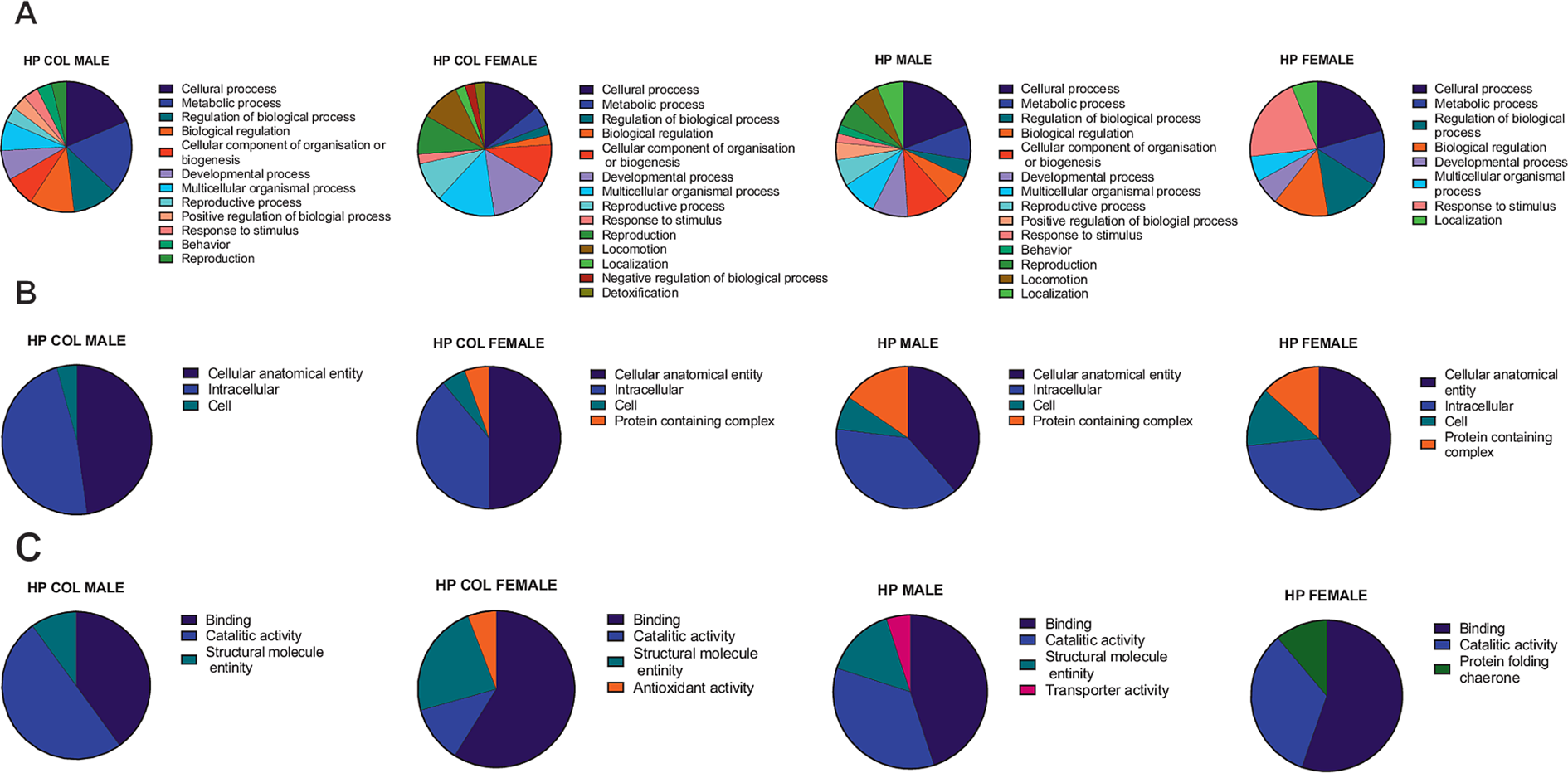
Immunogenic proteins identified in male L4 from mice with colitis were classified mostly to cellular processes (nine) or cellular component organization or biogenesis (five). For female L4 from mice with colitis, three proteins were assigned to cellular processes and three to responses to stimulus (Fig. 4A).
(Colour online) Comparison of gene ontology database analysis results for immunogenic proteins of male and female of H. polygyrus L4 stage isolated from mice with colitis or healthy mice. Identified proteins were analysed with Blast2Go program and based on the assigned biological process (A), cellular component (B) and molecular function (C). HP Male, antigen of male L4 H. polygyrus from control mice; HP Female, antigen of female L4 from control mice; HP Col Male, antigen of male L4 from DSS-treated mice; HP Col Female, antigen of female L4 from DSS-treated mice.
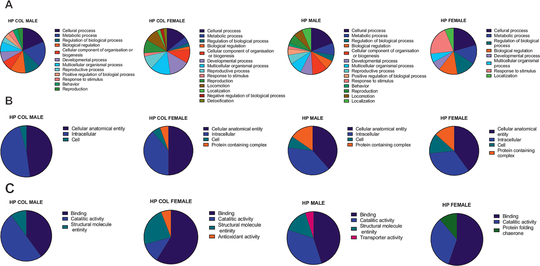
Immunogenic proteins identified in male L4 from control mice took part in 12 biological processes with five cellular and five metabolic processes. Proteins identified in female L4 from control mice were categorized to 14 processes; most of them were assigned to cellular processes (six), developmental processes (six) and multicellular organismal processes (six).
Studied proteins were also divided into three groups based on cellular components: cellular anatomical entity, intracellular, cells and protein containing complex. Most of immunogenic proteins in all groups were classified as cellular anatomical entity and intracellular (Fig. 4B).
All analysed proteins were divided into six various molecular functions. Proteins identified for male L4 from control mice had mainly catalytic activity (five) and binding function (four) similar to male L4 from mice with colitis where five proteins had protein binding function and three had catalytic activity. Proteins from female L4 from control mice were mainly characterized as binding (nine) or catalytic activity. Proteins from female L4 from mice with colitis were mainly assigned to binding function (10) (Fig. 4C).
Discussion
Helminths, including nematodes, may be a source of therapeutic agents in helminth therapy against autoimmune disorders. However, there is still a lack of knowledge on how changes induced by the disease in the host tissue influence live parasites. We have reported adaptation of H. polygyrus in mice with induced colitis (Donskow-Łysoniewska et al., Reference Donskow-Łysoniewska, Bien, Brodaczewska, Krawczak and Doligalska2013). Additionally, L4 H. polygyrus from mice with colitis had different immunomodulatory properties, specifically they switched activity of dendritic cells to a proinflammatory profile, compared to L4 from control mice which had curative properties. Heligmosomoides polygyrus L4 also influenced the dendritic cell profile (manuscript in preparation) and this could be due to changes in the production of excretory–secretory molecules by nematodes from mice with colitis.
In the current study, we examined the antibody response against male and female L4 of H. polygyrus. Using mass spectrometry, we identified proteins of the parasite that generate IgG1 and IgA antibodies. One of the important elements of the anti-helminthic host immune response is antibody production. Main classes of antibodies produced during infection with H. polygyrus are IgG1, IgE and IgA (Cypess et al., Reference Cypess, Lucia, Zidian and Rivera-Ortiz1977; Molinari et al., Reference Molinari, Ebersole and Cypess1978; McCoy et al., Reference McCoy, Stoel, Stettler, Merky, Fink, Senn, Schaer, Massacand, Odermatt, Oettgen, Zinkernagel, Bos, Hengartner, Macpherson and Harris2008). Primary infection is characterized by a high concentration of polyclonal and nonspecific IgG1 and IgE (McCoy et al., Reference McCoy, Stoel, Stettler, Merky, Fink, Senn, Schaer, Massacand, Odermatt, Oettgen, Zinkernagel, Bos, Hengartner, Macpherson and Harris2008). Specific IgG1 and IgA against H. polygyrus are present in mice serum and intestine (Molinari et al., Reference Molinari, Ebersole and Cypess1978). Based on our results, the most significant differences in the level of specific antibodies against L4 are observed in the small intestine. We observed higher levels of specific IgG1 and IgA during colitis. Female L4 antigen from mice with colitis is strongly recognized by specific IgE antibodies. IgG1 and IgA reduce the number of mature nematodes during normal infection with H. polygyrus (McCoy et al., Reference McCoy, Stoel, Stettler, Merky, Fink, Senn, Schaer, Massacand, Odermatt, Oettgen, Zinkernagel, Bos, Hengartner, Macpherson and Harris2008). However, our previous studies showed that inflammation of the colon induced by DSS increased survivability of parasites. This result suggests that changes in intestinal conditions can influence the antibody response, especially against female L4 (Donskow-Łysoniewska et al., Reference Donskow-Łysoniewska, Bien, Brodaczewska, Krawczak and Doligalska2013).
Analysis of Haemonchus contortus showed that most of the immunogenic proteins are specific to male or female worms (Yan et al., Reference Yan, Xu, Liu, Yan, Song and Li2010). We observed more spots of immunogenic actins in male L4 compared to female L4 from control mice. However, female L4 from mice with colitis or control mice both recognized several spots as actin. Actins are common in eukaryotes and form part of the cytoskeleton. They are also important in many cellular processes such as cell motility, cell division, cytokinesis, cell elements movement and cell signalling (Doherty and McMahon, Reference Doherty and McMahon2008). Parasite actins are employed in invasion and migration. Immunogenic actins have been found in somatic extract of other nematode species; H. contortus, Trichinella spiralis and Trichinella britovi (Yan et al., Reference Yan, Xu, Liu, Yan, Song and Li2010; Yang et al., Reference Yang, Pan, Sun, Zhao, Yuan, Sun, Huang and Zhu2015; Grzelak et al., Reference Grzelak, Moskwa and Bień2018).
In female L4 from control mice and mice with colitis, we identified galectins recognized by IgA. Mice with colitis recognized three galectin spots whereas control mice recognized only one. However, expression of those proteins based on silver staining was similar. There was no recognized galectin from male L4, but expression was lower, similar to Brugia malayi where galectins are produced primarily by female worms (Bennuru et al., Reference Bennuru, Semnani, Meng, Ribeiro, Veenstra and Nutman2009). Galectins are an important group of proteins involved in many processes of organisms including a role in both innate and adaptive immune responses (Brinchmann et al., Reference Brinchmann, Patel and Iversen2018). Although the role of galectins in mammalian immunity is well understood, knowledge about nematode-derived galectins is lacking. However, they may immunomodulate host–parasite interactions (Young and Meeusen, Reference Young and Meeusen2002). Galectins were also recognized in T. britovi and in both male and female adult Angiostrongylus costaricensis (Rebello et al., Reference Rebello, Barros, Mota, Carvalho, Perales, Lenzi and Neves-Ferreira2011; Grzelak et al., Reference Grzelak, Moskwa and Bień2018).
Another immunogenic protein was kettin, which was recognized in male and female L4 from control mice but not from mice with colitis. Kettin is essential for sarcomeric assembly of actin filaments in muscle cells in Caenorhabditis elegans (Ono et al., Reference Ono, Qin, Johnsen, Baillie and Ono2020).
Immunogenic succinate dehydrogenase, flavoprotein subunit was observed in male L4 from control mice and female L4 from mice with colitis. Succinate dehydrogenase is involved in oxidative phosphorylation and in the Krebs cycle. The protein is responsible for protection against oxidative stress, respiration and life span in C. elegans (Huang and Lemire, Reference Huang and Lemire2009).
One immunogenic protein identified only in male L4 from mice with colitis was intermediate filament protein. This protein builds the cytoskeleton of most eukaryotic cells. Intermediate filament proteins are found in muscle and epithelial tissues of many nematodes including Ascaris lumbricoides, Toxocara canis, Dirofilaria immitis, Anisakis simplex and T. britovi (Bartnik et al., Reference Bartnik, Osborn and Weber1896; Sato and Kamiya, Reference Sato and Kamiya2000). We examined the ultrastructure of nematodes using microscopy but we did not observe any differences in the surface structure of parasites from infected mice with or without colitis (data not shown).
Female nematodes from mice with colitis may develop faster (Donskow-Łysoniewska et al., Reference Donskow-Łysoniewska, Bien, Brodaczewska, Krawczak and Doligalska2013). We observed that nematodes were larger (data not shown). Larger nematodes could produce more proteins, that could influence development and reproduction processes. Furthermore, previous studies of egg production support this thesis: in control mice, 12 and 15 days after infection of mice with H. polygyrus, there is a rise in the number of eggs compared to mice with colitis (unpublished data).
The analysis of the differences in the antibody response against nematodes in mice with and without colitis could help to identify new parasite-derived compounds which may help the design of vaccines. In an era of increased resistance to anthelminthic drugs, new tools in protection against worms are necessary. In addition, some of the molecules identified in our study may have potential to treat various groups of disorders that result from inappropriately strong inflammatory responses.
Financial support
This work was supported by grants from the National Science Center, Poland no. 2013/09/B/NZ6/00653 and 2017/25/N/NZ6/01523.
Conflict of interest
None.
Ethical standards
The authors assert that all procedures contributing to this work comply with the ethical standards of the Polish Law on Animal Experimentation and Directive 2010/63/UE and approved by the First Warsaw Local Ethics Committee for Animal Experimentation with the approval ID 536/2014 and 570/2018.