DiGeorge syndrome was first described in 1968, as a spectrum of disorders resulting from dysmorphogenesis of the third and fourth pharyngeal pouches during embryonic development. Reference Kirkpatrick and DiGeorge1,Reference Conley, Beckwith and Mancer2 Microdeletion of a specific sequence of chromosome 22q11 is found in majority of cases. Characteristic features include aplasia or hypoplasia of the thymus and parathyroid gland, distinct facial features, and multiple conotruncal cardiac anomalies. CHD and aortic arch anomalies are associated in approximately 75–80% of the patients. Reference McDonald-McGinn, LaRossa and Goldmuntz3,Reference Ryan, Goodship and Wilson4 Unilateral pulmonary artery interruption in absence of intracardiac defects in 22q11 deletion is extremely rare. We present a case of unilateral interruption of the right pulmonary artery with aberrant right subclavian artery in an infant presenting with respiratory distress, who was subsequently diagnosed to have 22q11 deletion.
Case
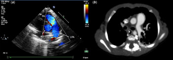
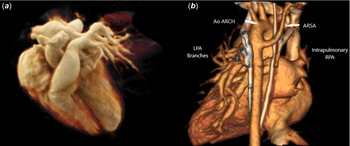
A 3-month-old infant presented to us with symptoms of respiratory distress over the last 1 month. There was no history suggestive of cyanotic spells, feeding difficulties, or stridor. On evaluation, there was respiratory distress with mild subcostal and intercostal recession. Room air saturation was 94% and cardiac examination revealed a soft ejection systolic murmur in left upper sternal border and a loud second heart sound. The rest of the systemic examination was normal. Chest X-ray showed normal bilateral lung fields with no cardiomegaly. Transthoracic echocardiography showed absent right pulmonary artery and the main pulmonary artery continuing solely into the left pulmonary artery (Fig 1a). The aortic arch was left-sided with aberrant origin of the right subclavian artery. Subsequent investigation revealed hypocalcemia (total calcium = 7.2 mg/dl, ionised calcium = 0.8 meq/dl, parathyroid hormone = 16.0 pg/ml, and phosphorus = 9.0 mg/dl) with normal magnesium levels (2.1 mg/dl). As a unit protocol in the presence of these abnormalities, 22q11 deletion genetic testing was done by fluorescence in situ hybridisation and found to be positive. CT cardiopulmonary angiography was done to delineate the vascular anatomy and collateral vasculature and rule out bronchial compression (Fig 1b; Fig 2a and b) and demonstrated the presence of an intrapulmonary right pulmonary artery. The patient also underwent barium swallow which was reported as normal. Subsequently diagnostic cardiac catherisation was undertaken and revealed precapillary moderate pulmonary hypertension with mean pulmonary artery pressures of 30 mmHg and pulmonary vascular resistance of 7.7 Wu/m2. Pulmonary venous wedge angiography was not performed. The baby was discharged home on pulmonary vasodilators with a plan for surgical repair to connect the pulmonary trunk to the hilar right pulmonary artery and dealing with the vascular ring in the future.
(a) Two-dimensional echocardiography in parasternal short-axis plane showing the main pulmonary artery (PA) continuing solely to the left pulmonary artery (LPA). (b) Axial contrast-enhanced arterial phase CT image demonstrating the main pulmonary artery continuing as the left pulmonary artery with absent mediastinal segment of the right pulmonary artery.

(a) Cinematic rendering image demonstrating the main pulmonary artery continuing as the left pulmonary artery with absent mediastinal segment of right pulmonary artery. (b) 3D volume-rendered image (posterior view) demonstrating the aortic arch and retroesophageal aberrant right subclavian artery (ARSA) as well as proximal interruption and intact intrapulmonary right pulmonary artery (RPA).

Discussion
DiGeorge syndrome was first described in 1968 by Dr Angelo DiGeorge as a constellation of disorders and was subsequently associated with the deletion of 22q11. Reference Kirkpatrick and DiGeorge1,Reference Goldmuntz5 Further molecular analysis revealed two other disorders velocardiofacial (or Shprintzen’s) and conotruncal anomaly face syndrome (or Takao) sharing common features with DiGeorge syndrome and all three abnormalities having a common genetic cause, deletion of 22q11. Reference Goldmuntz5 Evidence points towards developmental abnormalities in cephalic and cranial neural crest and its migration as the cause for associated anomalies in 22q11 deletion. The estimated prevalence of DiGeorge syndrome is 1 in 4000 live births with equal distribution between males and females. Reference Goldmuntz5,Reference Maggadottir and Sullivan6 The majority of the patients (nearly 90%) have a de novo mutation, whereas 10% are inherited from an affected parent. Reference Maggadottir and Sullivan6
Congenital heart malformations are common in 22q11 deletion with conotruncal defects being the most common (tetralogy of Fallot with or without pulmonary atresia, truncus arteriosus and interrupted aortic arch, more commonly with an associated right aortic arch). Other anomalies include transposition of great arteries, ventricular septal defect, and valvular pulmonary or aortic stenosis. Embryologically, neural crest cells are vital for the development of the aortic arch and its branches, and hence 22q11 deletion is associated with abnormalities of the aortic arch, its branches, and pulmonary arteries. Isolated aortic arch anomalies have been reported in almost 25% of patients with 22q11 deletion. Reference Hanneman, Newman and Chan7 Aortic arch anomalies can be myriad ranging from right aortic arch, cervical aortic arch, aberrant or isolated subclavian artery, double aortic arch, and interrupted aortic arch. The patient described here had a left aortic arch with aberrant right subclavian artery which is the most common congenital aortic arch anomaly. Reference Hanneman, Newman and Chan7 The aberrant right subclavian originates from the distal descending aorta taking an oblique retroesophageal course. It is generally an isolated finding and is usually asymptomatic. However, in nearly 10% of cases, extrinsic compression of the oesophagus may lead to dysphagia. Although in our case there was no history of suggestive of dysphagia and no features of compression were noted either on CT or barium study, it is possible that this contributed to the respiratory distress in our patient.
Pulmonary artery anomalies including discontinuous, hypoplastic, and isolated pulmonary artery have been well described in 22q11 deletion with associated conotruncal defects. However, pulmonary artery abnormalities in the absence of structural intracardiac lesions with 22q11 deletion are extremely rare. A recent study showed that patients with 22q11 deletion without cardiac defects had a smaller left pulmonary artery compared with healthy subjects. Reference Mastromoro, Calcagni and Versacci8 Proximal interruption of the left pulmonary artery in the absence of conotruncal abnormalities with 22q11 deletion has only been described in one case report in an adult patient. Reference Maldjian and Sanders9 To the best of our knowledge, this is the first case of an absent right pulmonary artery without CHD in the presence of 22q11 deletion being reported in an infant. Unilateral absence of a pulmonary artery is an extremely rare congenital malformation. Instead of absence of the pulmonary artery, “interruption of the proximal branch pulmonary artery” is now the preferred term when the intrapulmonary arterial network is intact Reference Kruzliak, Syamasundar, Novak, Pechanova and Kovacova10 . During embryological development, the extrapulmonary pulmonary artery arises from the proximal portion of the sixth aortic arch, distal intrapulmonary pulmonary arteries arise from their respective lung buds, and the main pulmonary artery is derived from the pulmonary arterial portion of the truncoaortic sac. The embryological basis of unilateral pulmonary arterial interruption is the abnormal resorption of the proximal sixth aortic arch and persistence of the connection of the intrapulmonary pulmonary artery to the distal sixth aortic arch. Reference Kruzliak, Syamasundar, Novak, Pechanova and Kovacova10 Distribution and origin of collateral supply to the affected lung is determined by the timing and extent of pulmonary arterial maldevelopment. Systemic collaterals from bronchial, internal mammary, innominate, subclavian vessels, or even coronary arteries have been documented to the involved lung vasculature. Reference Kruzliak, Syamasundar, Novak, Pechanova and Kovacova10 In our patient, the collaterals arising from descending aorta were supplying the right lung. Symptomatology varies from infants presenting with congestive heart failure and pulmonary hypertension to most adults being asymptomatic with occasional presentation with exercise intolerance or haemoptysis. Pulmonary hypertension seen in our patient can be explained by the fact that the whole cardiac output is directed to the contralateral lung leading to sheer stress in the endothelium with subsequent release of vasoconstrictive substances. Reference Kruzliak, Syamasundar, Novak, Pechanova and Kovacova10
Conclusion
Abnormalities of the pulmonary arteries in absence of CHD are extremely rare. This case highlights a novel cluster of vascular anomalies consisting of proximal right pulmonary arterial interruption and aberrant right subclavian artery in an infant with 22q11 deletion. Diagnosis of 22q11 deletion is imperative in presence of these vascular malformations as it is associated with long-term management issues.
Acknowledgments
None.
Financial support
This research received no specific grant from any funding agency, commercial or not-for-profit sectors.
Conflict of interest
None.